. 2014 Feb;88(3):1758-70.
doi: 10.1128/JVI.01949-13. Epub 2013 Nov 20.

Cryo-electron microscopy reconstruction shows poliovirus 135S particles poised for membrane interaction and RNA release

Affiliations

Cryo-electron microscopy reconstruction shows poliovirus 135S particles poised for membrane interaction and RNA release

Carmen Butan et al. J Virol. 2014 Feb.

Abstract

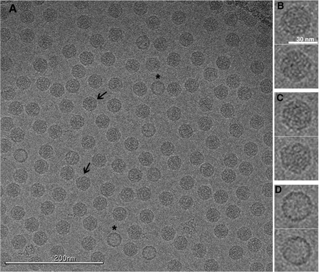
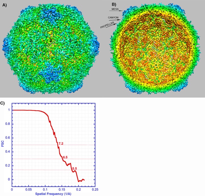
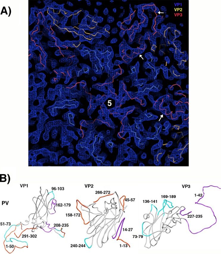
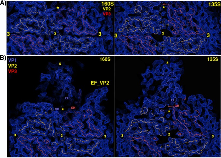
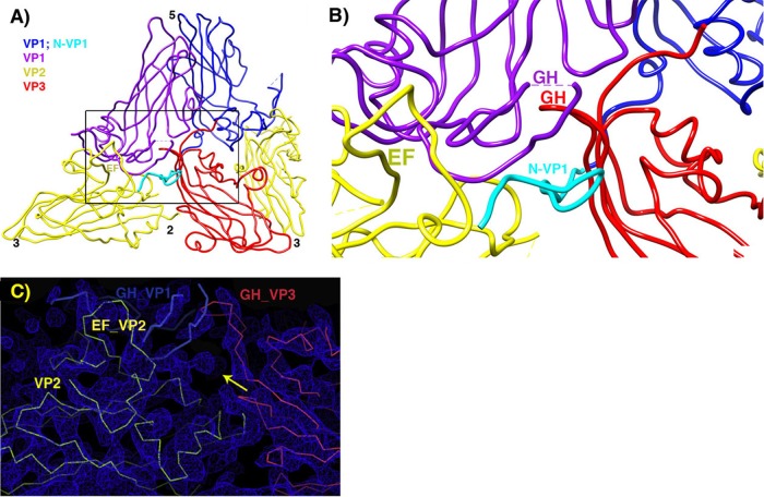
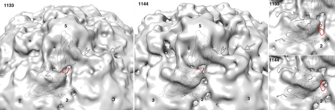
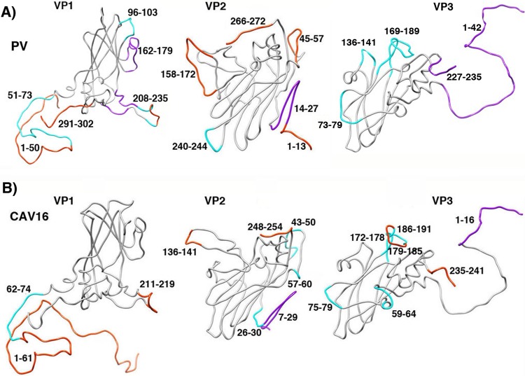
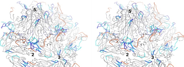
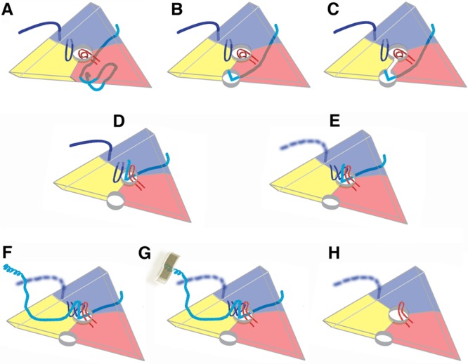
During infection, binding of mature poliovirus to cell surface receptors induces an irreversible expansion of the capsid, to form an infectious cell-entry intermediate particle that sediments at 135S. In these expanded virions, the major capsid proteins (VP1 to VP3) adopt an altered icosahedral arrangement to open holes in the capsid at 2-fold and quasi-3-fold axes, and internal polypeptides VP4 and the N terminus of VP1, which can bind membranes, become externalized. Cryo-electron microscopy images for 117,330 particles were collected using Leginon and reconstructed using FREALIGN. Improved rigid-body positioning of major capsid proteins established reliably which polypeptide segments become disordered or rearranged. The virus-to-135S transition includes expansion of 4%, rearrangements of the GH loops of VP3 and VP1, and disordering of C-terminal extensions of VP1 and VP2. The N terminus of VP1 rearranges to become externalized near its quasi-3-fold exit, binds to rearranged GH loops of VP3 and VP1, and attaches to the top surface of VP2. These details improve our understanding of subsequent stages of infection, including endocytosis and RNA transfer into the cytoplasm.


Figures

FIG 1
FIG 1
Cryo-electron micrograph showing a field of 135S particles in vitreous ice. The 135S particles are filled by RNA and, depending on orientation, can appear round (B) or angular (C and arrows in panel A). A small number of 80S empty capsids are present (D and asterisks in panel A). Scale bars are included. This representative CCD image showing 135S particles was recorded at −2.18-μm defocus and an ×62,000 nominal magnification, using a Tecnai F20 microscope (FEI).
FIG 2
FIG 2
Reconstruction of poliovirus 135S particles. (A and B) Surface rendering colored by radial position, showing the outer (A) and inner (B) surfaces. The map has been sharpened and displayed at high contour to emphasize higher-resolution details. In many places, individual polypeptide chains are resolved, which facilitates the rigid-body fitting of pseudoatomic models. Note the hole at the icosahedral 2-fold axis (center), where red-colored density is visible from the exterior. Labels indicate a mesa and a propeller tip, which are major projections from the outer surface. Separating the projections are canyons surrounding each 5-fold mesa and a saddle-shaped depression crossing the 2-fold axis. Symmetry axes are indicated by numbers. (C) A Fourier shell correlation (FSC) was calculated between randomly selected half-data sets. This curve suggests a resolution of ∼7 Å (at 0.5 correlation) and that there is meaningful information at ∼5.5 Å (the resolution at which FSC falls below 0.143).
FIG 3
FIG 3
The quality of the reconstructed density map makes rigid-body fitting of the major capsid proteins unambiguous. (A) Density features on the inner surface of the capsid are shown, with symmetry axes indicated by numbers and clipping planes applied for clarity. After rigid-body fitting of the major capsid proteins, segments of the backbone traces of VP1 (blue), VP2 (yellow), and VP3 (red) are clearly shape-similar to tubular features in the sharpened density map. Alpha-helices (arrows) are often well resolved and serve as fixed pivot points for positioning the beta sheets unambiguously. (B) Docked models from the crystal structure of mature (160S) poliovirus (PV) (PDB 1HXS) were compared with the reconstruction of 135S particles, in order to determine which polypeptide segments had become shifted (magenta), rearranged significantly (cyan), or disordered (orange) during virus expansion.
FIG 4
FIG 4
Upon poliovirus expansion, the topography of the outer surface changes dramatically. (A) A portion of the outer surface of the 135S reconstruction (right), viewed along a 2-fold axis, is compared with the corresponding density (left), calculated from the crystallographic model of mature poliovirus to 6 Å. Models of VP1, VP2, and VP3 are blue, yellow, and red, respectively, and symmetry axes are numbered. Shifts of the beta barrels cause a large hole to open at the 2-fold axis in 135S particles, as seen in a closeup comparison view. (B) The opening of a smaller hole at the quasi-3-fold (yellow asterisk) is associated with the shift or rearrangement nearby loops (as labeled). In the 135S reconstruction, in the upper right of the panel, the disordering of density for the VP2 propeller tip is dramatic.
FIG 5
FIG 5
Pseudoatomic model in the vicinity of the quasi-3-fold hole. (A) Overview, to provide context. (B) Zoomed-in view. VP2 is yellow, and VP3 is red. Two symmetry copies of VP1 are purple and blue, with the rebuilt N-terminal extension of the blue copy shown in cyan. Following the example of the crystal structure of the 135S-like particles of coxsackie virus A16, a short stretch of the N terminus of VP1 (labeled in cyan) forms a third beta strand alongside the two-stranded beta sheet (labeled in red) that is created by the rearrangement of the GH loop of VP3. The distal end of the GH loop of VP1 (labeled in purple) has become reoriented radially and leans rightward to contact the 3-stranded beta structure. Hypothetically, these three polypeptide segments may serve as a nucleus for building additional ordered structure atop the VP2 beta barrel, in order for the uncoating process to proceed further, upon membrane interaction. (C) In the vicinity of the quasi-3-fold axis, a section of the reconstruction shows that strong linear electron density features are present that correspond to proximal portions of the GH loops of VP3 (red) and VP1 (blue), which have both adopted an extended conformation that is rearranged relative to mature virus. The externalized N-terminal extension of VP1 extends toward the viewer through the indicated density feature (yellow arrow).
FIG 6
FIG 6
Lower-resolution maps validate the atomic model. To verify that the newly built conformation of the GH loop of VP1 is real and not an artifact of high-frequency noise, we displayed the pseudoatomic model, superimposed on two independent lower-resolution reconstructions of poliovirus 135S particles (EMDB entries 1133 and 1144) that were previously solved by Bubeck et al. at 10-Å resolution (17). Comfortably above the noise level, both 135S maps show extra density features (which are not present in 160S particles), located atop the VP2 beta barrel (arrow) and bridging across the canyon (open arrowhead). Importantly, these density features were more pronounced in the 1144 population of particles, though the two populations were produced using the same protocol and processed identically using PFT2 (42) and EM3DR2 (http://people.chem.byu.edu/belnap/). Observe that the 1144 map (middle panel) has a density projection that covers most of the newly built GH loop of VP1 (red). In contrast, coverage of the GH loop is poor in the 1133 map (left panel). For comparison (right), the 160S and 73S conformations of the VP1 GH loop (red), the VP1 C terminus (gray), and the tip of the EF loop of VP2 (gray) all clearly lie outside the isocontour surface in both maps.
FIG 7
FIG 7
The conformational changes seen in the expanded structures of other picornaviruses (such as the CAV16 135S particle [19]) (B) are less extensive than the ones seen in poliovirus 135S particles (A). Main chain traces from crystal structures of mature virions are shown for capsid proteins VP1, VP2, and VP3. The locations of polypeptide segments that become shifted, rearranged significantly, or disordered are colored magenta, cyan, and orange, respectively. The greater extent of changes in poliovirus 135S particles (A) might be the result of inherent lesser stability or simply greater heterogeneity in cryo-EM preparations, versus the preparations of expanded forms of other viruses that have been crystallized (15, 16, 19). In either case, our hypothesis is that much of the poliovirus population must be relatively further along the uncoating pathway. Panel A is Fig. 3B, repeated here for clarity.
FIG 8
FIG 8
Stereodiagram shows the location on the poliovirus surface of polypeptides that become disordered or rearranged during the 160S-to-135S expansion. A portion of the atomic model is shown. The parts of the structure that remain unchanged and are common in 160S and 135S particles are colored gray. Polypeptide segments in the 160S conformation that become disordered in the 135S particles are colored in orange, and those rearranged are shown in dark blue. Rearranged segments, as seen in 135S particles, are cyan. Observe that extensive changes occur in a continuous ring of altered polypeptides that surrounds the canyon, while the mesa shows few such changes. Rearranged polypeptides, displayed at high radius atop the VP2 beta barrel, may be poised for interaction with membranes and/or formation of the quasi-3-fold umbilicus (21) that is implicated in RNA transfer. Numbers indicate the positions of icosahedral symmetry axes.
FIG 9
FIG 9
A proposed sequence of polypeptide rearrangements during poliovirus uncoating is obtained by placing known picornavirus structures along a timeline. This simplified diagram represents a 5-3-3 icosahedral triangle, viewed from the outside of the virion, with the beta barrels of VP1, VP2, and VP3 shown in blue, yellow, and red, respectively. (A) In mature 160S virions, no hole is evident at the 2-fold axis, as capsid protein subunits fit together closely. The C terminus and GH loop of VP1 (both dark blue) bind atop VP2, on either side of the propeller. The quasi-3-fold hole (center) is blocked by the coiled GH loop of VP3 (red). The N terminus of VP1 (cyan) belongs to the symmetry-related VP1 whose beta barrel (not shown) is located to the right of the 5-3-3 triangle and binds to the inner surface of the capsid (2, 40). (B) In “breathing” expanded virions, subunits move apart reversibly. The N-terminal extensions of VP1 are temporarily exposed through the resulting 2-fold holes, where Fabs can trap them (39). (C) We surmise that the N-terminal extension of VP1 can shift from the 2-fold hole to the quasi-3-fold hole when the “gate” between the holes is open transiently. (D) The GH loop of VP3 (red) is rearranged to form a radially oriented hairpin. This opens the quasi-3-fold hole and, as seen in the structure reported here and in the coxsackievirus A16 135S structure (19), helps to form a binding site for the exposed N-terminal extension of VP1 to lock into place. At this 135S stage, RNA is still present, but VP4 has been released. (E) In poliovirus 135S particles, both the C terminus and the tips of the GH loop of VP1 (dark blue) become disordered. However, there is evidence in the density that at least some of the copies of the VP1 GH loop rearrange and bind to other rearranged polypeptides in the quasi-3-fold area, including the N terminus of VP1 and the exposed GH loop of VP3. This completely blocks the hole and may provide a nucleus for building asymmetric structures later on. (F) The long N terminus of VP1 migrates across the top of VP2. Its amphipathic helices, displayed at high radius at the propeller tip, are not well ordered and visible but are detectable using specific Fabs and V8-protease difference maps (23). This diagram represents the structure in most of the 60 icosahedrally related copies. (G) On the cell surface, one or two of the membrane-facing copies of the VP1 N terminus will bind to membrane. In our proposal, this tethering serves as a trigger for the asymmetric self-assembly of a large umbilical connection between virus and membrane, as previously visualized by Strauss et al. (21). The connector is believed to be involved in RNase-protected RNA transfer and most likely includes contributions from VP4 and various disordered N- and C-terminal extensions. (H) After some or all RNA exits, the expanded 80S empty capsid remains, showing 2-fold and quasi-3-fold holes. The GH loop of VP3 remains rearranged. In the absence of RNA and membranes, the N terminus of VP1 becomes disordered and retracted into the capsid interior (15, 16, 23). In the EV71 and HRV2 80S particles, the C terminus and GH loop of VP1 show a greater degree of order than they do in poliovirus (15, 16).

Similar articles

Cited by

References

    1. Tuthill TJ, Groppelli E, Hogle JM, Rowlands DJ. 2010. Picornaviruses. Curr. Top. Microbiol. Immunol. 343:43–89. 10.1007/82_2010_37 - DOI - PMC - PubMed
    1. Hogle JM, Chow M, Filman DJ. 1985. Three-dimensional structure of poliovirus at 2.9 A resolution. Science 229:1358–1365. 10.1126/science.2994218 - DOI - PubMed
    1. Hogle JM. 2002. Poliovirus cell entry: common structural themes in viral cell entry pathways. Annu. Rev. Microbiol. 56:677–702. 10.1146/annurev.micro.56.012302.160757 - DOI - PMC - PubMed
    1. Mendelsohn CL, Wimmer E, Racaniello VR. 1989. Cellular receptor for poliovirus: molecular cloning, nucleotide sequence, and expression of a new member of the immunoglobulin superfamily. Cell 56:855–865. 10.1016/0092-8674(89)90690-9 - DOI - PubMed
    1. Tsang SK, McDermott BM, Racaniello VR, Hogle JM. 2001. Kinetic analysis of the effect of poliovirus receptor on viral uncoating: the receptor as a catalyst. J. Virol. 75:4984–4989. 10.1128/JVI.75.11.4984-4989.2001 - DOI - PMC - PubMed

Publication types

Associated data

LinkOut - more resources