. 2014 Jan 17;289(3):1243-56.
doi: 10.1074/jbc.M113.522516. Epub 2013 Nov 20.

Overlapping and distinct roles of Aspergillus fumigatus UDP-glucose 4-epimerases in galactose metabolism and the synthesis of galactose-containing cell wall polysaccharides

Affiliations

Overlapping and distinct roles of Aspergillus fumigatus UDP-glucose 4-epimerases in galactose metabolism and the synthesis of galactose-containing cell wall polysaccharides

Mark J Lee et al. J Biol Chem. .

Abstract

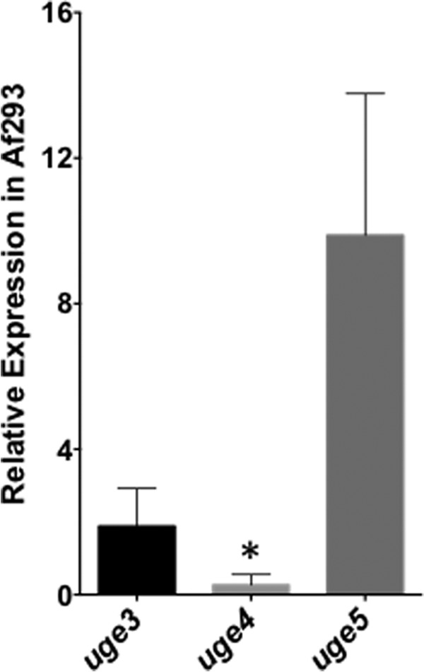
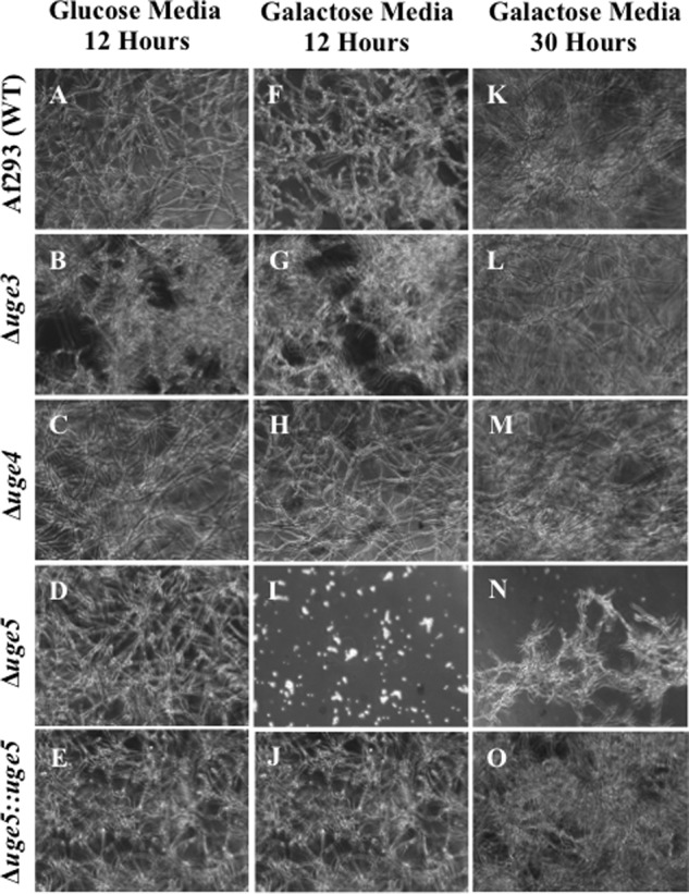
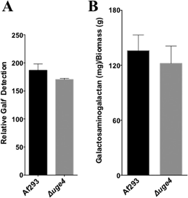
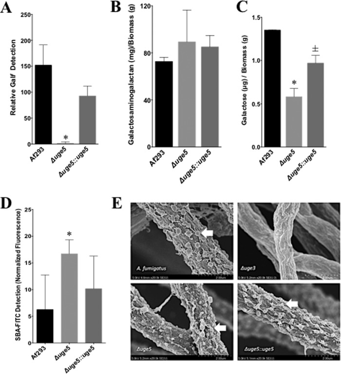
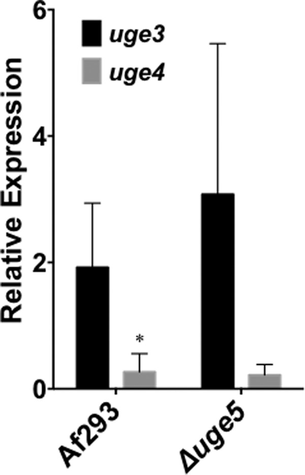
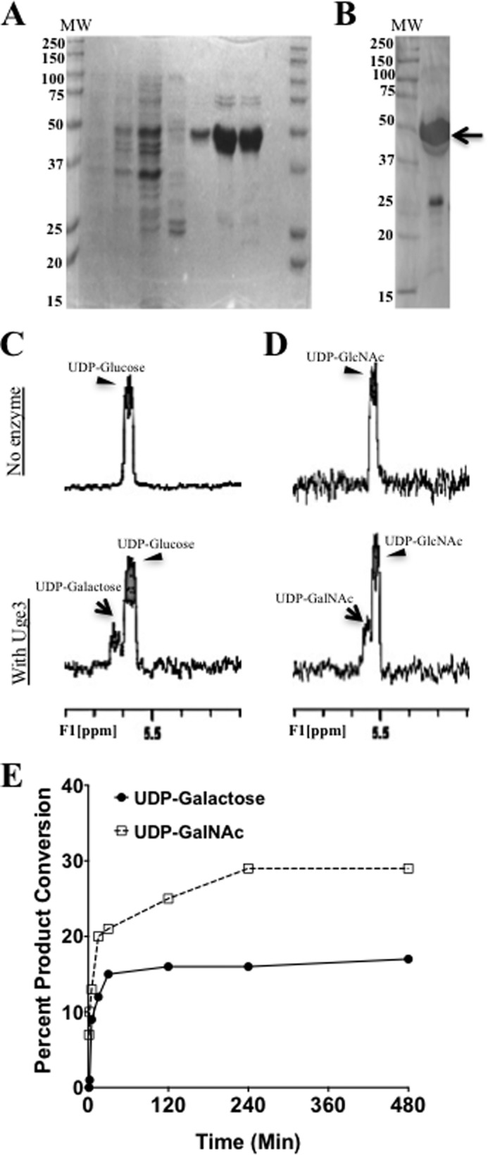
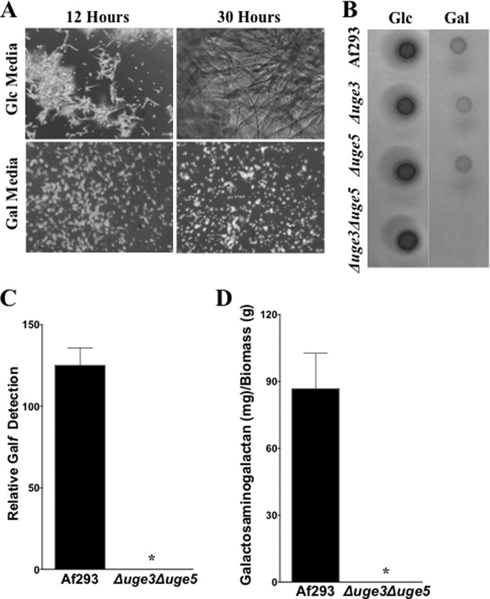
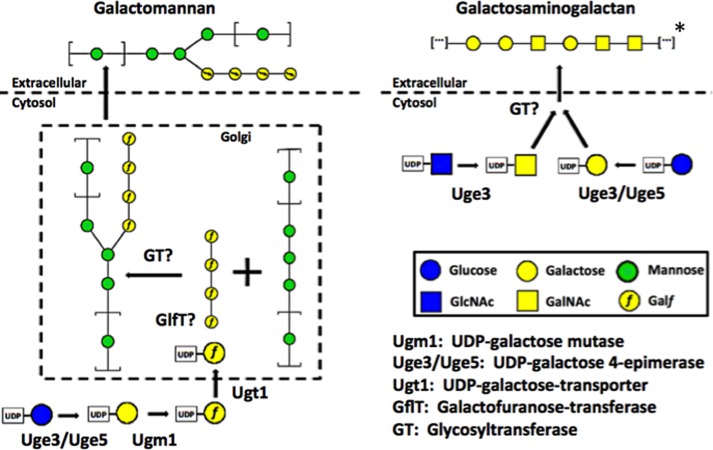
The cell wall of Aspergillus fumigatus contains two galactose-containing polysaccharides, galactomannan and galactosaminogalactan, whose biosynthetic pathways are not well understood. The A. fumigatus genome contains three genes encoding putative UDP-glucose 4-epimerases, uge3, uge4, and uge5. We undertook this study to elucidate the function of these epimerases. We found that uge4 is minimally expressed and is not required for the synthesis of galactose-containing exopolysaccharides or galactose metabolism. Uge5 is the dominant UDP-glucose 4-epimerase in A. fumigatus and is essential for normal growth in galactose-based medium. Uge5 is required for synthesis of the galactofuranose (Galf) component of galactomannan and contributes galactose to the synthesis of galactosaminogalactan. Uge3 can mediate production of both UDP-galactose and UDP-N-acetylgalactosamine (GalNAc) and is required for the production of galactosaminogalactan but not galactomannan. In the absence of Uge5, Uge3 activity is sufficient for growth on galactose and the synthesis of galactosaminogalactan containing lower levels of galactose but not the synthesis of Galf. A double deletion of uge5 and uge3 blocked growth on galactose and synthesis of both Galf and galactosaminogalactan. This study is the first survey of glucose epimerases in A. fumigatus and contributes to our understanding of the role of these enzymes in metabolism and cell wall synthesis.

Keywords: Carbohydrate Biosynthesis; Cell Wall; Galactose Metabolism; Glycobiology; Mycology; Polysaccharide.


Figures

FIGURE 1.
FIGURE 1.
uge5 gene is the most highly expressed of the three epimerase genes in A. fumigatus, whereas uge4 mRNA expression is barely detectable. Strain Af293 was grown in Brian medium for 18 h, and the levels of uge3, uge4, and uge5 mRNA were measured by real time RT-PCR. *, indicates significantly different expression as compared with tef1 reference gene (22).
FIGURE 2.
FIGURE 2.
Deletion of uge5 results in a partial galactose auxotrophy. The indicated strains were grown for the indicated time periods in Brian medium with either glucose (A–E) or galactose (F–O) as the sole carbon source. Bright field images at a magnification of ×200 are shown.
FIGURE 3.
FIGURE 3.
Deletion of uge4 does not alter galactosaminogalactan or Galf production. A, relative Galf detection by ELISA (EB-A2) in culture supernatant of Af293 or Δuge4 mutant after 72 h growth of indicated strains in Brian medium. B, galactosaminogalactan production by biomass from culture supernatant of Af293 or Δuge4 mutant after 72 h growth of the indicated strains in Brian medium.
FIGURE 4.
FIGURE 4.
Deletion of uge5 blocks Galf synthesis and results in production of galactosaminogalactan with a reduced galactose content. A, Galf content of culture supernatants as determined by ELISA. B, galactosaminogalactan content of culture supernatants, normalized to mycelia biomass. C, galactose content of galactosaminogalactan from the indicated strains as detected by gas chromatography followed by hexose and hexosamine quantification. D, FITC-tagged GalNAc-specific soybean agglutinin (SBA) lectin binding on 12-h, Brian medium-grown hyphae of indicated strains. Total fluorescence was measured with Spectramax® fluorescence microplate reader. E, scanning electron micrograph of hyphae of indicated strains after 24 h of growth at 37 °C, 5% CO2 in phenol-free RPMI 1640. Hyphae were fixed, sequentially dehydrated in ethanol, dried in CO2, coated in Pd-Au, and imaged under scanning electron microscope (Hitachi). Arrows indicate surface decorations associated with galactosaminogalactan production. *, significant reduction compared with Af293 wild-type, analysis of variance with pairwise comparison p < 0.05. ±, not statistically significant compared with Af293 but statistically significant compared with the Δuge5 mutant, p < 0.05. A–C, indicated strains were grown for 72 h in Brian medium.
FIGURE 5.
FIGURE 5.
Deletion of uge5 is not associated with significant up-regulation of expression of uge3 or uge4. The indicated strains were grown in Brian medium for 18 h, and the levels of uge3 (black bar) and uge4 (gray bar) mRNA from indicated strains were measured by real time RT-PCR. *, significant difference between uge3 and uge4 expression in the indicated strain as compared with tef1 reference gene, factorial analysis of variance with pairwise comparison, p < 0.05.
FIGURE 6.
FIGURE 6.
Uge3 and Uge5 are cytoplasmic. Af293 strains expressing uge3-gfp or uge5-rfp were grown in Brian medium for 12 h and imaged by confocal microscopy. For nuclear staining, Draq5® stain was used, and pseudocolor blue was red fluorescent protein at 543 nm, and Draq5® at 633 nm.
FIGURE 7.
FIGURE 7.
Substrate-binding pocket of Uge3 model aligns with hGalE. The hexagonal substrate-binding pocket of Uge3 model (green) was aligned to hGalE (cyan), with the six residues required for catalytic activity annotated. For Uge3 homology modeling, the N-terminal nonalignment regions were discarded, and resulting aligned sequences were modeled using Modeler Version 9.11. Bound UDP-GalNAc and NAD+ are indicated in gray at top and bottom, respectively.
FIGURE 8.
FIGURE 8.
Uge3 exhibits bifunctional UDP-Glc/UDP-Gal and UDP-GlcNAc/UDP-GalNAc epimerase activity. A, SDS-PAGE of lysate and purified His6-Uge3 stained with Commassie Blue from His6-Uge3 expressing BL21(DE) E. coli strain. Lanes are as follows: noninduced cells; cells grown in auto-induction medium; crude lysate; wash fraction eluted using 20 mm imidazole; and four fractions of His6-Uge3 eluted with 250 mm imidazole. B, Western blot of purified lysates for detection of His6-Uge3 using HRP-tagged anti-His6 antibody. Arrow indicates expected band for His6-Uge3. C and D, 1H NMR spectra were measured in a reaction mix containing either UDP-glucose (C) or UDP-GlcNAc (D) in phosphate buffer, in the presence or absence of Uge3. For all NMR experiments, products were detected after 1 h of co-incubation of Uge3 and respective substrates at 37 °C using Varian 500 MHz NMR spectroscopy. E, rate of product formation was measured using capillary electrophoresis over time in a reaction mix containing 20 pmol of Uge3 and 0.1 mm of either UDP-glucose or UDP-GlcNAc as substrate. Reactions took place at 37 °C in a total volume of 10 μl. Products were detected at 254 nm (UV) measuring UDP-moiety endogenous fluorescence. UDP-linked sugars were separated by borate adduct formation.
FIGURE 9.
FIGURE 9.
Deletion of uge3 and uge5 renders the resulting Δuge3Δuge5 double mutant deficient in Galf and galactosaminogalactan. A, indicated strains were grown for the indicated time periods in Brian medium using either glucose or galactose as the sole carbon source. Brightfield images at a magnification of 200x are shown. Glucose is abbreviated as Glc, and galactose as Gal. B, indicated strains were grown for 2 days on either glucose or galactose based Brian medium agar plates. C, relative Galf detection by ELISA in culture supernatant after 72 h growth in Brian medium of the indicated strains. D, galactosaminogalactan production after 72 h growth in Brian medium of the indicated strains. *, significant reduction compared with Af293 wild type and analysis of variance with pairwise comparison, p < 0.05.
FIGURE 10.
FIGURE 10.
Galactose-poor Δuge5 galactosaminogalactan retains normal virulence-associated functions. A, biofilm adherence of indicated strains after 24 h of growth on polystyrene plates, after several washes, and visualized by staining with crystal violet. B, adherence of germinated hyphae of the indicated strains to A549 epithelial cells after 30 min. C, detection of Fc-dectin-1 binding using immune staining. Conidia from respective strains were grown for 12 h in Brian medium, fixed, blocked, stained with Fc-dectin-1, and FITC-labeled F(ab) fragment, and total fluorescence was measured with Spectramax® fluorescence microplate reader. D, induction of bone marrow-derived dendritic cell apoptosis as determined by caspase-3 activity. Conidia from indicated strains were grown for 9 h in RPMI 1640 medium and then co-incubated with mouse bone marrow-derived dendritic cells at a multiplicity of infection of 10:1. Caspase-3 activity was measured by commercial assay following the manufacturer's instructions (Invitrogen).
FIGURE 11.
FIGURE 11.
Schematic of galactosaminogalactan and galactomannan pathways. Pathway diagrams showing common and distinct components in the biosynthesis of galactosaminogalactan and galactomannan. For Galf symbol representation, the “f” has been inserted to the galactose pyranose symbol. All other symbols follow common nomenclature convention (54). *, note that the depiction of galactosaminogalactan structure is representative of multiple potential combinations of galactose and GalNAc residues.

Similar articles

Cited by

References

    1. Garcia-Vidal C., Upton A., Kirby K. A., Marr K. A. (2008) Epidemiology of invasive mold infections in allogeneic stem cell transplant recipients: biological risk factors for infection according to time after transplantation. Clin. Infect. Dis. 47, 1041–1050 - PMC - PubMed
    1. Ostrosky-Zeichner L., Casadevall A., Galgiani J. N., Odds F. C., Rex J. H. (2010) An insight into the antifungal pipeline: selected new molecules and beyond. Nat. Rev. Drug Discov. 9, 719–727 - PubMed
    1. Latgé J.-P. (2010) Tasting the fungal cell wall. Cell. Microbiol. 12, 863–872 - PubMed
    1. Latge J. P. (2009) Galactofuranose containing molecules in Aspergillus fumigatus. Med. Mycol. 47, S104–S109 - PubMed
    1. Latgé J. P., Kobayashi H., Debeaupuis J. P., Diaquin M., Sarfati J., Wieruszeski J. M., Parra E., Bouchara J. P., Fournet B. (1994) Chemical and immunological characterization of the extracellular galactomannan of Aspergillus fumigatus. Infect. Immun. 62, 5424–5433 - PMC - PubMed

Publication types

MeSH terms