. 2014 Jan 3;289(1):1-12.
doi: 10.1074/jbc.M113.512368. Epub 2013 Nov 20.

CD4 and BST-2/tetherin proteins retro-translocate from endoplasmic reticulum to cytosol as partially folded and multimeric molecules

Affiliations

CD4 and BST-2/tetherin proteins retro-translocate from endoplasmic reticulum to cytosol as partially folded and multimeric molecules

Gianluca Petris et al. J Biol Chem. .

Abstract

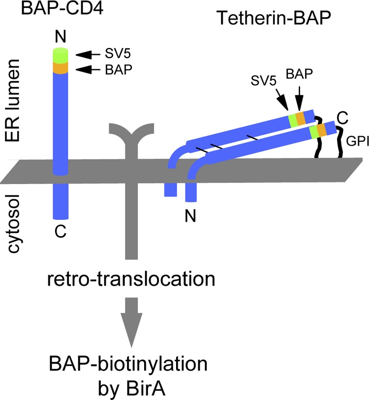
CD4 and BST-2/Tetherin are cellular membrane proteins targeted to degradation by the HIV-1 protein Vpu. In both cases proteasomal degradation following recruitment into the ERAD pathway has been described. CD4 is a type I transmembrane glycoprotein, with four extracellular immunoglobulin-like domains containing three intrachain disulfide bridges. BST-2/Tetherin is an atypical type II transmembrane glycoprotein with an N-terminal transmembrane domain and a C-terminal glycophosphatidylinositol anchor, which dimerizes through three interchain bridges. We investigated spontaneous and Vpu-induced retro-translocation of CD4 and BST-2/Tetherin using our novel biotinylation technique in living cells to determine ER-to-cytosol retro-translocation of proteins. We found that CD4 retro-translocates with oxidized intrachain disulfide bridges, and only upon proteasomal inhibition does it accumulate in the cytosol as already reduced and deglycosylated molecules. Similarly, BST-2/Tetherin is first exposed to the cytosol as a dimeric oxidized complex and then becomes deglycosylated and reduced to monomers. These results raise questions on the required features of the putative retro-translocon, suggesting alternative retro-translocation mechanisms for membrane proteins in which complete cysteine reduction and unfolding are not always strictly required before ER to cytosol dislocation.

Keywords: BST-2; Biotin; Biotinylation; CD4; Disulfide; ER-associated Degradation; ERAD; Oxidation-Reduction; Retro-translocation; Tetherin.


Figures

FIGURE 1.
FIGURE 1.
Scheme of CD4 and Tetherin tagged with BAP in ER-luminal positions. The 11-amino acid-long SV5 tag is also shown. Only retro-translocated BAP-tagged molecules are biotinylated by cytosolic BirA (cyt-BirA).
FIGURE 2.
FIGURE 2.
Biotinylation of BAP-tagged CD4. A, WB-ra of BAP-tagged CD4. Extracts from HEK293T cells were co-transfected with BAP-CD4 and, where indicated, Vpu in the absence or presence of MG132 (5 μm for 12 h), and developed with anti-SV5. Biotinylated molecules appear as retarded bands when run with StrAv. B, glycosylation status of biotinylated CD4. WB shows b-CD4 from extracts of cells co-expressing Vpu in the presence of MG132 (5 μm for 12 h), immunoprecipitated with anti-SV5, treated with Endo-H or PNGase, and developed with HRP-conjugated StrAv. C, BirA lack of interference with CD4 retro-translocation. WB (developed with anti-SV5) shows BAP-CD4 from cells co-expressing Vpu, treated with MG132 (10 μm) for 4 h in the presence or absence of cyt-BirA. The deglycosylated band (de-glyc) is indicated. D, [35S]methionine labeling. Cells co-transfected with CD4 and cyt-BirA and with or without Vpu, were [35S]methionine-labeled for 15 min in the presence of MG132 (10 μm). CD4 was then immunoprecipitated (IP) with anti-SV5, run in a reducing PAGE retardation assay, and developed by autoradiography. Filled arrowheads indicate deglycosylated material, whereas open arrowheads indicate nonbiotinylated CD4 isoforms (observed in the presence of Vpu). E, trypsin-sensitivity assay. WB-ra of microsome-containing lysates (microsomes) derived from cells expressing BAP-CD4 and Vpu and treated with MG132 (5 μm) for 12 h before lysis, were incubated with (+) or without (−) trypsin for 1 h at 37 °C. Nonidet P-40 indicates the same microsomes-containing lysates treated with 0.5% Nonidet P-40 to make ER luminal proteins accessible to trypsin. Open arrowhead indicates CD4 with trypsin-digested cytosolic C-terminal tail; filled arrowhead indicates deglycosylated CD4. F, plasma membrane CD4. Membrane-displayed CD4 in MG132-treated cells was immunoprecipitated with anti-SV5 and analyzed in a WB-ra developed with anti-SV5. G, cellular fractionation. WB-ra shows total lysates of cells co-transfected with BAP-CD4 and cyt-BirA (tot) and of the pellet (ER) and supernatant (cytosolic, cyt) fractions obtained by ultracentrifugation. Calnexin and actin were used as ER and cytosol markers, respectively. WB of anti-EGFP was performed as loading and transfection control. Filled arrowheads indicate deglycosylated CD4.
FIGURE 3.
FIGURE 3.
Biotinylation of BAP-tagged Tetherin. A, WB-ra of BAP-tagged Tetherin. Extracts from HEK293T cells were co-transfected with Tetherin-BAP and, where indicated, Vpu in the absence or presence of MG132 (5 μm for 12 h) and developed with anti-SV5. Open and filled arrowheads indicate ER-like glycosylated (ER-glyc) and deglycosylated (de-glyc) Tetherin, respectively, whereas open and filled arrows indicate the corresponding biotinylated (retarded) fractions. B, plasma membrane displayed Tetherin in MG132-treated cells. Membrane-displayed Tetherin was immunoprecipitated with anti-SV5, and the resulting material was digested with Endo-H or PNGase and analyzed in a WB-ra developed with anti-SV5. Cells were co-transfected with Vpu. C, biotinylation of glycosylated and deglycosylated Tetherin. WB-ra shows lysates from cells expressing Tetherin treated with MG132 (5 μm for 12 h). Lysates were treated with Endo-H or PNGase, and the blot was developed with anti-SV5. Open and filled arrowheads and arrows are as in A. D, cyt-BirA biotinylating only retro-translocated molecules. WB-ra shows extracts from cells co-expressing Tetherin-BAP and a scFv BAP-tagged control protein, together with cyt-BirA and treated with MG132 (5 μm) for 12 h. Tetherin was detected with anti-SV5 and the scFv with the anti-roTag. E, cellular fractionation. WB-ra shows total lysates (tot) of cells co-transfected with Tetherin-BAP and cyt-BirA and treated with MG132 for 12 h before lysis and of the pellet (ER) and supernatant (cytosolic, cyt) fractions obtained by ultracentrifugation. Derlin1 and actin were used as ER and cytosolic markers, respectively. F, biotinylation as a non-postlysis event. Cells expressing only Tetherin-BAP (treated with 5 μm MG132 for 12 h) were mixed with cells co-transfected with cyt-BirA and a C-terminal BAP-tagged MHC-Iα (in its cytosolic tail) before mechanical lysis as performed in the cell fractionation experiments. Postnuclear supernatants corresponding to microsomal containing lysates were analyzed in WB-ra. Tetherin was revealed with anti-SV5 and MHC-Iα with anti-roTag. G, WB-ra shows lysates derived from Tetherin-BAP-transfected cells treated with MG132 (10 μm) or chloroquine (50 μm) for 4 h, as indicated. WB of anti-EGFP was used as loading and transfection control.
FIGURE 4.
FIGURE 4.
Cytosolic exposure of CD4 and Tetherin luminal domains. A, scheme of CD4 and Tetherin with the BAP tag positioned, in both cases, proximal to the transmembrane domains. The position of the SV5 tag is also shown. B, WB-ra of membrane-proximal BAP-tagged CD4 (CD4-BAP). Lysates were obtained from cells co-transfected with or without Vpu and in absence or presence of MG132 (5 μm for 12 h). C, WB-ra of membrane-proximal BAP-tagged Tetherin (BAP-Teth). Lysates were obtained from cells co-transfected with or without Vpu in the presence of MG132 (5 μm for 12 h.) D, quantification of retro-translocated fractions of membrane-proximal and membrane-distal BAP-tagged molecules. Comparison shows relative levels of retro-translocated CD4 (left panel) and Tetherin (right panel) for molecules with the BAP tag in membrane-distal or membrane-proximal position, co-expressed with or without Vpu (in all cases in the presence of MG132, 10 μm for 4 h), expressed as percentage of total protein. In both panels colors represent different experiments.
FIGURE 5.
FIGURE 5.
Retro-translocation of CD4 with oxidized disulfide bonds. A, alkylation of CD4-free cysteines. Cells co-transfected with BirA, BAP-CD4, and Vpu were incubated for 5 min at 4 °C in PBS in the presence or absence of 20 mm NEM and rinsed in SDS-lysis buffer. Samples were then run in reducing SDS-PAGE, blotted, and developed with anti-SV5. B, nonreducing and reducing WB-ra of BAP-tagged CD4. Lysates from cells co-transfected with Vpu and treated with MG132 (5 μm for 12 h) were run in nonreducing (left) and reducing (right) conditions and developed with anti-SV5. Biotinylated and nonbiotinylated fractions of glycosylated and deglycosylated CD4 and their oxidation status are indicated with corresponding open and filled arrowheads and arrows. C, WB of biotinylated BAP-tagged CD4. CD4 was immunoprecipitated (IP) with anti-SV5 from cells expressing Vpu in the presence of MG132 (5 μm for 12 h), analyzed in nonreducing (left) and reducing (right) conditions, and developed with HRP-conjugated StrAv. CD4 glycosylation and oxidation status are indicated with open and filled arrows. D, ER-like glycosylation of biotinylated BAP-tagged CD4. WB shows reducing conditions of CD4 immunoprecipitated with anti-SV5 from cells expressing Vpu in the presence or absence of MG132 (5 μm for 12 h) and treated with or without Endo-H. Blot was developed with HRP-conjugated StrAv. E, [35S]methionine labeling of biotinylated CD4. Biotinylated CD4 fraction was immunoprecipitated from cells expressing Vpu after 2-h labeling with [35S]methionine in the presence of MG132 (10 μm), first with anti-SV5 and then with StrAv-conjugated beads, and analyzed in nonreducing (left) and reducing (right) conditions and developed by autoradiography. The glycosylation and oxidation status of biotinylated CD4 bands is indicated.
FIGURE 6.
FIGURE 6.
Retro-translocation of dimeric Tetherin. A, nonreducing and reducing WB-ra of BAP-tagged Tetherin. Lysates from cells co-transfected with or without Vpu in the absence and presence of MG132 (5 μm for 12 h) were run in nonreducing (left) and reducing (right) conditions and developed with anti-SV5. Arrowheads indicate the deglycosylated fractions of Tetherin monomers and dimers. B and C, bidimensional (nonreducing/reducing) analysis of Tetherin. Lysates from cells co-expressing Tetherin and BirA treated (C) or not treated (B) with MG132 (5 μm for 12 h) were analyzed in a two-dimensional (nonreducing/reducing) WB developed with anti-SV5 or HRP-conjugated StrAv, as indicated. The uppermost panels show the same sample after the first (nonreducing) dimension. Vertical lanes show the same sample separated only through the reducing dimension. The different Tetherin glycosylation species of monomers and dimers are indicated. D, Z-VAD-fmk treatment confirming the presence of deglycosylated Tetherin dimers. Nonreducing WB of Tetherin from cells treated with MG132 (10 μm) and the PNGase inhibitor Z-VAD-fmk (100 μm) for 4 h is indicated. To better observe dimeric and monomeric glycosylation isoforms, diverse exposure times from the same gel are shown. Boxes highlight Tetherin molecules accumulated during proteasomal inhibition, which were sensitive to the concomitant presence of the PNGase inhibitor. E, [35S]methionine pulse-chase labeling. Cells co-transfected with Tetherin-BAP were labeled with a 15-min pulse of [35S]methionine and then chased for 2 h. MG132 (10 μm) was present from the beginning of the pulse. Tetherin was immunoprecipitated (IP) with anti-SV5, run in a nonreducing PAGE retardation assay, and developed by autoradiography. F, nonreducing WB-ra of cell extracts form cells expressing WT or N65A,N92A Tetherin mutant after MG132 treatment (10 μm, 4 h) is shown. The nonglycosylated monomer and the shifted biotinylated monomer and dimers are indicated. G, alkylation of Tetherin-free cysteines. Cells co-transfected with cyt-BirA and Tetherin-BAP were incubated 5 min at 4 °C in PBS in the presence or absence of 20 mm NEM and lysed in SDS-lysis buffer. Samples were run in nonreducing WB-ra and developed with anti-SV5. H, Tetherin ectodomain oxidation state. Extracts from cells transfected with full-length Tetherin (Teth), the cytosolically expressed Tetherin ectodomain (cyt-ecto), or the ER/secretory version (sec-ecto) of the same domain were analyzed in reducing and nonreducing WBs developed with anti-SV5.

Similar articles

Cited by

References

    1. Ghaemmaghami S., Huh W. K., Bower K., Howson R. W., Belle A., Dephoure N., O'Shea E. K., Weissman J. S. (2003) Global analysis of protein expression in yeast. Nature 425, 737–741 - PubMed
    1. Rapoport T. A. (2007) Protein translocation across the eukaryotic endoplasmic reticulum and bacterial plasma membranes. Nature 450, 663–669 - PubMed
    1. Vembar S. S., Brodsky J. L. (2008) One step at a time: endoplasmic reticulum-associated degradation. Nat. Rev. Mol. Cell Biol. 9, 944–957 - PMC - PubMed
    1. Afshar N., Black B. E., Paschal B. M. (2005) Retrotranslocation of the chaperone calreticulin from the endoplasmic reticulum lumen to the cytosol. Mol. Cell. Biol. 25, 8844–8853 - PMC - PubMed
    1. Geiger R., Andritschke D., Friebe S., Herzog F., Luisoni S., Heger T., Helenius A. (2011) BAP31 and BiP are essential for dislocation of SV40 from the endoplasmic reticulum to the cytosol. Nat. Cell Biol. 13, 1305–1314 - PubMed

Publication types

MeSH terms

Substances