. 2014 Jan 3;289(1):335-45.
doi: 10.1074/jbc.M113.479386. Epub 2013 Nov 20.

Metformin suppresses expression of the selenoprotein P gene via an AMP-activated kinase (AMPK)/FoxO3a pathway in H4IIEC3 hepatocytes

Affiliations

Metformin suppresses expression of the selenoprotein P gene via an AMP-activated kinase (AMPK)/FoxO3a pathway in H4IIEC3 hepatocytes

Hiroaki Takayama et al. J Biol Chem. .

Abstract

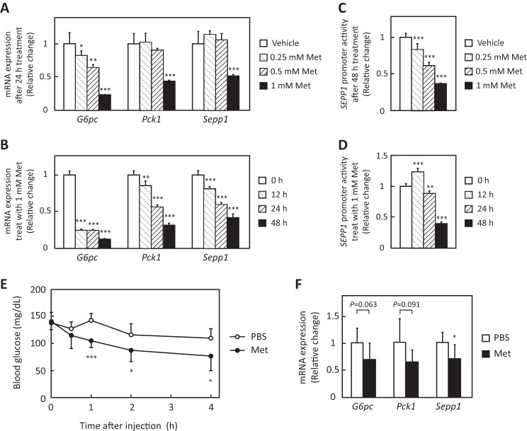
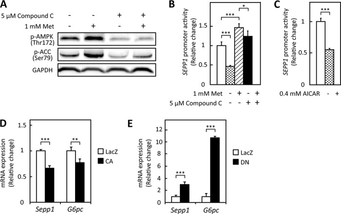
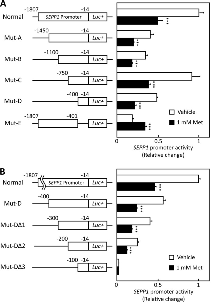
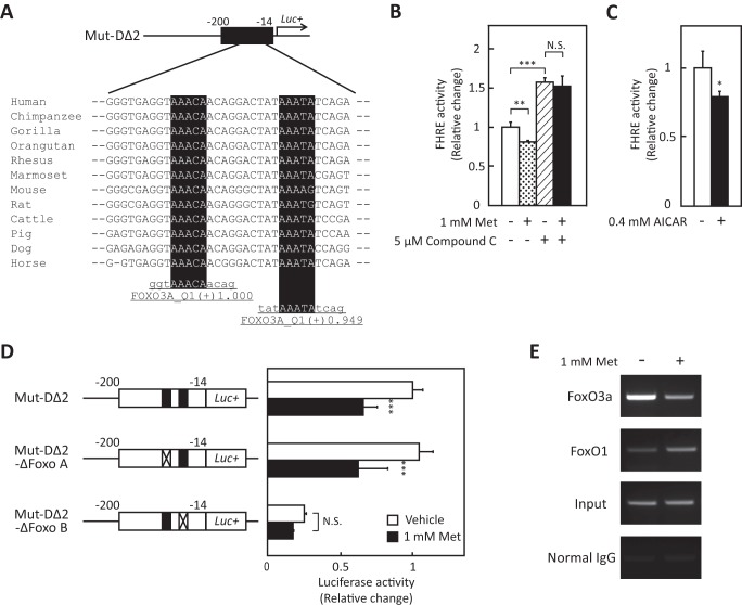
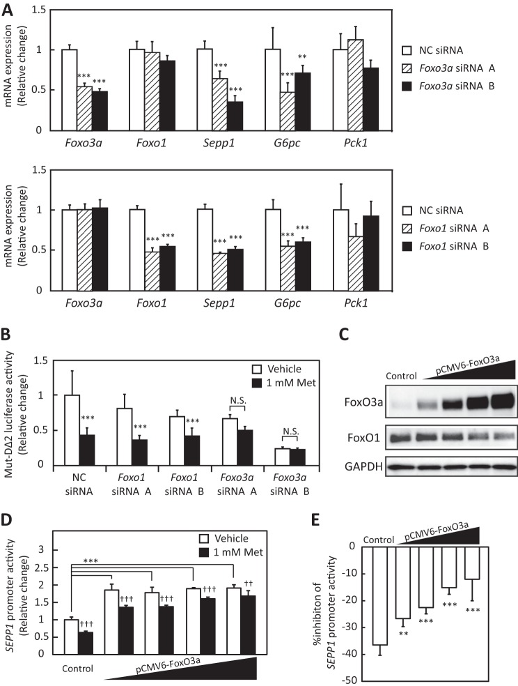
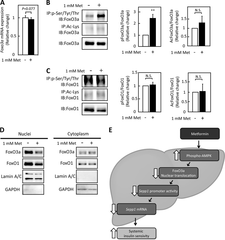
Selenoprotein P (SeP; encoded by SEPP1 in humans) is a liver-derived secretory protein that induces insulin resistance in type 2 diabetes. Suppression of SeP might provide a novel therapeutic approach to treating type 2 diabetes, but few drugs that inhibit SEPP1 expression in hepatocytes have been identified to date. The present findings demonstrate that metformin suppresses SEPP1 expression by activating AMP-activated kinase (AMPK) and subsequently inactivating FoxO3a in H4IIEC3 hepatocytes. Treatment with metformin reduced SEPP1 promoter activity in a concentration- and time-dependent manner; this effect was cancelled by co-administration of an AMPK inhibitor. Metformin also suppressed Sepp1 gene expression in the liver of mice. Computational analysis of transcription factor binding sites conserved among the species resulted in identification of the FoxO-binding site in the metformin-response element of the SEPP1 promoter. A luciferase reporter assay showed that metformin suppresses Forkhead-response element activity, and a ChIP assay revealed that metformin decreases binding of FoxO3a, a direct target of AMPK, to the SEPP1 promoter. Transfection with siRNAs for Foxo3a, but not for Foxo1, cancelled metformin-induced luciferase activity suppression of the metformin-response element of the SEPP1 promoter. The overexpression of FoxO3a stimulated SEPP1 promoter activity and rescued the suppressive effect of metformin. Metformin did not affect FoxO3a expression, but it increased its phosphorylation and decreased its nuclear localization. These data provide a novel mechanism of action for metformin involving improvement of systemic insulin sensitivity through the regulation of SeP production and suggest an additional approach to the development of anti-diabetic drugs.

Keywords: AMP-activated Kinase (AMPK); FoxO3a; Gene Expression; Hepatocyte; Hepatokine; Insulin Resistance; Metformin; Selenoprotein P; Transcription Promoter.


Figures

FIGURE 1.
FIGURE 1.
Metformin suppressed Sepp1 gene expression in H4IIEC3 hepatocytes and livers of C57BL/6J mice. A and B, metformin suppressed Sepp1 mRNA expression in a concentration- and time-dependent manner. H4IIEC3 cells were treated with the indicated concentrations of metformin for the indicated times. Expression values were normalized to Actb mRNA. Data represent means ± S.D. (error bars) (n = 4). *, p < 0.05; **, p < 0.01; ***, p < 0.001 versus vehicle-treated cells or 0 h. C and D, SEPP1 promoter activity was suppressed in a concentration- and time-dependent manner. H4IIEC3 cells were co-transfected with the SEPP1 promoter reporter vector and control reporter vector. 24 h later, the cells were treated with the indicated concentrations of metformin for the indicated times. Values were normalized to the activity of the control luciferase vector. Data represent means ± S.D. (n = 4). **, p < 0.01; ***, p < 0.001 versus vehicle-treated cells or 0 h. E and F, metformin suppressed Sepp1 mRNA expression in livers of C57BL/6J mice. Following fasting for 4 h, 12-week-old female C57BL/6J mice were administrated 300 mg/kg metformin. 4 h after metformin administration, mice were sacrificed, and liver mRNA expression was examined. Expression values were normalized to Actb mRNA. Data represent means ± S.D. (n = 7). *, p < 0.05 versus PBS-injected mice.
FIGURE 2.
FIGURE 2.
Metformin suppressed SEPP1 promoter activity via AMPK pathway in H4IIEC3 hepatocytes. A, metformin-induced AMPK phosphorylation in the absence or presence of compound C. H4IIEC3 cells were treated with the indicated concentrations of metformin and compound C for 24 h. AMPK phosphorylation was examined by Western blotting. B, compound C treatment recovered metformin-induced suppression of the SEPP1 promoter. H4IIEC3 cells were co-transfected with the SEPP1 promoter reporter vector and control reporter vector at 24 h and then treated with the indicated concentrations of metformin and compound C for 48 h. Signals were normalized to the control reporter vector. Data represent means ± S.D. (error bars) (n = 4). ***, p < 0.001. C, AICAR suppressed SEPP1 promoter activity. H4IIEC3 cells were co-transfected with the SEPP1 promoter reporter vector and control reporter vector at 24 h and then treated with 0.4 mm AICAR for 24 h. Signals were normalized to the control reporter vector. Data represent means ± S.D. (n = 4). ***, p < 0.001. D and E, influence of adenoviruses carrying constitutive active (CA) or dominant negative (DN) AMPK. H4IIEC3 cells were infected with adenoviruses encoding CA-AMPK, DN-AMPK, or LacZ. Expression values were normalized to Actb mRNA. Data represent means ± S.D. (n = 4). **, p < 0.01; ***, p < 0.001.
FIGURE 3.
FIGURE 3.
SEPP1 promoter activity of deletion mutants. A and B, structure and luciferase activity of promoter-deletion mutants. The sequences deleted within the constructs are shown as thin lines. The remaining parts of the SEPP1 promoter were fused to a luciferase reporter gene. H4IIEC3 cells were co-transfected with each reporter vector and control reporter vector at 24 h and then treated with the indicated concentrations of metformin for 48 h. Signals were normalized to the control reporter vector. Data represent means ± S.D. (error bars) (n = 4). ***, p < 0.001 versus vehicle-treated cells.
FIGURE 4.
FIGURE 4.
Activation of the AMPK suppressed FoxO activity. A, putative FoxO3a binding sites of Mut-DΔ2 sequence. Detection of the conserved TFBSs was performed using multiple-genome alignments and the highlighted putative transcriptional factor binding sites. B, FoxO activity in the absence or presence of metformin and compound C. H4IIEC3 cells were co-transfected with the FHRE-Luc vector and control reporter vector at 24 h and then treated with the indicated concentrations of metformin and compound C for 48 h. Signals were normalized to the control reporter vector. Data represent means ± S.D. (error bars) (n = 4). **, p < 0.01; ***, p < 0.001. N.S., not significant. C, FoxO activity in the absence or presence of AICAR. H4IIEC3 cells were co-transfected with the FHRE-Luc vector and control reporter vector at 24 h and then treated with the indicated concentrations of AICAR for 24 h. Signals were normalized to the control reporter vector. Data represent means ± S.D. (n = 4). *, p < 0.05 versus vehicle-treated cells. D, deficiency of putative FoxO binding site cancelled metformin-induced suppression of SEPP1 promoter activity. H4IIEC3 cells were co-transfected with each reporter vector and control reporter vector at 24 h and then treated with the indicated concentrations of metformin for 24 h. Signals were normalized to the control reporter vector. Data represent means ± S.D. (n = 4). ***, p < 0.001 versus vehicle-treated cells. E, chromatin immunoprecipitation assay of HepG2 cells treated with metformin. HepG2 cells were treated with metformin for 6 h. Chromatin samples precipitated with anti-FoxO3a, anti-FoxO1, or normal IgG were amplified using primers for the Mut-DΔ2 region of the human SEPP1 promoter.
FIGURE 5.
FIGURE 5.
Metformin suppressed SeP expression via FoxO3a. A, efficiency of Foxo3a siRNA and Foxo1 siRNA. H4IIEC3 cells were transfected with Foxo3a siRNAs or Foxo1 siRNAs or a negative control (NC) siRNA at 48 h. Knockdown efficiency was assessed by real-time PCR. Expression values were normalized to Actb mRNA. Data represent means ± S.D. (error bars) (n = 4). **, p < 0.01; ***, p < 0.001 versus negative control siRNA-treated cells. B, luciferase activity of Mut-DΔ2 treated with Foxo3a or Foxo1 siRNA and metformin. H4IIEC3 cells were transfected with Foxo3a siRNAs or Foxo1 siRNAs or negative control siRNA at 24 h and then co-transfected with Mut-DΔ2 vector and control reporter vector. 24 h after transfection, cells were treated with the indicated concentrations of metformin for 24 h. Signals were normalized to the control reporter vector. Data represent means ± S.D. (n = 4). ***, p < 0.001 versus vehicle-treated cells. N.S., not significant. C, protein levels in the presence of the FoxO3a overexpression vector. H4IIEC3 cells were transfected with the pCMV-FoxO3a vector or pCMV empty vector at 24 h. FoxO3a protein levels were then assessed by Western blotting. D, SEPP1 promoter activity transfected with the FoxO3a overexpression vector. H4IIEC3 cells were co-transfected with the expression vectors for FoxO3a, SEPP1 promoter reporter and control reporter at 24 h and then treated with the indicated concentrations of metformin for 48 h. Data represent means ± S.D. (n = 3–4). ***, p < 0.001 versus control; ††, p < 0.01; †††, p < 0.001 versus vehicle-treated cells. E, percentage inhibition of SEPP1 promoter activity by metformin. Suppression ratios of SEPP1 promoter activity were calculated based on the data in E. Data represent means ± S.D. (n = 3–4). **, p < 0.01; ***, p < 0.001 versus control.
FIGURE 6.
FIGURE 6.
Metformin treatment did not suppress FoxO3a expression but did suppress its activity. A, FoxO3a mRNA expression in H4IIEC3 hepatocytes treated with metformin for 6 h. Expression values were normalized to Actb mRNA. Data represent means ± S.D. (n = 5–6). B and C, modification of FoxOs proteins by metformin treatment. Proteins were extracted after 6 h of metformin treatment. Immunoblotting was performed using anti-FoxO3a antibody (B) or anti-FoxO1 antibody (C). Data represent means ± S.D. (n = 3). **, p < 0.01. N.S., not significant. IP, immunoprecipitation; IB, immunoblot. D, intracellular localization of FoxO3a and FoxO1 in H4IIEC3 hepatocytes upon treatment with metformin. Proteins were extracted after 6 h of metformin treatment. E, scheme of SeP suppression by metformin in the liver. FoxO3a positively regulates SEPP1 promoter activity. Metformin suppresses FoxO3a activity via AMPK activation, resulting in suppression of SeP expression. Thus, the hypoglycemic effects of metformin may be mediated at least in part by SeP suppression in the liver.

Similar articles

Cited by

References

    1. Burk R. F., Hill K. E. (2005) Selenoprotein P. An extracellular protein with unique physical characteristics and a role in selenium homeostasis. Annu. Rev. Nutr. 25, 215–235 - PubMed
    1. Carlson B. A., Novoselov S. V., Kumaraswamy E., Lee B. J., Anver M. R., Gladyshev V. N., Hatfield D. L. (2004) Specific excision of the selenocysteine tRNA[Ser]Sec (Trsp) gene in mouse liver demonstrates an essential role of selenoproteins in liver function. J. Biol. Chem. 279, 8011–8017 - PubMed
    1. Schomburg L., Schweizer U., Holtmann B., Flohé L., Sendtner M., Köhrle J. (2003) Gene disruption discloses role of selenoprotein P in selenium delivery to target tissues. Biochem. J. 370, 397–402 - PMC - PubMed
    1. Hill K. E., Zhou J., McMahan W. J., Motley A. K., Atkins J. F., Gesteland R. F., Burk R. F. (2003) Deletion of selenoprotein P alters distribution of selenium in the mouse. J. Biol. Chem. 278, 13640–13646 - PubMed
    1. Misu H., Takamura T., Takayama H., Hayashi H., Matsuzawa-Nagata N., Kurita S., Ishikura K., Ando H., Takeshita Y., Ota T., Sakurai M., Yamashita T., Mizukoshi E., Yamashita T., Honda M., Miyamoto K., Kubota T., Kubota N., Kadowaki T., Kim H.-J., Lee I., Minokoshi Y., Saito Y., Takahashi K., Yamada Y., Takakura N., Kaneko S. (2010) A liver-derived secretory protein, selenoprotein P, causes insulin resistance. Cell Metab. 12, 483–495 - PubMed

Publication types

MeSH terms