. 2014 Jun;71(12):2359-71.
doi: 10.1007/s00018-013-1520-0. Epub 2013 Nov 21.

Induction of recombination between diverged sequences in a mammalian genome by a double-strand break

Affiliations

Induction of recombination between diverged sequences in a mammalian genome by a double-strand break

Vikram Bhattacharjee et al. Cell Mol Life Sci. 2014 Jun.

Abstract

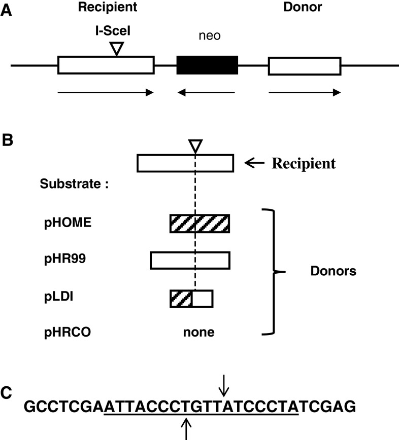
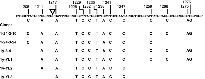
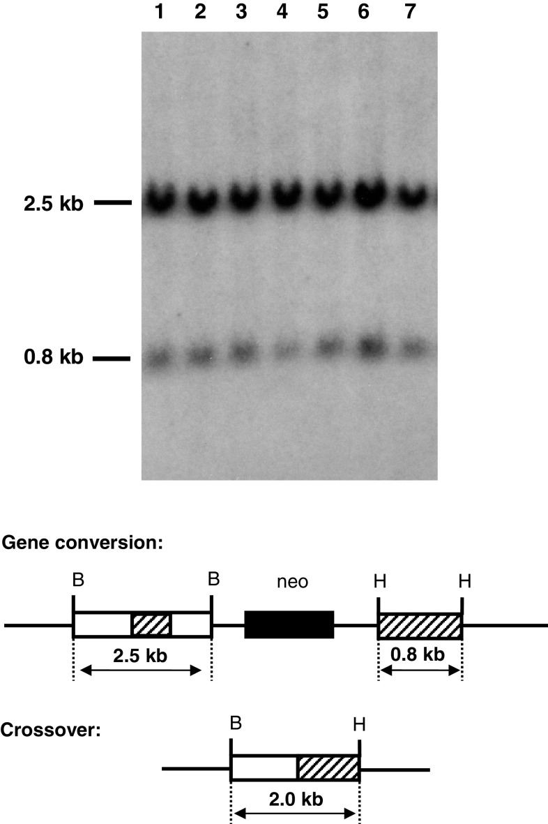
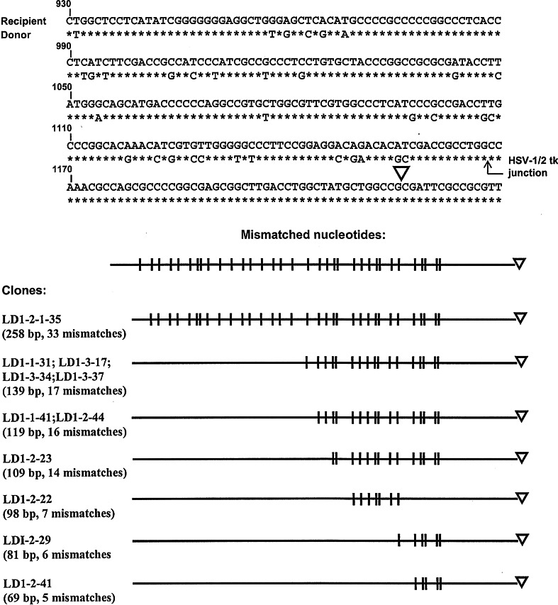
To investigate whether mammalian cells can carry out recombinational double-strand break (DSB) repair between highly diverged sequences, mouse fibroblasts were transfected with DNA substrates that contained a "recipient" thymidine kinase (tk) gene disrupted by the recognition site for endonuclease I-SceI. Substrates also contained a linked "donor" tk gene sequence. Following DSB induction by I-SceI, selection for tk-expressing clones allowed recovery of repair events occurring by nonhomologous end-joining or recombination with the donor sequence. Although recombinational repair was most efficient when donor and recipient shared near-perfect homology, we recovered recombination events between recipient and donor sequences displaying 20 % nucleotide mismatch. Recombination between such imperfectly matched ("homeologous") sequences occurred at a frequency of 1.7 × 10(-7) events per cell and constituted 3 % of the DSB repair events recovered with the pair of homeologous sequences. Additional experiments were done with a substrate containing a donor sequence comprised of a region sharing high homology with the recipient and an adjacent region homeologous to the recipient. Recombinational DSB repair tracts initiating within high homology propagated into homeology in 11 of 112 repair events. These collective results contrasted with our earlier work in which spontaneous recombination (not intentionally induced by a DSB) between homeologous sequences occurred at an undetectable frequency of less than 10(-9) events per cell, and in which events initiating within high homology propagated into adjoining homeology in one of 81 events examined. Our current work suggests that homology requirements for recombination are effectively relaxed in proximity to a DSB in a mammalian genome.


Figures

Fig. 1
Fig. 1
Recombination and repair substrates. a The general structure of all substrates. The recipient tk gene in all substrates is disrupted by a 30-bp oligonucleotide (inverted triangle) containing the 18-bp recognition site for endonuclease I-SceI. The orientations of recipient and donor tk sequences, as well as the neo gene used to establish stable transfectants, are indicated by arrows. b Substrates pHOME, pHR99, and pLD1 each contain a donor tk sequence; these donors are shown aligned beneath the recipient tk sequence. Substrate pHRCO does not contain a donor sequence. Open rectangles represent HSV-1 tk sequences, striped rectangles represent HSV-2 tk sequences. HSV-1 and HSV-2 tk sequences are homeologous to one another in that they display about 80 % sequence identity. c The sequence of the 30-bp oligonucleotide that disrupts the recipient tk gene is shown, with the 18-bp I-SceI recognition site underlined. The sites of staggered cleavage by I-SceI are indicated
Fig. 2
Fig. 2
DSB-induced HeR events recovered from cell lines containing pHOME. Presented at the top of the figure is the nucleotide sequence in the vicinity of the I-SceI recognition site (denoted by an inverted triangle) in the recipient tk gene from pHOME. Shown immediately below the recipient sequence are “marker” nucleotides from the donor tk sequence from pHOME that do not match the recipient sequence; the nucleotide positions at which these mismatches occur are indicated above the recipient sequence. The clones listed arose from HeR in the form of gene conversions and were recovered from cell lines containing pHOME following DSB induction with I-SceI. For each clone, the donor marker nucleotides present in the gene conversion tract are shown. Clones 1-24-2-10 and 1-24-3-24 were recovered from cell line HOME-1-24 while the other four clones were recovered from cell line HOME-1y
Fig. 3
Fig. 3
Southern-blot analysis of HeR events recovered from cell lines containing pHOME. Genomic DNA was isolated from 183 HATR clones recovered from cell lines HOME-1-24 and HOME-1y following DSB induction. DNA samples (8 μg each) were digested with BamHI and HindIII and displayed on a Southern blot using a mixture of probes specific for HSV-1 and HSV-2 tk. Shown (lanes 2–7) is the blot analysis for HeR clones 1-24-2-10, 1-24-3-24, 1y-8-4, 1y-YL1, 1y-YL2, and 1y-YL3. DNA from parent cell line HOME-1y is displayed in lane 1. As illustrated below the blot, clones that arose through a gene conversion event were each expected to display a 2.5-kb and a 0.8-kb band, while any clone that arose via a crossover was expected to display a single 2.0-kb band. The parent cell line and any clone that arose via NHEJ with a small deletion were each expected to display the same pattern as a gene conversion. All clones analyzed displayed a 2.5-kb and a 0.8-kb band, confirming that all six recovered HeR events were gene conversions and that the remaining 177 clones arose from NHEJ events with small deletions. No gross rearrangements were found
Fig. 4
Fig. 4
DSB-induced HeR events recovered from cell lines containing pLD1. Shown at the top of the figure is the recipient tk sequence from pLD1 with mismatched marker nucleotides from the donor tk sequence aligned immediately below the recipient sequence. Also indicated is the position of the junction between HSV-1 and HSV-2 tk sequences in the donor, as well as the position of the I-SceI recognition site (inverted triangle) in the recipient. In the lower portion of the figure is a list of clones recovered from cell lines containing pLD1 that arose from HeR events in the form of gene conversions. Schematic representations of the donor marker nucleotides present in the conversion tracts are shown. The number of mismatched marker nucleotides contained in each conversion tract is shown, as is the length of each conversion tract. The tract length is the number of nucleotides from the most upstream marker nucleotide in the conversion tract to the I-SceI site inserted after nucleotide position 1,215

Similar articles

Cited by

References

    1. O’Driscoll M, Jeggo PA. The role of double-strand break repair—insights from human genetics. Nat Rev Genet. 2006;7:45–54. doi: 10.1038/nrg1746. - DOI - PubMed
    1. Sonoda E, Hochegger H, Saberi A, Taniguchi Y, Takeda S. Differential usage of non-homologous end-joining and homologous recombination in double-strand break repair. DNA Repair. 2006;5:1021–1029. doi: 10.1016/j.dnarep.2006.05.022. - DOI - PubMed
    1. Hartlerode AJ, Scully R. Mechanisms of double-strand break repair in somatic mammalian cells. Biochem J. 2009;423:157–168. doi: 10.1042/BJ20090942. - DOI - PMC - PubMed
    1. Pardo B, Gómez-González B, Aguilera A. DNA double-strand break repair: how to fix a broken relationship. Cell Mol Life Sci. 2009;66:1039–1056. doi: 10.1007/s00018-009-8740-3. - DOI - PMC - PubMed
    1. Shrivastav M, De Haro LP, Nickoloff JA. Regulation of DNA double-strand break repair pathway choice. Cell Res. 2009;18:134–147. doi: 10.1038/cr.2007.111. - DOI - PubMed

Publication types

Substances