. 2014 Jan;25(2):290-301.
doi: 10.1091/mbc.E13-08-0448. Epub 2013 Nov 20.

Lipid droplet autophagy in the yeast Saccharomyces cerevisiae

Affiliations

Lipid droplet autophagy in the yeast Saccharomyces cerevisiae

Tim van Zutphen et al. Mol Biol Cell. 2014 Jan.

Abstract

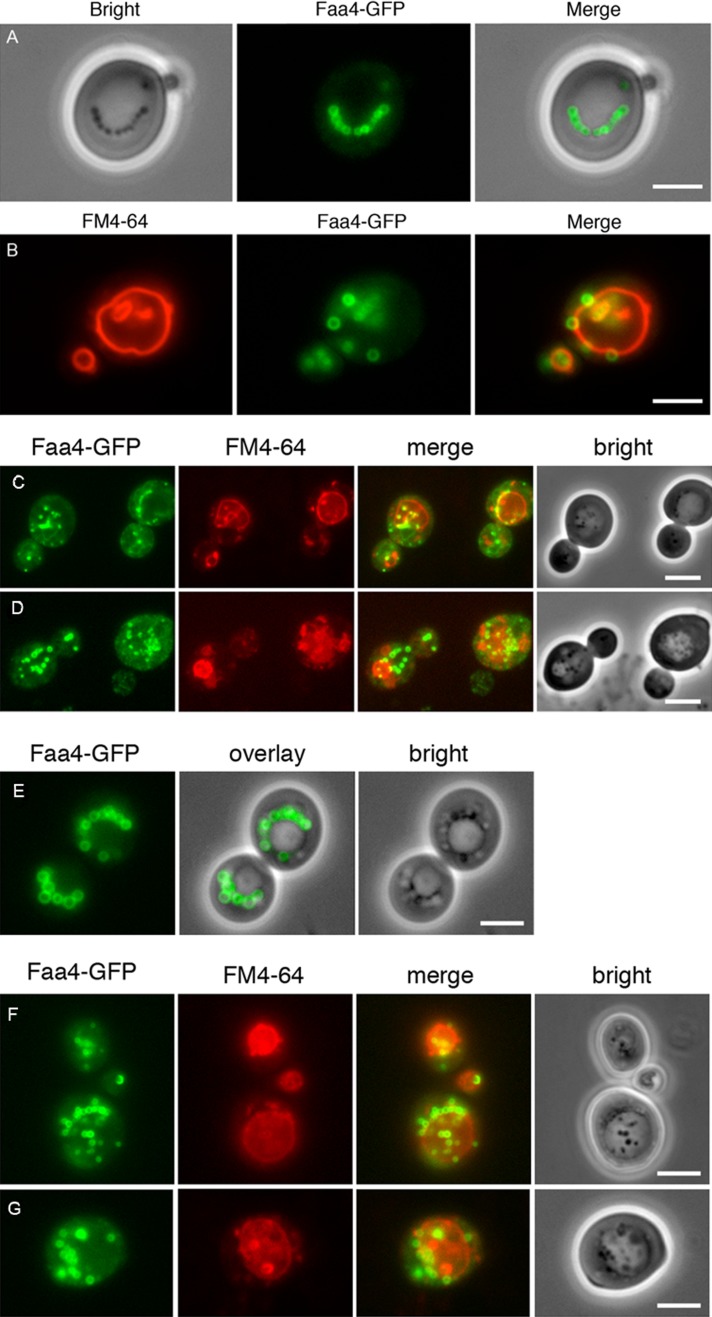
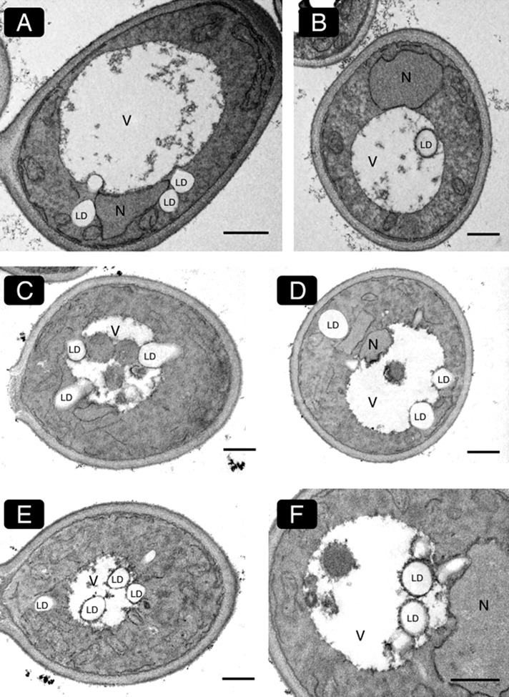
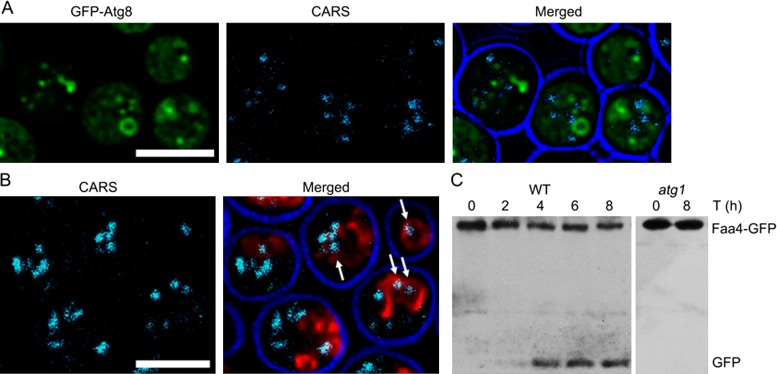
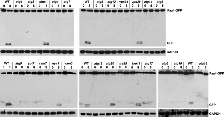
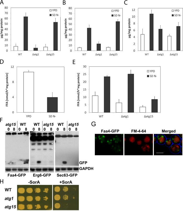
Cytosolic lipid droplets (LDs) are ubiquitous organelles in prokaryotes and eukaryotes that play a key role in cellular and organismal lipid homeostasis. Triacylglycerols (TAGs) and steryl esters, which are stored in LDs, are typically mobilized in growing cells or upon hormonal stimulation by LD-associated lipases and steryl ester hydrolases. Here we show that in the yeast Saccharomyces cerevisiae, LDs can also be turned over in vacuoles/lysosomes by a process that morphologically resembles microautophagy. A distinct set of proteins involved in LD autophagy is identified, which includes the core autophagic machinery but not Atg11 or Atg20. Thus LD autophagy is distinct from endoplasmic reticulum-autophagy, pexophagy, or mitophagy, despite the close association between these organelles. Atg15 is responsible for TAG breakdown in vacuoles and is required to support growth when de novo fatty acid synthesis is compromised. Furthermore, none of the core autophagy proteins, including Atg1 and Atg8, is required for LD formation in yeast.


Figures

FIGURE 1:
FIGURE 1:
Lipid droplet–vacuole interaction and uptake in glucose- and oleate-grown yeast cells. LDs are labeled with endogenously expressed Faa4-GFP in cells grown on 0.5% glucose for 21 h (A) and 46 h (B). LDs are typically localized in strings adjacent to the vacuole (A) or randomly distributed in the cytosol. They are also frequently observed inside the vacuole, especially in the stationary phase of growth (absence of glucose; B). Cells expressing Faa4-GFP were pregrown on glucose and subsequently shifted to oleate-containing media. After 6 (C) and 12 (D) h of incubation, LDs are massively induced in the cytosol and are also present inside the vacuoles. In stationary phase (28 h of incubation) distinct LDs are no longer detectable in the vacuole (E). After shift of these cells to fresh oleic acid–containing medium lacking a nitrogen source, LDs are rapidly incorporated into the vacuole: after 1 h (F) and 5 h (G). Vacuolar membranes are stained with FM4-64. Scale bar, 5 μm.
FIGURE 2:
FIGURE 2:
Electron microscopy of vacuolar lipid droplet internalization. Cells were grown in the absence of a nitrogen source (A, B) or for 5 h in oleic acid–containing media (C–F) and processed for electron microscopy. Both conditions lead to a stimulated internalization of LDs into the vacuole. Various stages of LD internalization are shown. Lipid droplets that enter the vacuole are partially covered by an electron-dense vacuolar membrane (B, E; higher magnification in F). These morphological characteristics suggest that LD internalization into the vacuole occurs via microautophagy in yeast. Scale bar, 1 μm.
FIGURE 3:
FIGURE 3:
Lipid droplets are degraded in the yeast vacuole upon induction of autophagy. (A) ypt7 cells expressing GFP-Atg8 show the accumulation of autophagosomes that lack LDs. (B) Detection of LDs inside the vacuole of wild-type cells with CARS imaging; vacuolar membranes are labeled with FM4-64. Cells were shifted to nitrogen starvation medium for 8 h in the presence of PMSF before microscopy to induce autophagy. Scale bar, 5 µm. (C) Western blot of cell extracts of wild-type cells expressing the LD marker Faa4-GFP, using an anti-GFP antibody. Late exponential cells grown in rich medium were shifted for 8 h to medium lacking a nitrogen source. The appearance of one or two bands at ∼27 kDa is indicative of vacuolar proteolytic processing of the Faa4-GFP fusion protein. This band is absent in atg1 cells.
FIGURE 4:
FIGURE 4:
Lipid droplet autophagy requires the core autophagy machinery and additional factors. Western blots were prepared from crude extracts of the indicated mutant cells, which were grown to the late logarithmic growth phase in rich medium and shifted to synthetic minimal medium lacking nitrogen for 8 h. Blots were decorated with anti-GFP and anti-GAPDH antibodies.
FIGURE 5:
FIGURE 5:
Lipid droplet autophagy requires tubulin. (A) atg4-, atg7-, and atg11-mutant cells expressing Faa4-GFP were shifted to synthetic minimal medium lacking nitrogen for 8 h. LDs are closely associated with the cytoplasmic site of the vacuolar membrane (labeled with FM4-64). Scale bar, 5 μm. (B) Western blots were prepared from crude extracts of wild-type cells expressing either Faa4-GFP or Om45-GFP or no marker, as indicated. Cells were incubated in synthetic minimal medium lacking nitrogen supplemented with 15 μg/ml nocodazole for 4 or 8 h. Blots were decorated with anti-GFP, anti–aminopeptidase I, or anti-GAPDH antibodies. Faa4-GFP degradation is strongly reduced, suggesting that nocodazole treatment inhibits LD internalization into the vacuole. In contrast, processing of Om45-GFP is not affected, consistent with previous results that tubulin is not required for mitophagy (Kanki et al., 2009). (C) Western blot of cell extracts prepared from Faa4-GFP–expressing elo1 and elo3 mutant cells, which display highly fragmented vacuoles (Kohlwein et al., 2001). Cells were grown to the late logarithmic growth phase in rich medium and shifted to synthetic minimal medium lacking nitrogen for 8 h. Both, elo1 and elo3 mutants show normal Faa4-GFP processing, indicating that vacuolar fragmentation does not affect LD autophagy. Blots were decorated with anti-GFP and anti-GAPDH antibodies.
FIGURE 6:
FIGURE 6:
Lipid droplet autophagy requires selective adapters and differs from ER-phagy. (A) Protein extracts of various mutant cells expressing Faa4-GFP were grown to the late logarithmic growth phase in rich medium and shifted to synthetic minimal medium lacking nitrogen for the indicated time intervals. This analysis shows the requirement for Vac8 and a partial requirement for Atg11 for Faa4-GFP cleavage. Blots were decorated with anti-GFP and anti-GAPDH antibodies. (B) Quantification of cleaved Faa4-GFP at different time points after the shift to starvation medium in wild-type and atg11 mutant cells expressing Faa4-GFP relative to the GAPDH loading control. (C) CARS images of atg11-mutant cells shifted to nitrogen starvation medium for 8 h in the presence of PMSF. LDs are internalized into vacuoles of atg11 cells that are labeled with FM4-64. (D) Protein extracts from various mutant cells expressing the ER marker Sec63-GFP analyzed by Western blotting. Cells were grown to the late logarithmic growth phase in rich medium and shifted to synthetic minimal medium lacking nitrogen for indicated times. Blots were decorated with anti-GFP and anti-GAPDH antibodies. This analysis shows that LD autophagy is distinct from ER-phagy. See the text for details.
FIGURE 7:
FIGURE 7:
The yeast vacuole has lipase activity that depends on Atg15. Steryl ester (A), triacylglycerol (B), and free fatty acid (C) content of vacuolar fractions of wild-type, atg1, and atg15 cells grown on either rich (YPD) or autophagy-inducing (SD N) media. Lipase activity in isolated lipid droplet (D) and vacuole fractions (E). Western blot (F) of proteins in crude extracts of wild-type and atg15 cells expressing either Faa4-GFP or Erg6-GFP to analyze lipid droplet autophagy or Sec63-GFP to determine ER-phagy. Cells were grown to the end of the logarithmic growth phase and shifted to SD N medium for 8 h. Single optical sections (G) of atg15-mutant cells expressing Faa4-GFP (green) and labeled with FM4-64. Cells were cultivated in SD N for 8 h, showing accumulation of GFP in the vacuole lumen. Scale bar, 5 µm. Lack of the vacuolar lipase Atg15 renders cells sensitive to the inhibitor soraphen A, which blocks de novo fatty acid synthesis (H).

Similar articles

Cited by

References

    1. Athenstaedt K, Daum G. Tgl4p and Tgl5p, two triacylglycerol lipases of the yeast Saccharomyces cerevisiae are localized to lipid particles. J Biol Chem. 2005;280:37301–37309. - PubMed
    1. Backues SK, Klionsky DJ. Atg11: a Rab-dependent, coiled-coil membrane protein that acts as a tether for autophagy. Autophagy. 2012;8:1275–1278. - PMC - PubMed
    1. Bernales S, McDonald KL, Walter P. Autophagy counterbalances endoplasmic reticulum expansion during the unfolded protein response. PLOS Biol. 2006;4:e423. - PMC - PubMed
    1. Cheong H, Klionsky DJ. Biochemical methods to monitor autophagy-related processes in yeast. Methods Enzymol. 2008;451:1–426. - PubMed
    1. Cusi K. Role of obesity and lipotoxicity in the development of nonalcoholic steatohepatitis: pathophysiology and clinical implications. Gastroenterology. 2012;142:711–725. - PubMed

Publication types

MeSH terms