. 2013 Nov 20;11(91):20130850.
doi: 10.1098/rsif.2013.0850. Print 2014 Feb 6.

Mechanosensitive channels and bacterial cell wall integrity: does life end with a bang or a whimper?

Affiliations

Mechanosensitive channels and bacterial cell wall integrity: does life end with a bang or a whimper?

Marcel Reuter et al. J R Soc Interface. .

Abstract

Mechanogated channels are fundamental components of bacterial cells that enable retention of physical integrity during extreme increases in cell turgor. Optical tweezers combined with microfluidics have been used to study the fate of individual Escherichia coli cells lacking such channels when subjected to a bursting stress caused by increased turgor. Fluorescence-activated cell sorting and electron microscopy complement these studies. These analyses show that lysis occurs with a high probability, but the precise path differs between individual cells. By monitoring the loss of cytoplasmic green fluorescent protein, we have determined that some cells release this protein but remain phase dark (granular) consistent with the retention of the majority of large proteins. By contrast, most cells suffer cataclysmic wall failure leading to loss of granularity but with the retention of DNA and overall cell shape (protein-depleted ghosts). The time span of these events induced by hypo-osmotic shock varies but is of the order of milliseconds. The data are interpreted in terms of the timing of mechanosensitive channel gating relative to osmotically induced water influx.

Keywords: (bacterial) cell wall; bacterial stress response; fluorescence-activated cell sorting; mechanosensitive channels; microfluidics; optical tweezers.


Figures

Figure 1.
Figure 1.
FACS analysis correlating cell size and cytoplasmic granularity (forward and side scatter, FS and SC) and uptake of propidium iodide (PI) into damaged cells that have retained the nucleoid. PI positive cells are coloured red. (a) Unshocked E. coli FRAG1 parental cells; (b) hypo-osmotically shocked E. coli FRAG1 cells; (c) unshocked E. coli MJF465 channel mutant cells; (d) hypo-osmotically shocked E. coli MJF465 cells. (e) and (f) overlay of data from control (e) and shock (f) samples (pale blue dots, FRAG1; dark blue dots, MJF465). In both cell populations, a significant number of particles with high FS and low SC were also observed in the high osmolarity samples and were found to be non-biological material arising from the buffer solutions (see the electronic supplementary material, figure S2). (a–c) Approximately 30 000 events are within the gate (see §2), but in (d), this falls to approximately 19 000, owing to cell lysis yielding material of the same size as background (see electronic supplementary material, figure S2a,b).
Figure 2.
Figure 2.
Phase-contrast microscopy time series for the transfer of an optically trapped E. coli MJF465 cell from LB + 0.5 M NaCl into distilled water via a third (middle) microfluidic channel containing 50 μM hydrogen peroxide. (a) A ‘burster’ cell that completely vanishes upon hypo-osmotic shock. No remnants could be observed in the entire field of view (fivefold larger). The full movie file is available as electronic supplementary material, movie S1. (b) A ‘leaker’ cell whose contrast decreases upon hypo-osmotic shock. Large cell wall ruptures (>20 nm) allow proteins to escape, which causes the image of the cell to fade. (The green cross and the red circle indicate the vicinity of the optical trap, for entire film see the electronic supplementary material, movie S2.) For this type of experiment, 40 E. coli MJF465 and 40 parent (E. coli FRAG1) cells were investigated. For MJF465, 17 cells lysed, 12 cells survived and the remaining 11 cells had an ambiguous outcome. By contrast, seven FRAG1 cells lysed, 19 cells survived and 14 cells had an ambiguous outcome.
Figure 3.
Figure 3.
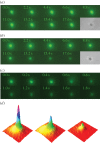
Fluorescence microscopy time series of optically trapped GFP-expressing E. coli MJF465(DE3) cells. (a) A control cell not subjected to downshock. Slow loss in GFP fluorescence occurs as a result of photobleaching. After complete GFP bleaching (300 s), the cell still appeared dark in phase-contrast mode (see electronic supplementary material, movie S4). (b) A cell subjected to hypo-osmotic downshock (LB + 0.5 M NaCl into distilled water via a third microfluidic channel containing 50 μM hydrogen peroxide). Owing to lesions, GFP leaks out of the cell resulting in a fast decrease in GFP fluorescence. After complete GFP leakage (30 s), the cell still appeared dark in phase-contrast mode (see electronic supplementary material, movie S5). The vast majority of cells (>95%) subjected to this treatment showed exactly this behaviour. For this experiment, 14 cells were subjected to hypo-osmotic shock and eight cells to control conditions, respectively. (c) A cell subjected to hypo-osmotic shock showing cell wall rupturing at 0.6 s. A burst of fluorescence could be detected moving away to the top right corner of this image (see electronic supplementary material, movie S6). This is an extremely rare event (<5%). (d) Individual frames from the movie in (c) were used to construct a three-dimensional plot where x- and y-axes are coordinates for each pixel and z-axis is the pixel intensity. Frames at t = 0 s, 1.80 s and 3.20 s, left to right, respectively, were plotted The plots are rotated 180° with respect to the frames presented in (c) to allow visualization of the GFP cloud, which would otherwise be hidden by the peak.
Figure 4.
Figure 4.
Fractional changes in intensity for the different classes of cellular events. Bursting of E. coli MJF465 cells with subsequent vanishing of all debris (black triangle) occurs in less than 200 ms (shown as a single point). Leakage of large material from mutant cells (green triangles) happens fast, on a timescale of 1 s. The bursting of a GFP-expressing E. coli MJF465(DE3) cell (orange triangles) was rare and occurred on the 1 s timescale. GFP leakage (black circles) and GFP photobleaching (blue circles) from MJF465(DE3) cells were characterized by a mono-exponential decay function. Individual traces for MJF465(DE3) cells that were hypo-osmotically shocked without hydrogen peroxide support (red circles) exhibited an initial rapid decrease in GFP fluorescence (leakage from cell) followed by a slower decay, consistent with fluorescence bleaching of the retained GFP. Out of 14 shocked cells, five cells displayed this biphasic behaviour, whereas the remaining shocked cells behaved like control cells (nine cells tested), and thus did not release GFP.

References

    1. Mitchell P, Moyle J. 1956. Osmotic function and structure in bacteria. In Bacterial anatomy (eds Spooner E, Stocker B.), pp. 150–180. Cambridge, UK: Cambridge University Press.
    1. Kung C. 2005. A possible unifying principle for mechanosensation. Nature 436, 647–654. (10.1038/nature03896) - DOI - PubMed
    1. Booth IR, Cairney J, Sutherland L, Higgin CF. 1988. Enteric bacteria and osmotic stress: an integrated homeostatic system. Soc. Appl. Bacteriol. Symp. Ser. 17, 35S–49S. - PubMed
    1. Mallo RC, Ashby MT. 2006. AqpZ-mediated water permeability in Escherichia coli measured by stopped-flow spectroscopy. J. Bacteriol. 188, 820–822. (10.1128/JB.188.2.820-822.2006) - DOI - PMC - PubMed
    1. Pilizota T, Shaevitz JW. 2012. Fast, multiphase volume adaptation to hyperosmotic shock by Escherichia coli. PLoS ONE 7, e35205 (10.1371/journal.pone.0035205) - DOI - PMC - PubMed

Publication types

MeSH terms

LinkOut - more resources