. 2014;29(5):1269-82.
doi: 10.1002/jbmr.2148.

Stem cell-derived endochondral cartilage stimulates bone healing by tissue transformation

Affiliations

Stem cell-derived endochondral cartilage stimulates bone healing by tissue transformation

Chelsea S Bahney et al. J Bone Miner Res. 2014.

Abstract

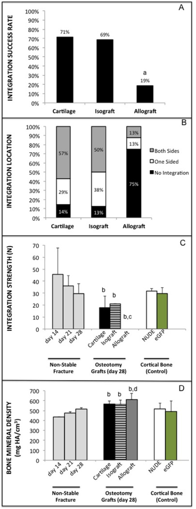
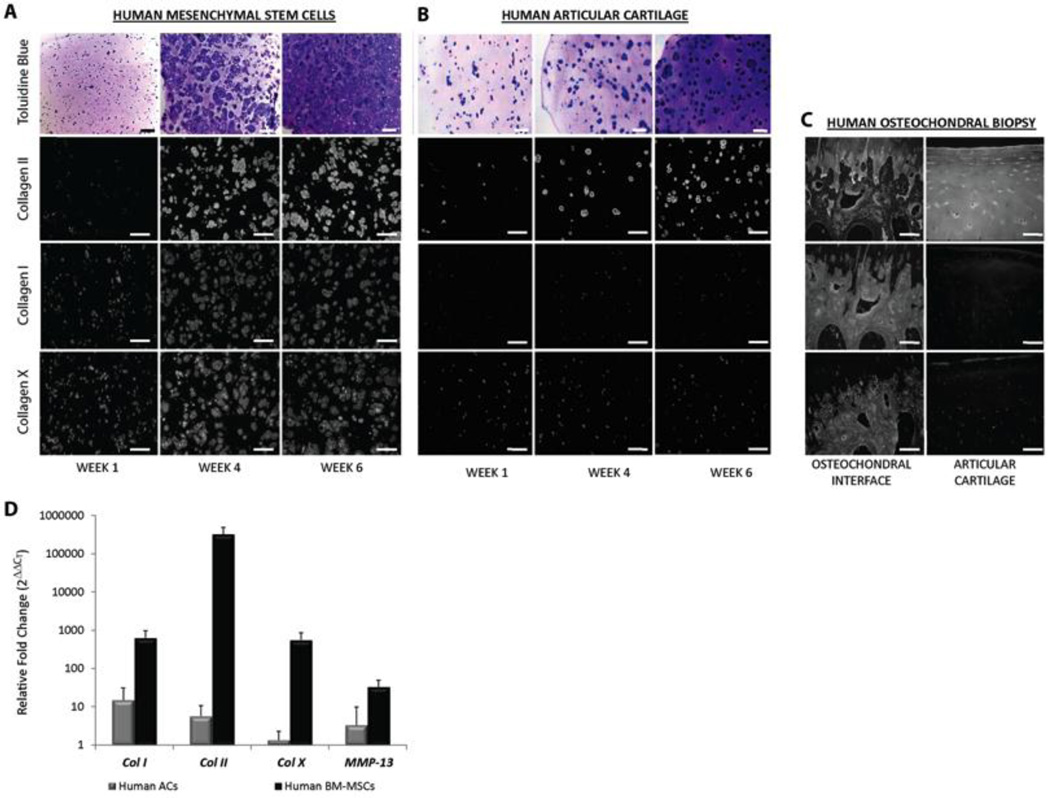
Although bone has great capacity for repair, there are a number of clinical situations (fracture non-unions, spinal fusions, revision arthroplasty, segmental defects) in which auto- or allografts attempt to augment bone regeneration by promoting osteogenesis. Critical failures associated with current grafting therapies include osteonecrosis and limited integration between graft and host tissue. We speculated that the underlying problem with current bone grafting techniques is that they promote bone regeneration through direct osteogenesis. Here we hypothesized that using cartilage to promote endochondral bone regeneration would leverage normal developmental and repair sequences to produce a well-vascularized regenerate that integrates with the host tissue. In this study, we use a translational murine model of a segmental tibia defect to test the clinical utility of bone regeneration from a cartilage graft. We further test the mechanism by which cartilage promotes bone regeneration using in vivo lineage tracing and in vitro culture experiments. Our data show that cartilage grafts support regeneration of a vascularized and integrated bone tissue in vivo, and subsequently propose a translational tissue engineering platform using chondrogenesis of mesenchymal stem cells (MSCs). Interestingly, lineage tracing experiments show the regenerate was graft derived, suggesting transformation of the chondrocytes into bone. In vitro culture data show that cartilage explants mineralize with the addition of bone morphogenetic protein (BMP) or by exposure to human vascular endothelial cell (HUVEC)-conditioned medium, indicating that endothelial cells directly promote ossification. This study provides preclinical data for endochondral bone repair that has potential to significantly improve patient outcomes in a variety of musculoskeletal diseases and injuries. Further, in contrast to the dogmatic view that hypertrophic chondrocytes undergo apoptosis before bone formation, our data suggest cartilage can transform into bone by activating the pluripotent transcription factor Oct4A. Together these data represent a paradigm shift describing the mechanism of endochondral bone repair and open the door for novel regenerative strategies based on improved biology.

Keywords: BIOENGINEERING; CARTILAGE BIOLOGY; CHONDROCYTES; INJURY/FRACTURE HEALING; MOLECULAR PATHWAYS; REMODELING; THERAPEUTICS.


Conflict of interest statement

Disclosures

There are no competing or conflicts of interest to report related to the work presented in this manuscript for any of the authors. Author contribution to the paper was as follows: study design: CSB, DPH, TM, RSM; study conduct and data collection: CSB, DPH, AJT, FF; Data interpretation: CSB, DPH, AJT, FF, BJ, TM, RSM; Drafting manuscript: CSB; Revising Manuscript: CSB, RM; Approval of final version of manuscript: CSB, DPH, AJT, FF, BJ, TM, RSM. CSB take responsibility for the integrity of the data.

Figures

FIGURE 1
FIGURE 1. Cartilage Grafts from Fracture Callus
(A) X-ray image of unstabilized mid-diaphysial tibia fracture (arrow) created by three point bending. (B) Safranin-O staining of fracture callus 7 days following injury. Grafts were isolated ex-vivo, boxed region represents the approximate area from where grafts were harvested. (C) Grafts were transplanted into 2mm segmental defects created in the mid-diaphysis of externally stabilized murine tibia. (D) Safranin-O staining of the cartilage graft (top), followed by DIG-probe in situ hybridization for sox-9, col2, col10, and osteocalcin (oc). (E) Quantitative RT-PCR shows gene expression in the cartilage graft (n=6) relative to the whole fracture callus (n=5), data represents mean ± 95% confidence, with significance (*) of p < 0.005.
FIGURE 2
FIGURE 2. Cartilage grafts produce an integrated and vascularized bone regenerate in a segmental bone defect
(A–C) Safranin-O or (E–G) Masson’s Trichrome staining at days (A&E) 7, (B&F) 14, or (C&G–D) 28 after implantation of the cartilage graft. (D) µCT image of tibia defect 4 weeks post-surgically. “cb” = cortical bone (host), “graft” = transplanted fracture callus cartilage.
FIGURE 3
FIGURE 3. Integration of cartilage grafts is equivalent to isograft and superior to allograft after 4 weeks of healing
(A) Overall percentage that cartilage, isograft, or allograft integrated with the host tissue based on two potential integration sites per graft (7–8 animals, 14–16 integration opportunities; a = statistically different than cartilage and isograft, p < 0.005). (B) Graphical representation of integration for cartilage, isograft, and allograft. (C) Ultimate failure of grafts by three-point bending. Ultimate failure of uninjured cortical bone for both the grafts (eGFP) and host mice (immunocompromised/nude) are provided as a positive control alongside failure of non-stabilized fractures for reference (b = statistically different than d14 fx, c = different than cortical bone, P < 0.05). (D) Bone mineral density (BMD) of the fracture callus, grafts and cortical bone (d = statistically different than d21 fx, p < 0.02).
FIGURE 4
FIGURE 4. Donor (blue) versus host contribution to the bone regenerate
Cartilage grafts were obtained from Lac-Z+/+-Rosa26 reporter mice and transplanted into immunocompromised mice (SCID Beige) mice. Donor cells were labeled using x-gal staining to detect β-galactosidase activity from the Rosa26 mice at (A) 7, (B–E) 28, or (F–H) 42 days after cartilage engraftment. Special characters (D) donor osteocytes (yellow star), donor chondrocytes (yellow arrow), host chondrocytes (green triangle). “cb” = cortical bone (host), “graft” = transplanted fracture callus cartilage from LacZ+/+ mouse.
FIGURE 5
FIGURE 5. Human MSC-derived cartilage pellets transplanted into segmental bone defect regenerate bone
(A–C) hMSC pellets following 3 weeks of in vitro culture in chondrogenic conditions stained with (A) Safranin-O, or antibodies to (B) collagen II and (C) collagen X. (D–E) Masson’s Trichrome staining of segmental defect 4 weeks following transplantation of hMSC-derived cartilage pellets. (F) hMSC-dervied pellet with antibody staining for human mitochondria in the trichrome positive bone.
FIGURE 6
FIGURE 6. Engineering endochondral cartilage for tissue engineering
Human MSCs (A) or healthy human articular chondrocytes (hACs) (B) were photoencapsulated into PEGDA based scaffolds and cultured in vitro with chondrogenic medium for six weeks. (C) Antibody specificity was verified using articular cartilage and the osteochondral interface. (D) Gene expression of the hMSC (n=6) or hAC (n=6) derived tissue engineered cartilage following six weeks of in vitro culture was compared to gene expression of each tissue at day 0. Graph represents mean ± 95% confidence.
FIGURE 7
FIGURE 7. HUVEC conditioned medium is sufficient for producing mineralized cartilage in vitro
Alizarin red and safranin-o/fast green staining of cartilage obtained from the day-7 fracture callus and cultured in vitro in basal medium containing 10 nM dexamethasone and 100 µg/ml A2P for 2 weeks; then either maintained in that basal condition (A–C), or transferred to osteogenic supplements (rhBMP2,βGP,A2P) (D–F), HUVEC-CM (G–I), or HUVEC-CM with osteogenic supplements (J–L). Alizarin red and safranin-o/fast green staining of cartilage grafts (M). Quantification of the percentage of mineralization (gray) and proteoglycan (black) in cartilage explants following 4-weeks of in vitro culture. Graph represents mean ± 95% confidence, with significance (*) of p < 0.005.
FIGURE 8
FIGURE 8. Cartilage to bone transition zone in fracture callus
(A–D) Hall and Brunt Quadruple stain (HBQ, bone = red, cartilage = blue) of non-stabilized fracture callus 10-days post-injury. (E–F) Osteocalcin immunohistochemistry of non-stabilized fracture callus 10-days post-injury. (G–H) In situ cell death detection (GFP) merged with Safranin-O (cartilage = red, bone = blue) staining of day (G) 7 or (H) day 10 fracture callus. (I–J) Oct4A immunohistochemistry of day 10 fracture callus. Arrows point to Oct4A positive hypertrophic chondrocytes, “BV” = blood vessel or bone marrow space.

Similar articles

Cited by

References

    1. Lim SS, Vos T, Flaxman AD, Danaei G, Shibuya K, Adair-Rohani H, et al. A comparative risk assessment of burden of disease and injury attributable to 67 risk factors and risk factor clusters in 21 regions, 1990–2010: a systematic analysis for the Global Burden of Disease Study 2010. Lancet. 2012;380(9859):2224–2260. Epub 2012/12/19. doi: 10.1016/S0140-6736(12)61766-8 S0140-6736(12)61766-8 [pii]. PubMed PMID: 23245609. - PMC - PubMed
    1. Agency for Healthcare Research and Quality. Available from: http://www.ahrq.gov/news/nn/nn012506.htm. - PubMed
    1. The Burden of Musculoskeletal Diseases in the United States. Rosemont, IL: American Academy of Orthopaedic Surgeons; 2008.
    1. Nguyen LH, Annabi N, Nikkhah M, Bae H, Binan L, Park S, et al. Vascularized bone tissue engineering: approaches for potential improvement. Tissue engineering Part B, Reviews. 2012;18(5):363–382. PubMed PMID: 22765012; PubMed Central PMCID: PMC3458624. - PMC - PubMed
    1. Brigman BE, Hornicek FJ, Gebhardt MC, Mankin HJ. Allografts about the Knee in Young Patients with High-Grade Sarcoma. Clinical orthopaedics and related research. 2004;(421):232–239. Epub 2004/05/05. PubMed PMID: 15123953. - PubMed

Publication types