

Cisplatin inhibits bone healing during distraction osteogenesis
- PMID: 24259375
- PMCID: PMC4080883
- DOI: 10.1002/jor.22527
Cisplatin inhibits bone healing during distraction osteogenesis
Abstract
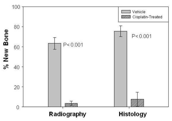
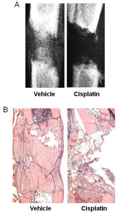
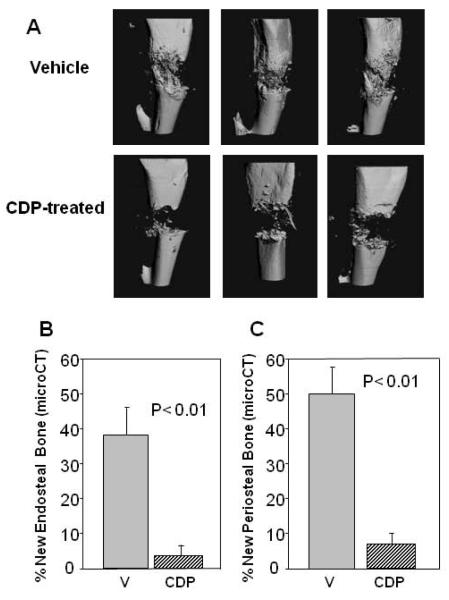
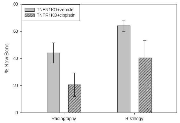
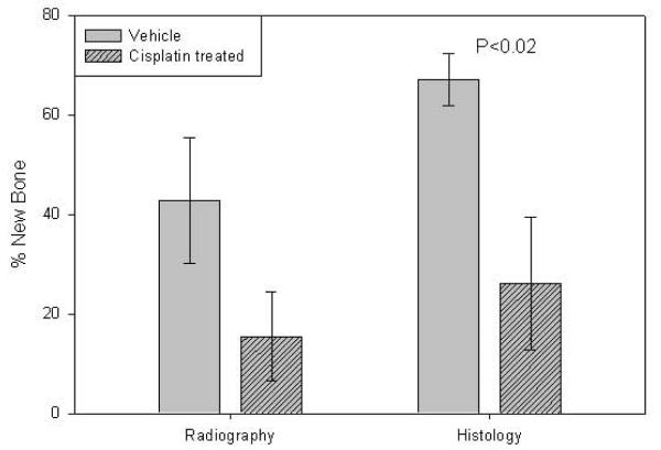
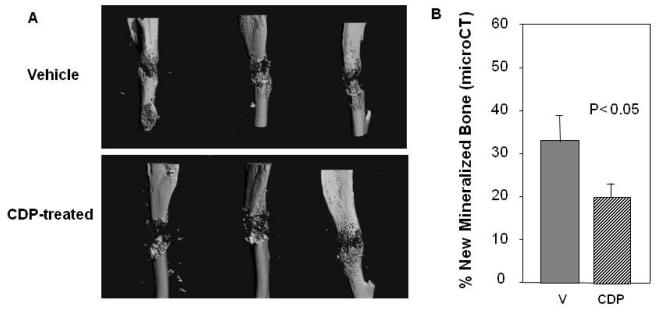
Osteosarcoma (OS) is the most common malignant bone tumor affecting children and adolescents. Many patients are treated with a combination of chemotherapy, resection, and limb salvage protocols. Surgical reconstructions after tumor resection include structural allografts, non-cemented endoprostheses, and distraction osteogenesis (DO), which require direct bone formation. Although cisplatin (CDP) is extensively used for OS chemotherapy, the effects on bone regeneration are not well studied. The effects of CDP on direct bone formation in DO were compared using two dosing regimens and both C57BL/6 (B6) and tumor necrosis factor receptor 1 knockout (TNFR1KO) mice, as CDP toxicity is associated with elevated TNF levels. Detailed evaluation of the five-dose CDP regimen (2 mg/kg/day), demonstrated significant decreases in new bone formation in the DO gaps of CDP treated versus vehicle treated mice (p < 0.001). Further, no significant inhibitory effects from the five-dose CDP regimen were observed in TNFR1KO mice. The two-dose regimen significantly inhibited new bone formation in B6 mice. These results demonstrate that CDP has profound short term negative effects on the process of bone repair in DO. These data provide the mechanistic basis for modeling peri-operative chemotherapy doses and schedules and may provide new opportunities to identify molecules that spare normal cells from the inhibitory effects of CDP.
Keywords: chemotherapy; cisplatin; distraction osteogenesis; limb salvage; mouse.
© 2013 Orthopaedic Research Society. Published by Wiley Periodicals, Inc.
Figures
References
-
- Messerschmitt PJ, Garcia RM, Abdul-Karim FW, et al. Osteosarcoma. J Am Acad Orthop Surg. 2009;17:515–27. Review. - PubMed
-
- Bilariki K, Anagnostou E, Masse V, et al. Low bone mineral density and high incidences of fractures and vitamin D deficiency in 52 pediatric cancer survivors. Horm Res Paediatric. 2010;74:319–27. - PubMed
-
- Bertoldo F, Pancheri S, Zenari S, Boldini S. Emerging drugs for the management of cancer treatment induced bone loss. Expert Opin Emerg Drugs. 2010;15:323–42. Review. - PubMed
-
- Bundred N. Antiresorptive therapies in oncology and their effects on cancer progression. Cancer Treat Rev. 2012;38:776–86. - PubMed
Publication types
MeSH terms
Substances
Grants and funding
LinkOut - more resources
Full Text Sources
Other Literature Sources
