. 2014 Feb;55(2):247-57.
doi: 10.1194/jlr.M043422. Epub 2013 Nov 20.

LXRα fuels fatty acid-stimulated oxygen consumption in white adipocytes

Affiliations

LXRα fuels fatty acid-stimulated oxygen consumption in white adipocytes

Lea Dib et al. J Lipid Res. 2014 Feb.

Abstract

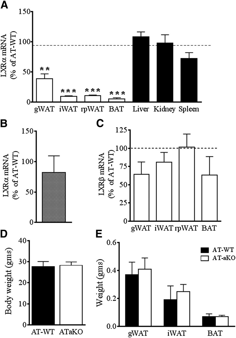
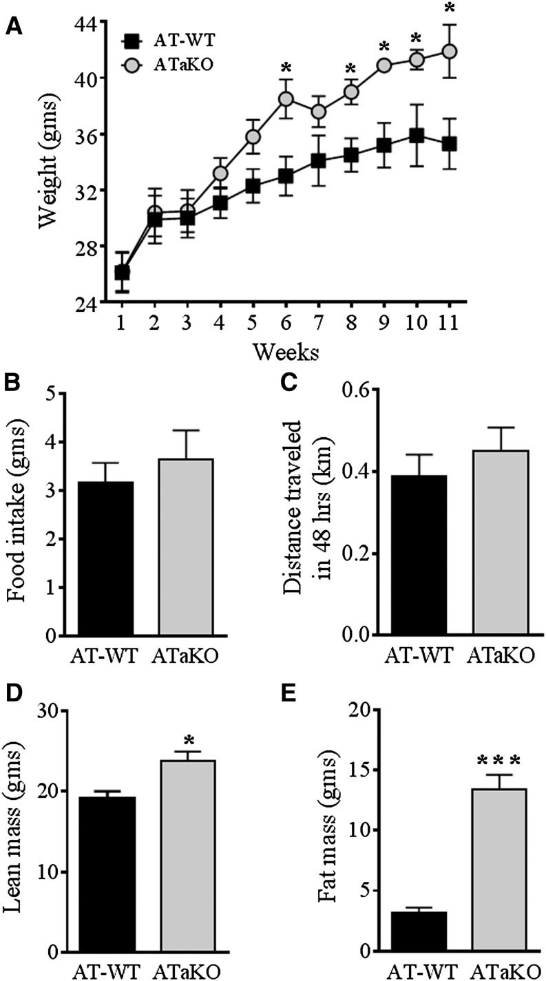
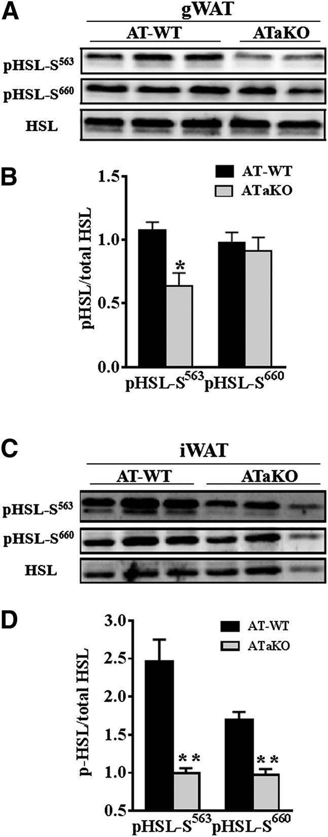
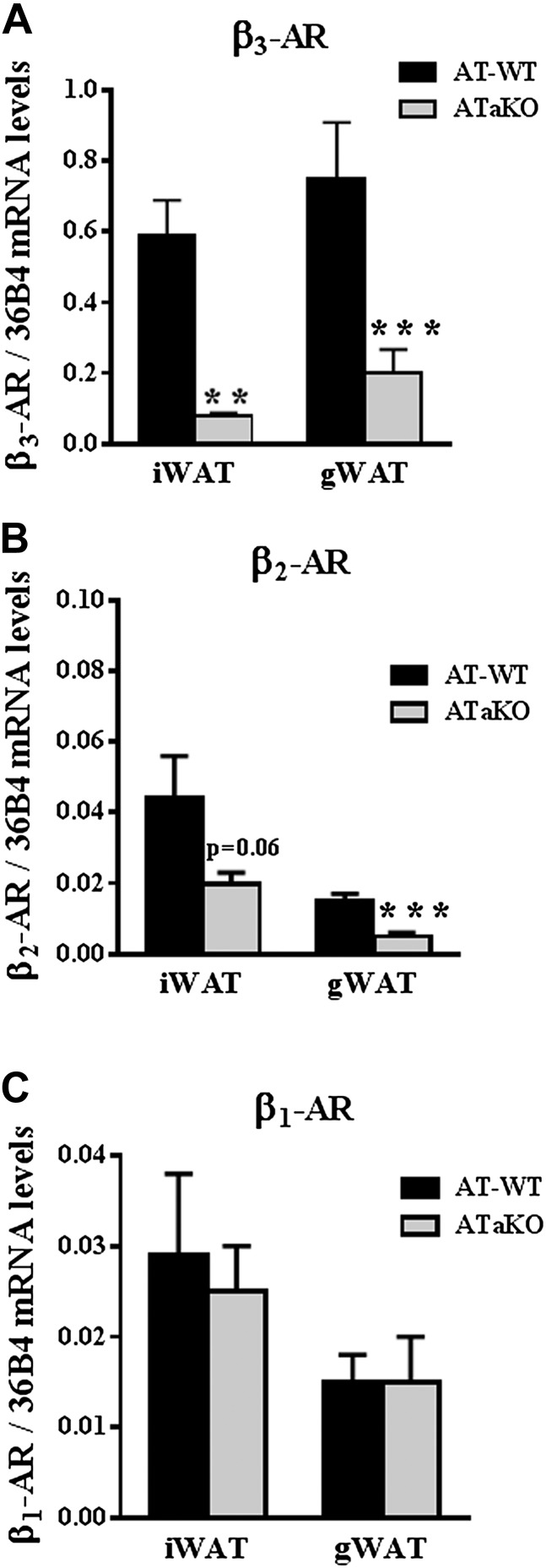
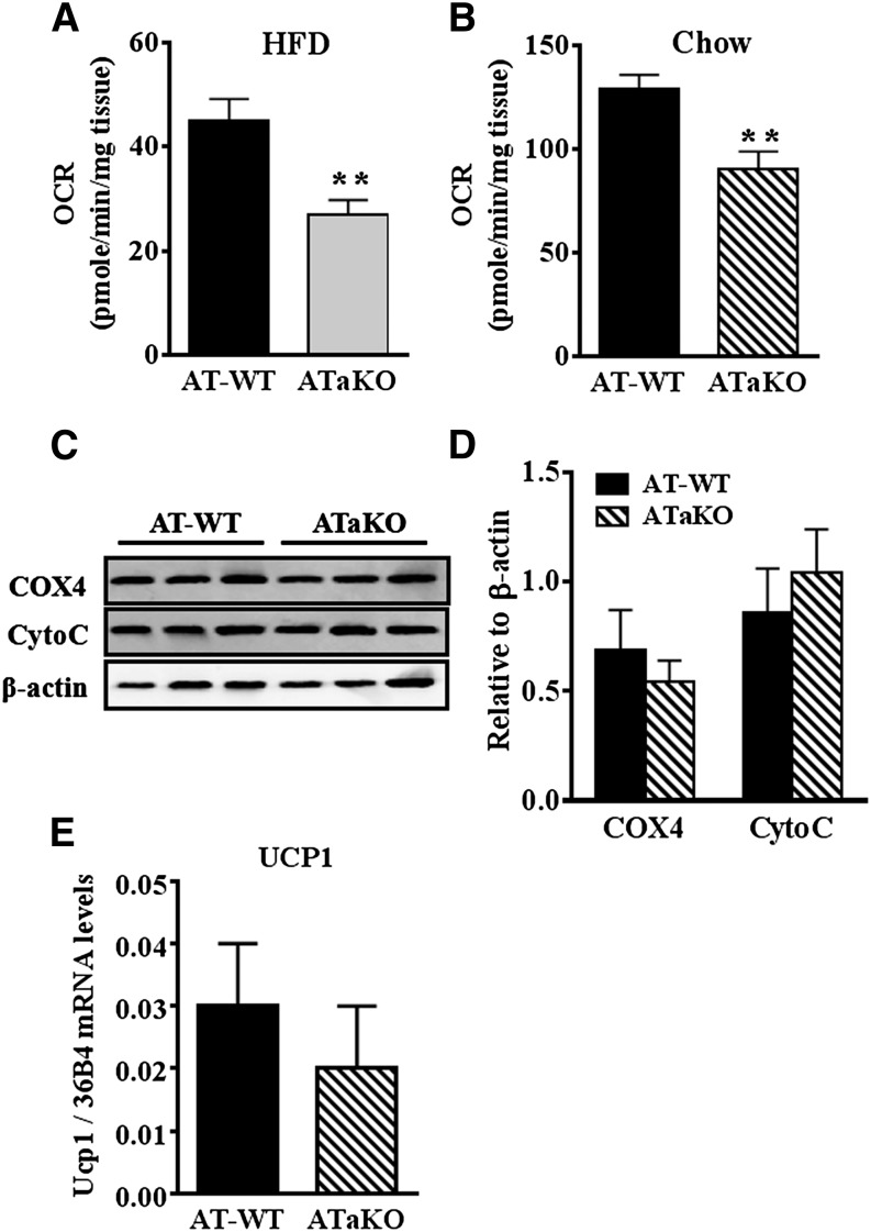
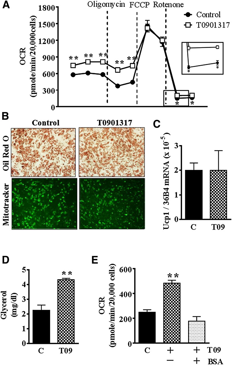
Liver X receptors (LXRs) are transcription factors known for their role in hepatic cholesterol and lipid metabolism. Though highly expressed in fat, the role of LXR in this tissue is not well characterized. We generated adipose tissue LXRα knockout (ATaKO) mice and showed that these mice gain more weight and fat mass on a high-fat diet compared with wild-type controls. White adipose tissue (WAT) accretion in ATaKO mice results from both a decrease in WAT lipolytic and oxidative capacities. This was demonstrated by decreased expression of the β2- and β3-adrenergic receptors, reduced level of phosphorylated hormone-sensitive lipase, and lower oxygen consumption rates (OCRs) in WAT of ATaKO mice. Furthermore, LXR activation in vivo and in vitro led to decreased adipocyte size in WAT and increased glycerol release from primary adipocytes, respectively, with a concomitant increase in OCR in both models. Our findings show that absence of LXRα in adipose tissue results in elevated adiposity through a decrease in WAT oxidation, secondary to attenuated FA availability.

Keywords: adipose tissue; lipolysis; mitochondria; oxidation.


Figures

Fig. 1.
Fig. 1.
ATaKO have similar anthropometric characteristic as the LXRαfl/fl control mice on a chow diet. Percentage expression of LXRα mRNA in LXRα-expressing tissues (A) and macrophages (B) and percentage expression of LXRβ in adipose tissue depots (C) in ATaKO mice compared with controls as analyzed by real-time quantitative PCR. Body weight (D) and adipose tissue pad weights (E) of male AT-WT and ATaKO mice on chow diet. Values are mean ± SEM. **P < 0.01, ***P < 0.001; n = 3–7 animals/group.
Fig. 2.
Fig. 2.
ATaKO mice gain more weight than controls (AT-WT) on a HFD. Body weights (A), food intake (B), physical activity (C), and lean (D) and fat mass (E) of ATaKO and AT-WT after 12 weeks of a HFD. Values are mean ± SEM. *P < 0.05, **P < 0.01, ***P < 0.001; n = 4–5 animals/group.
Fig. 3.
Fig. 3.
ATaKO mice display larger adipocytes compared with controls (AT-WT) after 12 weeks of HFD. Adipose tissue morphology in representative H and E staining sections (A), mean adipocytes area (B), and adipocyte size distribution (%) in gonadal (gWAT) (C) and inguinal (iWAT) (D) fat depots of ATaKO and AT-WT mice on a HFD. Values are mean ± SEM. *P < 0.05; n = 4–5 animals/group.
Fig. 4.
Fig. 4.
ATaKO mice have decreased levels of phospho-HSL (pHSL) in their adipose tissue after 12 weeks of HFD compared with controls (AT-WT). Gonadal (gWAT) (A, B) and inguinal (iWAT) (C, D) adipose tissue protein levels and quantification relative to total HSL of pHSL563, pHSL660 in ATaKO and AT-WT mice after a HFD. Values are mean ± SEM. *P < 0.05, ***P < 0.001; n = 2–3 animals/group.
Fig. 5.
Fig. 5.
ATaKO mice have depressed expression of β3- and β2-adrenergic receptors (βAR) after 12 weeks of HFD compared with controls (AT-WT). Gene expression relative to 36B4 of β3-AR (A), β2-AR (B), and β1-AR (C) in ATaKO and AT-WT mice in gonadal (gWAT) and inguinal (iWAT) adipose tissues on HFD. Values are mean ± SEM. **P < 0.01, ***P < 0.001; n = 4–5 animals/group.
Fig. 6.
Fig. 6.
Inguinal fat of ATaKO mice have depressed OCR compared with controls (AT-WT) independent of mitochondrial markers and UCP1 expression. OCR was measured ex vivo in inguinal tissue from ATaKO and AT-WT mice on HFD (A) or chow diet (B). Western blots (C) and quantification relative to β-actin (D) of mitochondrial proteins COX4 and CytoC in inguinal fat pads of chow-fed ATaKO and AT-WT mice. Ucp1 gene expression in inguinal fat depots of ATaKO and AT-WT mice (E). Values are mean ± SEM. **P < 0.01; n = 3 animals/group.
Fig. 7.
Fig. 7.
Treatment of mice with T0901317 increases adipose tissue OCR. OCR in WAT of WT mice treated or not with 1 µM T0901317 (A). Measurement of fasting serum glycerol in mice treated with T0901317 (50 µg/g body weight) or saline for 7 days (B). Representative images of H and E-stained gonadal (gWAT), inguinal (iWAT), and liver of T0901317-treated or control mice on chow and HFD (C). Relative gene expression of SREBP1c on chow and HFD (D) and UCP1 on chow (E) in inguinal adipose tissue of T0901317-treated and control mice. Veh, vehicle; T09, T0901317. Values are mean ± SEM. **P < 0.01, ***P < 0.001; n = 3–4 animals/group.
Fig. 8.
Fig. 8.
T0901317 treatment of primary adipocytes increases OCR and lipolysis. OCR measurement at baseline and after oligomycin (1 µM), FCCP (0.25 µM), and rotenone (2 µM) injections (A); Oil Red O and MitoTracker staining (B); Ucp1 gene expression levels (C); and glycerol release (D) in primary inguinal adipocytes treated or not with 1 µM T0901317 for 72 h. Baseline OCR in cells treated or not with 1 µM T0901317 for 72 h with or with 1 h pretreatment with 4% FA free BSA (E). C, control; T09, T0901317. Values are mean ± SEM. **P < 0.01, n = 3/group; each n is a pooled sample from two mice.

Similar articles

Cited by

References

    1. Yach D., Stuckler D., Brownell K. D. 2006. Epidemiologic and economic consequences of the global epidemics of obesity and diabetes. Nat. Med. 12: 62–66 - PubMed
    1. Shoelson S. E., Herrero L., Naaz A. 2007. Obesity, inflammation, and insulin resistance. Gastroenterology. 132: 2169–2180 - PubMed
    1. Pi-Sunyer F. X. 1993. Medical hazards of obesity. Ann. Intern. Med. 119: 655–660 - PubMed
    1. Sethi J. K., Vidal-Puig A. J. 2007. Thematic review series: adipocyte biology. Adipose tissue function and plasticity orchestrate nutritional adaptation. J. Lipid Res. 48: 1253–1262 - PMC - PubMed
    1. Strawford A., Antelo F., Christiansen M., Hellerstein M. K. 2004. Adipose tissue triglyceride turnover, de novo lipogenesis, and cell proliferation in humans measured with 2H2O. Am. J. Physiol. Endocrinol. Metab. 286: E577–E588 - PubMed

Publication types

MeSH terms

LinkOut - more resources