. 2013 Nov 20;33(47):18553-65.
doi: 10.1523/JNEUROSCI.2005-13.2013.

Functional subpopulations of V3 interneurons in the mature mouse spinal cord

Affiliations

Functional subpopulations of V3 interneurons in the mature mouse spinal cord

Joanna Borowska et al. J Neurosci. .

Abstract

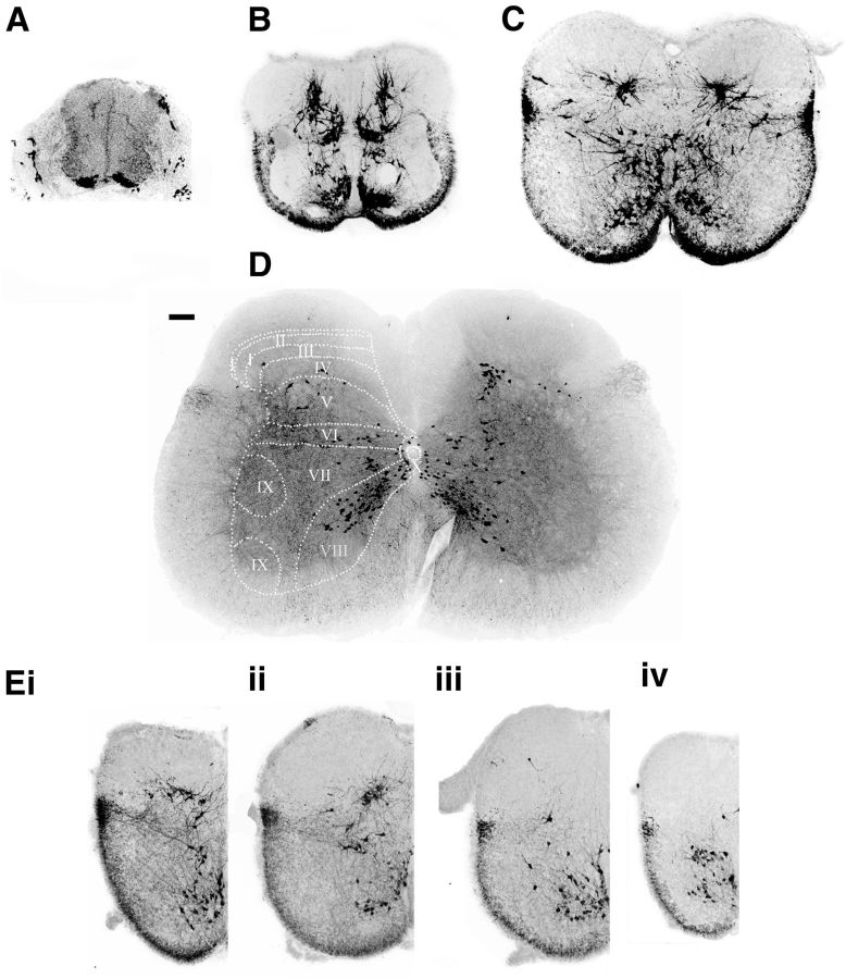
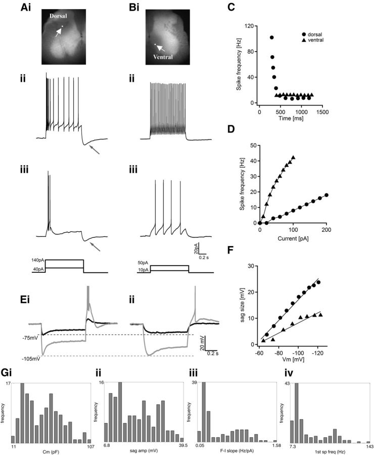
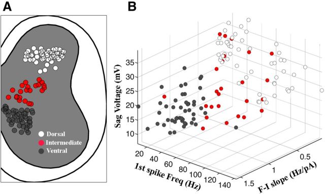
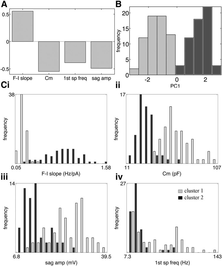
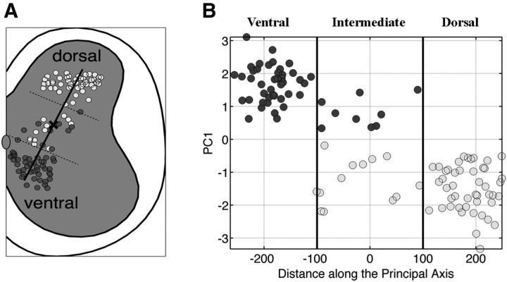
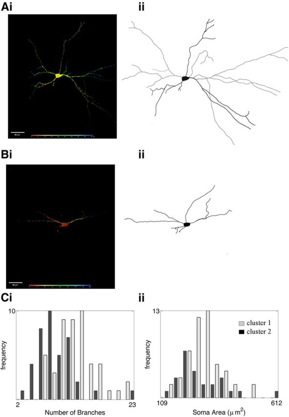
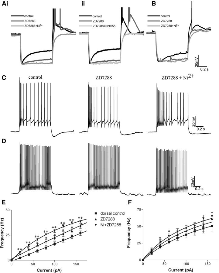
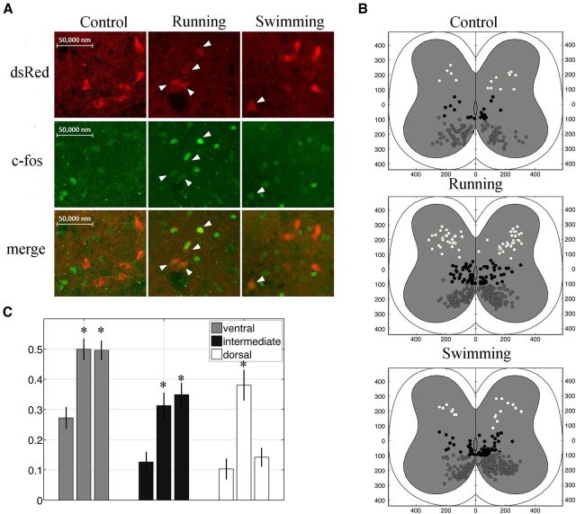
V3 interneurons (INs) are a major group of excitatory commissural interneurons in the spinal cord, and they are essential for producing a stable and robust locomotor rhythm. V3 INs are generated from the ventral-most progenitor domain, p3, but migrate dorsally and laterally during postmitotic development. At birth, they are located in distinctive clusters in the ventral horn and deep dorsal horn. To assess the heterogeneity of this genetically identified group of spinal INs, we combined patch-clamp recording and anatomical tracing with cluster analysis. We examined electrophysiological and morphological properties of mature V3 INs identified by their expression of tdTomato fluorescent proteins in Sim1(Cre/+); Rosa(floxstop26TdTom) mice. We identified two V3 subpopulations with distinct intrinsic properties and spatial distribution patterns. Ventral V3 INs, primarily located in lamina VIII, possess a few branching processes and were capable of generating rapid tonic firing spikes. By contrast, dorsal V3 INs exhibited a more complex morphology and relatively slow average spike frequency with strong adaptation, and they also displayed large sag voltages and post-inhibitory rebound potentials. Our data suggested that hyperpolarization-activated cation channel currents and T-type calcium channel currents may account for some of the membrane properties of V3 INs. Finally, we observed that ventral and dorsal V3 INs were active in different ways during running and swimming, indicating that ventral V3 INs may act as premotor neurons and dorsal V3 INs as relay neurons mediating sensory inputs. Together, we detected two physiologically and topographically distinct subgroups of V3 INs, each likely playing different roles in locomotor activities.


Figures

Figure 1.
Figure 1.
Distribution patterns of V3 INs in the mouse spinal cord. A–D, The 30 μm transverse sections from lumbar segments of spinal cords from Sim1Cre/+; Rosafloxstop26TdTommice, at E10.5 (A), E14.5 (B), P0 (C), and P21 (D). Scale bar, 100 μm. The dashed lines and numbers indicate the Rexed's laminae. E, The 30 μm half-transverse sections from a P0 spinal cord at thoracic (i), upper lumbar (ii), lower lumbar (iii), and sacral (iv) regions.
Figure 2.
Figure 2.
Heterogeneous electrophysiological properties of spinal V3 interneurons. A, B, representative traces from two V3 INs responding to 1 s suprathreshold current steps. A, A dorsal V3 IN labeled by neurobiotin (Ai) displays initial bursts at low (Aiii, 40 pA) and high (Aii, 140 pA) injected currents. B, A ventral V3 IN (Bi) maintains tonic firing with 10 pA (Biii) and 50 pA (Bii) injected currents. White arrows in Ai and Bi point to the recorded cells; neuron's bodies are labeled with a white dot to show their position. Black arrows in Aii and iii point to the afterhyperpolarization potential after the depolarization pulses. The schematic diagrams show the injected currents. C, individual spike frequency is plotted versus time during 1 s injected current on dorsal cell (circles) and ventral cell (triangles). D, Average spike frequency of dorsal cells (circles) and ventral cells (triangles) was plotted versus injected currents. E, Representative traces from two V3 neurons hyperpolarized to −75 mV (black) and −105 mV (gray). The dashed lines indicate the membrane potentials. F, Plots of the size of sag voltage versus membrane potentials of the two cells. G, Histograms showing the distribution frequency of various electrophysiological properties of all recorded V3 neurons: Cm (i), sag voltage amplitude (ii), slope of f–I plot (iii), and frequency of first spike (first sp freq, iv).
Figure 3.
Figure 3.
Spatial distribution of recorded V3 neurons. A, All recorded V3 INs were mapped on a half-cross section of spinal cord, and divided into ventral (dark gray), intermediate (red) and dorsal (white) groups). B, The slope of the f–I plot was plotted versus the first spike frequency and sag voltage amplitude.
Figure 4.
Figure 4.
Cluster analysis of V3 neurons based on electrophysiological properties. A, The first eigenvector of four attributes (f–I slope, Cm, first spike frequency, and sag amplitude at −120 mV) that produced the best criterion. B, Histogram of PC1 exhibits two well defined modes corresponding to positive and negative values representing cluster 1 (PC1 <0, light gray) and cluster 2 (PC1 >0, dark gray). Ci–iv, Histogram of distribution of the four main attributes (A) with cells in cluster 1 (light gray) and cluster 2 (dark gray).
Figure 5.
Figure 5.
Electrophysiological clusters of V3 INs are topologically distributed in the spinal cord. A, Cross-section map of V3 neurons divided into cluster 1 (light gray) and cluster 2 (dark gray). The dark line indicates the principal axis of the location of cells in the cross section, and a bold “x” labels the center of the axis. Dashed lines are the presumed borders of ventral, intermediate, and dorsal separations. B, Plot of the value of PC1 as a function of distance along the axis. The dashed lines in A and the dark lines in B indicate the arbitrary borders to separate the ventral, intermediate, and dorsal populations.
Figure 6.
Figure 6.
The morphological properties of dorsal and ventral V3 clusters. Ai, Bi, Confocal images of representative V3 INs from clusters 1 (Ai) and 2 (Bi). Colors reflect the depth of the image from the surface (red) to deep into the section (blue). Aii, Bii, Reconstruction of corresponding cells in Ai and Bi. Ci, Distribution histogram of primary branches of cells in clusters 1 (light gray) and 2 (dark gray). Cii, Distribution histogram of soma areas of cells in clusters 1 (light gray) and 2 (dark gray).
Figure 7.
Figure 7.
The effects of different channel blockers on the electrophysiological properties of V3 INs. A, Effects of ZD7288 (dark gray) and ZD7288 + nickel (light gray, Ai) or ZD7288 + NNC 55–0396 (Aii) on the sag voltage and rebound reactions of representative dorsal V3 INs from cluster 1. B, Effects of ZD7288 (dark gray) and ZD7288 + nickel (light gray) on cluster2. C, D, Effects of ZD7288 and ZD7288 + Ni on the spike patterns of representative cells from cluster 1 (C) and cluster 2 (D) responding to 1 s suprathreshold currents. E, F, f–I plots of cells from cluster 1 (E) and cluster 2 (F). *p < 0.05; **p < 0.01.
Figure 8.
Figure 8.
Differential recruitment of V3 INs in running and swimming behaviors. A, Confocal image of dorsal V3 neurons (red) expressing c-Fos protein (green) at rest or after running and swimming. B, c-Fos-positive V3 INs are mapped onto cross sections of lumbar cords from control (Bi), running (Bii), and swimming (Biii) mice. Cells are divided into ventral (gray), intermediate (black), and dorsal (white). C, Histogram showing proportion of c-Fos-positive V3 INs relative to the total number V3 INs at different positions. *p < 0.01.

Similar articles

Cited by

References

    1. Alvarez FJ, Benito-Gonzalez A, Siembab VC. Principles of interneuron development learned from Renshaw cells and the motoneuron recurrent inhibitory circuit. Ann N Y Acad Sci. 2013;1279:22–31. doi: 10.1111/nyas.12084. - DOI - PMC - PubMed
    1. Arber S. Motor circuits in action: specification, connectivity, and function. Neuron. 2012;74:975–989. doi: 10.1016/j.neuron.2012.05.011. - DOI - PubMed
    1. Ascoli GA, Alonso-Nanclares L, Anderson SA, Barrionuevo G, Benavides-Piccione R, Burkhalter A, Buzsáki G, Cauli B, Defelipe J, Fairén A, Feldmeyer D, Fishell G, Fregnac Y, Freund TF, Gardner D, Gardner EP, Goldberg JH, Helmstaedter M, Hestrin S, Karube F, et al. Petilla terminology: nomenclature of features of GABAergic interneurons of the cerebral cortex. Nat Rev Neurosci. 2008;9:557–568. doi: 10.1038/nrn2402. - DOI - PMC - PubMed
    1. Baccei ML, Fitzgerald M. Intrinsic firing properties of developing rat superficial dorsal horn neurons. Neuroreport. 2005;16:1325–1328. doi: 10.1097/01.wnr.0000175612.08560.10. - DOI - PubMed
    1. Blacklaws J. Dalhousie University; 2013. The development of V3 interneurons in the mouse spinal cord. PhD thesis.

Publication types

MeSH terms

LinkOut - more resources