. 2014 Jan 15;127(Pt 2):411-21.
doi: 10.1242/jcs.136028. Epub 2013 Nov 20.

The Smad7-Skp2 complex orchestrates Myc stability, impacting on the cytostatic effect of TGF-β

Affiliations

The Smad7-Skp2 complex orchestrates Myc stability, impacting on the cytostatic effect of TGF-β

Tae-Aug Kim et al. J Cell Sci. .

Abstract

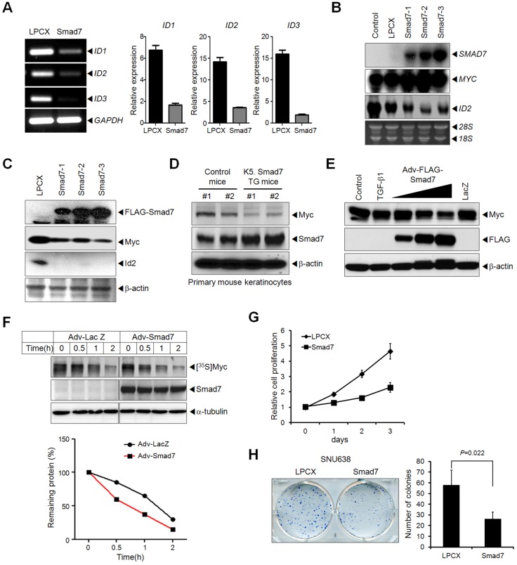
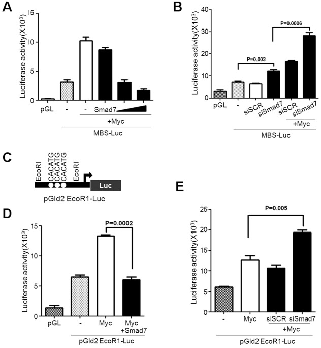
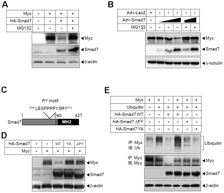
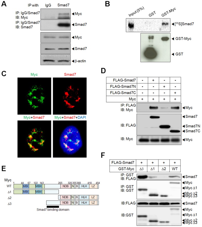
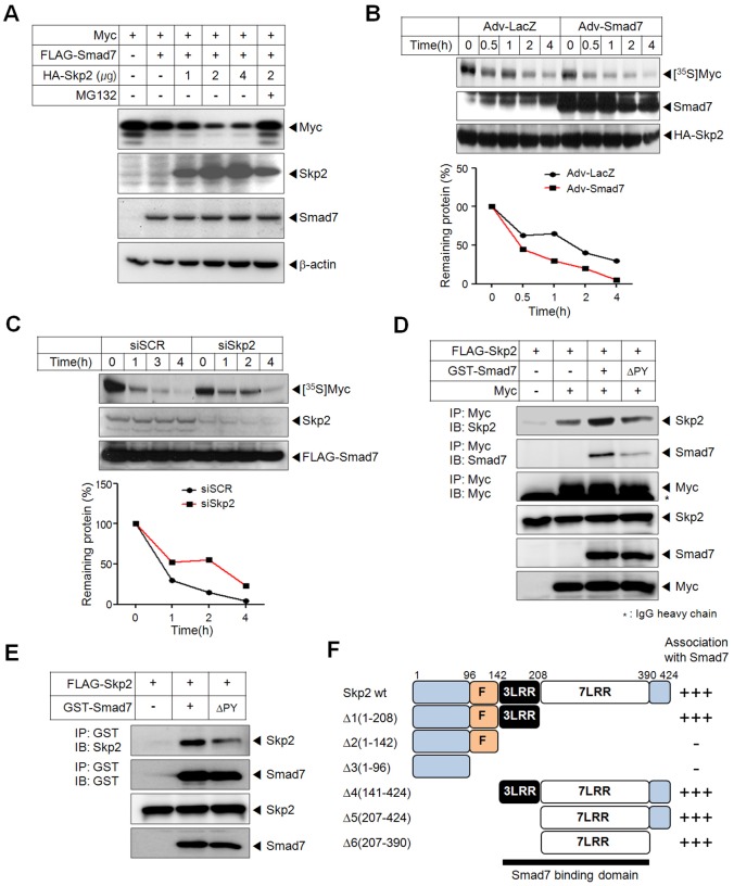
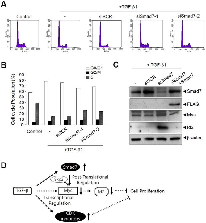
In most human cancers the Myc proto-oncogene is highly activated. Dysregulation of Myc oncoprotein contributes to tumorigenesis in numerous tissues and organs. Thus, targeting Myc stability could be a crucial step for cancer therapy. Here we report Smad7 as a key molecule regulating Myc stability and activity by recruiting the F-box protein, Skp2. Ectopic expression of Smad7 downregulated the protein level of Myc without affecting the transcription level, and significantly repressed its transcriptional activity, leading to inhibition of cell proliferation and tumorigenic activity. Furthermore, Smad7 enhanced ubiquitylation of Myc through direct interaction with Myc and recruitment of Skp2. Ablation of Smad7 resulted in less sensitivity to the growth inhibitory effect of TGF-β by inducing stable Myc expression. In conclusion, these findings that Smad7 functions in Myc oncoprotein degradation and enhances the cytostatic effect of TGF-β signaling provide a possible new therapeutic approach for cancer treatment.

Keywords: Myc; Protein stability; Skp2; Smad7; Ubiquitylation.


Figures

Fig. 1.
Fig. 1.
Regulation of ID family genes and Myc expression by Smad7. (A) Semi-quantitative and quantitative RT-PCR analysis was performed in the SNU638-LPCX and -Smad7 cell lines using primers for ID family genes. GAPDH was used as an internal control. (B) The expression of ID2, MYC and Smad7 mRNA was examined by northern blot analysis. 18S and 28S rRNAs were used as the internal loading control. (C) Immunoblot analysis showed the expression of Id2 and Myc. β-actin was used as a loading control. (D) Regulation of Myc expression by induction of Smad7 in primary epidermal keratinocytes isolated from neonatal mouse skin of two K5.Smad7 transgenic and two control mice. Expression of Myc and Smad7 were examined by western blot analysis. (E) After infection with increased amount of adenoviral FLAG-Smad7 or control lacZ, HaCaT-tetoffMyc cells were cultured for 24 hours without doxycycline. A group was treated with TGF-β1 (5 ng/ml) for 24 hours. The level of proteins in each group was checked by immunoblotting. (F) A pulse–chase experiment was performed using HaCaT cells infected with an adenovirus expressing either FLAG-Smad7 or β-galactosidase (LacZ). 35S-labeled Myc proteins were immunoprecipitated and analyzed by autoradiography (upper panel) and quantified (lower panel). (G) The effect of Smad7 on cell proliferation was examined by the comparison of the number of viable SNU638-LPCX and SNU638-Smad7 cells. Relative cell proliferation was calculated as the number of cells at the indicated time divided by that at day 0. (H) Inhibition of a tumorigenic activity by Smad7, which was measured using a colony forming assay in the SNU638 cell line. Graph represents quantification of colonies from triplicate samples (right panel; P = 0.022).
Fig. 2.
Fig. 2.
Regulation of Myc transcriptional activity by Smad7. (A) HaCaT cells were transfected with a Myc responsive reporter gene construct (MBS-Luc) and a β-galactosidase expression construct (β-gal) without or with Myc and Smad7. A luciferase assay showed Myc transcriptional activity. (B) After transfection of the MBS-Luc and β-gal constructs with Myc and/or Smad7 siRNA (40 nM) or scrambled siRNA (siSCR; 40 nM), equal amounts of cell lysate was subjected to a luciferase assay. (C) Schematic representation of the human Id2 promoter reporter construct pGId2EcoR1-Luc. (D) To examine the effect of Smad7 on Myc-mediated activation of the Id2 promoter, the pGId2EcoR1-Luc plasmid was transfected into HaCaT cells, together with Myc in the absence or presence of FLAG-Smad7. (E) HaCaT cells were transfected with pGId2EcoR1-Luc together with Myc and Smad7 siRNA (40 nM) or siSCR (40 nM). Luciferase activity was measured 2 days after transfection. All experiments were performed three times independently and β-galactosidase activity was measured to normalize luciferase activity.
Fig. 3.
Fig. 3.
Smad7 induces degradation of Myc through the ubiquitin-proteasomal pathway. (A) Myc and Smad7 constructs were transfected into HEK 293T cells. The cells were then treated with the proteasome inhibitor, MG132 (2 µM) for 4 hours before harvesting. Total cell lysates was subjected to western blot analysis. (B) HaCaT tetonMyc cells were infected with the adenoviral lacZ or Smad7 constructs (Adv-Lacz and Adv-Smad7). To induce Myc expression, cells were treated with doxycycline (1 µg/ml) for 24 hours. Expression level of Myc, Smad7 and α-tubulin were examined by western blotting. (C) A schematic diagram of Smad7 domains. (D) HEK 293T cells were transfected with HA-Smad7 wild-type (WT) or mutants [ΔPY and Y211A (YA)], together with Myc. (E) Cells were co-transfected with Myc and HA-Smad7 constructs and/or pcDNA3-ubiquitin as indicated. Immunoprecipitation using anti-Myc antibodies was performed, and the blot was probed with the indicated antibodies.
Fig. 4.
Fig. 4.
Smad7 directly interacts with Myc. (A) Interaction between endogenous Myc and Smad7 protein was examined in HaCaT cells by immunoprecipitation with anti-Smad7 antibody. (B) 35S-labeled Smad7 proteins were pulled down with the indicated GST fusions and visualized by autoradiography (upper panel). 5% of 35S-labeled Smad7 input proteins were loaded as positive control. (C) Confocal analysis revealed co-localization of Smad7 and Myc in the nucleus. After inducing Myc expression, HaCaT tetonMyc cells were immunostained with anti-Smad7 (red) and anti-Myc (green) antibodies. The nuclei were stained with DAPI. (D) HEK 293T cells were transfected with Myc together with full-length, N-terminal (aa 1–203) and C-terminal (aa 204–427) FLAG-Smad7 plasmids. Cell extracts were subjected to immunoprecipitation with anti-FLAG antibody, followed by immunoblotting. (E) A schematic diagram of Myc constructs and mapping of Smad7-binding domains in Myc. MBI, Myc box I; MBII, Myc box II; NDB, non-specific DNA binding; N, nuclear localization signal; B, basic domain; HLH, helix-loop-helix domain, LZ, leucine-zipper domain. (F) GST-Myc constructs together with full-length Flag-Smad7 were transfected into HEK 293T cells. Total cell lysates were prepared from cells treated with MG132 and immunoprecipitated with anti-GST antibody.
Fig. 5.
Fig. 5.
Smad7 recruits the F-box protein, Skp2, for Myc degradation. (A) HEK 293T cells were transfected with Myc together with Smad7 in the absence or presence of an increasing amount of Skp2, as indicated. The cells were then treated with MG132 (2 µM) for 4 hours. (B) A pulse–chase experiment was performed in cells transfected with Skp2, infected with adenovirus carrying lacZ or FLAG-Smad7 and labeled with [35S]methionine. The remaining proteins were quantified as indicated (bottom). (C) HaCaT cells were transfected with Skp2 siRNA (siSkp2, 40 nM) or siSCR, followed by infection with Smad7 adenovirus. A pulse–chase experiment was performed as described above. (D) HEK 293T cells were transfected with FLAG-Skp2 and/or Myc, together with GST-Smad7 WT or ΔPY. Immunoprecipitation was performed with anti-Myc antibody, followed by western blotting with the indicated antibodies. (E) Interaction of Smad7 with Skp2 was demonstrated by transfection of Skp2 with the Smad7 WT or ΔPY mutant into HEK 293T cells. GST pull-down showed that the Smad7 ΔPY mutant diminished the interaction of Skp2 with Smad7. (F) Schematic diagram of the Smad7-binding domain in Skp2, which was confirmed by immunoprecipitation (supplementary material Fig. S8C,D). F, F-box; 3LRR, 3 leucine-rich repeats; 7LRR, 7 leucine-rich repeats.
Fig. 6.
Fig. 6.
Cells lacking Smad7 are less sensitive to the cytostatic effect of TGF-β. (A) Flow cytometric analysis of HaCaT cells transfected with Smad7 siRNA 24 hours after treatment with TGF-β1 (5 ng/ml). (B) Quantification of the cell cycle profile, as above. (C) HaCaT cells transfected with Smad7 siRNA or siSCR were treated with TGF-β1 for 24 hours. Equal amounts of cell extracts were analyzed by western blotting with the indicated antibodies. (D) Schematic model of Smad7-mediated growth control in TGF-β signaling.

References

    1. Adhikary S., Marinoni F., Hock A., Hulleman E., Popov N., Beier R., Bernard S., Quarto M., Capra M., Goettig S. et al. (2005). The ubiquitin ligase HectH9 regulates transcriptional activation by Myc and is essential for tumor cell proliferation. Cell 123, 409–421 10.1016/j.cell.2005.08.016 - DOI - PubMed
    1. Albihn A., Johnsen J. I., Henriksson M. A. (2010). MYC in oncogenesis and as a target for cancer therapies. Adv. Cancer Res. 107, 163–224 10.1016/S0065--230X(10)07006--5 - DOI - PubMed
    1. Aragón E., Goerner N., Xi Q., Gomes T., Gao S., Massagué J., Macias M. J. (2012). Structural basis for the versatile interactions of Smad7 with regulator WW domains in TGF-β Pathways. Structure 20, 1726–1736 10.1016/j.str.2012.07.014 - DOI - PMC - PubMed
    1. Bahram F., von der Lehr N., Cetinkaya C., Larsson L. G. (2000). c-Myc hot spot mutations in lymphomas result in inefficient ubiquitination and decreased proteasome-mediated turnover. Blood 95, 2104–2110 - PubMed
    1. Bhatia K., Huppi K., Spangler G., Siwarski D., Iyer R., Magrath I. (1993). Point mutations in the c-Myc transactivation domain are common in Burkitt's lymphoma and mouse plasmacytomas. Nat. Genet. 5, 56–61 10.1038/ng0993--56 - DOI - PubMed

Publication types

MeSH terms