Review
. 2013 Nov 14;19(42):7247-57.
doi: 10.3748/wjg.v19.i42.7247.

Ultrasonography in diagnosing chronic pancreatitis: new aspects

Affiliations
Review

Ultrasonography in diagnosing chronic pancreatitis: new aspects

Georg Dimcevski et al. World J Gastroenterol. .

Abstract

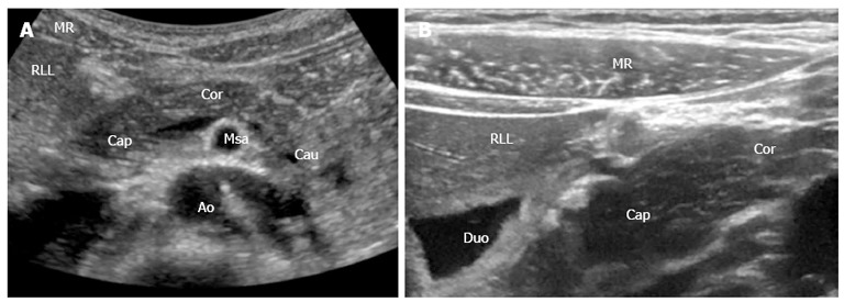
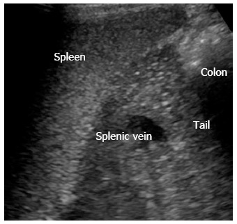
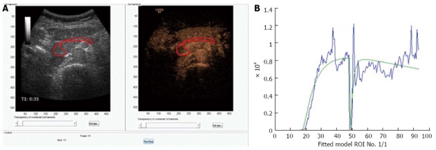
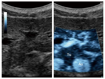
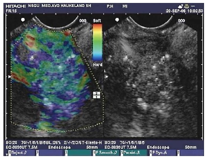
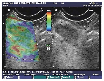
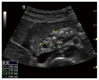
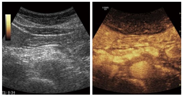
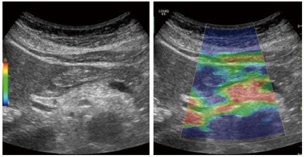
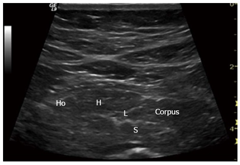
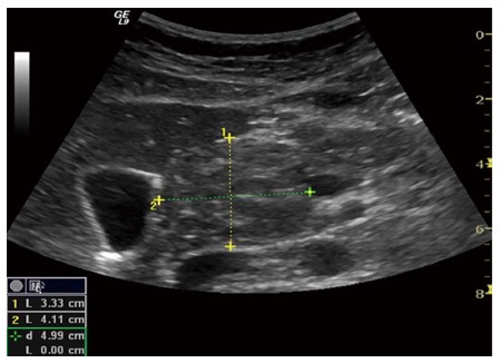
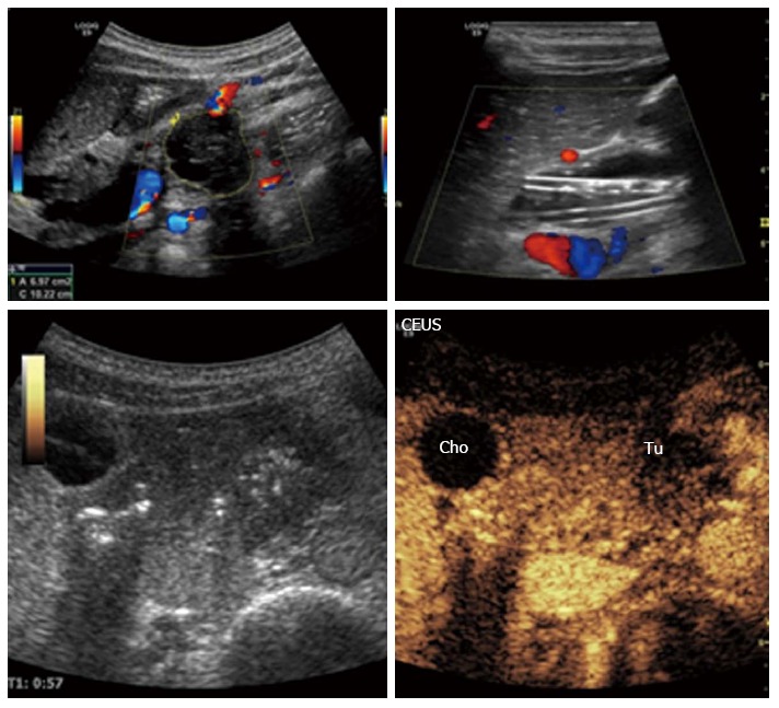
The course and outcome is poor for most patients with pancreatic diseases. Advances in pancreatic imaging are important in the detection of pancreatic diseases at early stages. Ultrasonography as a diagnostic tool has made, virtually speaking a technical revolution in medical imaging in the new millennium. It has not only become the preferred method for first line imaging, but also, increasingly to clarify the interpretation of other imaging modalities to obtain efficient clinical decision. We review ultrasonography modalities, focusing on advanced pancreatic imaging and its potential to substantially improve diagnosis of pancreatic diseases at earlier stages. In the first section, we describe scanning techniques and examination protocols. Their consequences for image quality and the ability to obtain complete and detailed visualization of the pancreas are discussed. In the second section we outline ultrasonographic characteristics of pancreatic diseases with emphasis on chronic pancreatitis. Finally, new developments in ultrasonography of the pancreas such as contrast enhanced ultrasound and elastography are enlightened.

Keywords: Chronic pancreatitis; Contrast enhanced ultrasonography; Elastography; Medical imaging technique; Pancreas; Strain imaging; Transabdominal ultrasound; Ultrasonography.


Figures

Figure 1
Figure 1
Pancreas and the surrounding anatomical landmarks. A: B-mode image (1-5 MHz); B: B-mode image with a 12-15 MHz transducer. Details shown with high resolution. MR: Musculus rectus abdominis; RLL: Right liver lobe; Cap: Caput pancreatis; Cor: Corpus pancreatis; Cau: Cauda pancreatis; Msa: Superior mesenteric artery; Duo: Duodenum; Ao: Aorta.
Figure 2
Figure 2
Left lateral side scan shows the pancreatic tail (cauda) using the spleen as acoustic window.
Figure 3
Figure 3
Perfusion analysis of the pancreas. A: Dual view of contrast-enhanced ultrasonography examination of the pancreas in a healthy volunteer. 1.5 mL bolus of Sonovue was given as a bolus and after approximately 45 s the area of interest was exposed to high MI ultrasound bursting the bubbles in the imaging plane; B: A motion correcting analyzing software was used (DCE-US, http://www.isibrno.cz/perfusion/). A region of interest have been drawn including the head and body of the pancreas (unpublished data).
Figure 4
Figure 4
Ultrasound targeted treatment of pancreatic cancer using combined microbubbles and a chemotherapeutic. Left: B-mode frame. Right: The main tumor with spicules, clearly demarked, using the sonoporation settings. Tu: Tumor; Ao: Aorta.
Figure 5
Figure 5
Endoscopic ultrasound with elastography of chronic pancreatitis. Endoscopic ultrasonography B-mode sonogram (right) and an elastogram superimposed a sonogram (left). In this image of the pancreatic head, hyperechoic foci and strands are seen in the parenchyma, as well as inhomogeneous echogenecity, which are signs of chronic pancreatitis. The elastogram shows predominantly a blue, indicating harder tissue, and green representing intermediate hardness in a honeycomb pattern over the pancreatic tissue.
Figure 6
Figure 6
Endoscopic ultrasonography B-mode sonogram and elastogram of lymph node in chronic pancreatitis. The sonogram (right) shows a lymph node as a hypoechoic oval shape surrounded by more echogenic tissue in the liver hilum. This lymph node approximately 18 mm × 10 mm, appeared in the liver hilum of a patient with chronic pancreatitis. On the left, the lymph node is not harder than the surrounding tissue as the predominant color hue is green. This finding is frequent in reactive lymph nodes, and may be a sign of benign etiology.
Figure 7
Figure 7
Advanced chronic pancreatitis. Classical signs in advanced chronic pancreatitis: main pancreatic duct dilatation in an atrophic organ with sharp, irregular contours, calcifications and small cysts. The pancreatic head is outlined.
Figure 8
Figure 8
An example of contrast enhanced ultrasound in advanced chronic pancreatitis. The parenchymal enhancement is clearly irregular reflecting the parenchymal heterogeneity, calcifications and focal inflammation.
Figure 9
Figure 9
Elastography of the pancreas in moderate chronic pancreatitis. The colors show tissue hardness; The scale on the left defines the color code: Blue is hard, red is soft, yellow and green are intermediate. The elastogram shows predominantly soft (red) tissue with parts of green and yellow, indicating harder pancreatic tissue.
Figure 10
Figure 10
Early chronic pancreatitis. Typical signs in early chronic pancreatitis: lobularity (L), stranding (S), hyperechoic foci (H) and honeycombing (Ho). Pancreatic body (Corpus). This subtle changes are usually only seen in endoscopic ultrasonography.
Figure 11
Figure 11
Autoimmune pancreatitis. Enlarged pancreas with a tumorous formation in the head. The inflammatory lesion is isoechoic to the rest of the pancreas but still clearly visible.
Figure 12
Figure 12
Malignant tumor of the pancreatic body: Ductal adenocarcinoma. The tumor is clearly visible on B-mode, left upper image. Contrast enhanced ultrasonography: The characteristics are hypoechogenicity and diffuse contours. The plastic biliary stents are clearly noticeable on B-mode, right upper image. Cho: Biliary duct (ductus choledochus); Tu: Tumor; CEUS: Contrast-enhanced ultrasonography.

Similar articles

Cited by

References

    1. Gilja OH, Hatlebakk JG, Odegaard S, Berstad A, Viola I, Giertsen C, Hausken T, Gregersen H. Advanced imaging and visualization in gastrointestinal disorders. World J Gastroenterol. 2007;13:1408–1421. - PMC - PubMed
    1. Rösch T, Schusdziarra V, Born P, Bautz W, Baumgartner M, Ulm K, Lorenz R, Allescher HD, Gerhardt P, Siewert JR, et al. Modern imaging methods versus clinical assessment in the evaluation of hospital in-patients with suspected pancreatic disease. Am J Gastroenterol. 2000;95:2261–2270. - PubMed
    1. Swobodnik W, Meyer W, Brecht-Kraus D, Wechsler JG, Geiger S, Malfertheiner P, Junge U, Ditschuneit H. Ultrasound, computed tomography and endoscopic retrograde cholangiopancreatography in the morphologic diagnosis of pancreatic disease. Klin Wochenschr. 1983;61:291–296. - PubMed
    1. Rettenmaier G. [Methods and results of ultrasonography in the diagnosis of pancreatic diseases] Leber Magen Darm. 1972;2:88–90. - PubMed
    1. Russell JG, Vallon AG, Braganza JM, Howat HT. Ultrasonic scanning in pancreatic disease. Gut. 1978;19:1027–1033. - PMC - PubMed

LinkOut - more resources