

Differentiation between dysplastic nodule and early-stage hepatocellular carcinoma: the utility of conventional MR imaging
- PMID: 24259975
- PMCID: PMC3831226
- DOI: 10.3748/wjg.v19.i42.7433
Differentiation between dysplastic nodule and early-stage hepatocellular carcinoma: the utility of conventional MR imaging
Abstract
Aim: To elucidate the variety of ways early-stage hepatocellular carcinoma (HCC) can appear on magnetic resonance (MR) imaging by analyzing T1-weighted, T2-weighted, and gadolinium-enhanced dynamic studies.
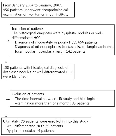
Methods: Seventy-three patients with well-differentiated HCC (wHCC) or dysplastic nodules were retrospectively identified from medical records, and new histological sections were prepared and reviewed. The tumor nodules were categorized into three groups: dysplastic nodule (DN), wHCC compatible with Edmondson-Steiner grade I HCC (w1-HCC), and wHCC compatible with Edmondson-Steiner grade II HCC (w2-HCC). The signal intensity on pre-contrast MR imaging and the enhancing pattern for each tumor were recorded and compared between the three tumor groups.
Results: Among the 73 patients, 14 were diagnosed as having DN, 40 were diagnosed as having w1-HCC, and 19 were diagnosed as having w2-HCC. Hyperintensity measurements on T2-weighted axial images (T2WI) were statistically significant between DNs and wHCC (P = 0.006) and between DN and w1-HCC (P = 0.02). The other imaging features revealed no significant differences between DN and wHCC or between DN and w1-HCC. Hyperintensity on both T1W out-phase imaging (P = 0.007) and arterial enhancement on dynamic study (P = 0.005) showed statistically significant differences between w1-HCC and w2-HCC. The other imaging features revealed no significant differences between w1-HCC and w2-HCC.
Conclusion: In the follow-up for a cirrhotic nodule, increased signal intensity on T2WI may be a sign of malignant transformation. Furthermore, a noted loss of hyperintensity on T1WI and the detection of arterial enhancement might indicate further progression of the histological grade.
Keywords: Dysplastic nodule; Hepatocellular carcinoma; Histological grading; Magnetic resonance imaging; Well-differentiated hepatocellular carcinoma.
Figures
References
-
- Parkin DM, Bray F, Ferlay J, Pisani P. Global cancer statistics, 2002. CA Cancer J Clin. 2005;55:74–108. - PubMed
-
- Donato F, Tagger A, Chiesa R, Ribero ML, Tomasoni V, Fasola M, Gelatti U, Portera G, Boffetta P, Nardi G. Hepatitis B and C virus infection, alcohol drinking, and hepatocellular carcinoma: a case-control study in Italy. Brescia HCC Study. Hepatology. 1997;26:579–584. - PubMed
-
- Kudo M. Multistep human hepatocarcinogenesis: correlation of imaging with pathology. J Gastroenterol. 2009;44 Suppl 19:112–118. - PubMed
-
- Szklaruk J, Silverman PM, Charnsangavej C. Imaging in the diagnosis, staging, treatment, and surveillance of hepatocellular carcinoma. AJR Am J Roentgenol. 2003;180:441–454. - PubMed
Publication types
MeSH terms
Substances
LinkOut - more resources
Full Text Sources
Other Literature Sources
Medical
