Case Reports
. 2013 Nov 14;19(42):7480-6.
doi: 10.3748/wjg.v19.i42.7480.

Successful treatment of multiple hepatocellular adenomas with percutaneous radiofrequency ablation

Affiliations
Case Reports

Successful treatment of multiple hepatocellular adenomas with percutaneous radiofrequency ablation

Sun Young Ahn et al. World J Gastroenterol. .

Abstract

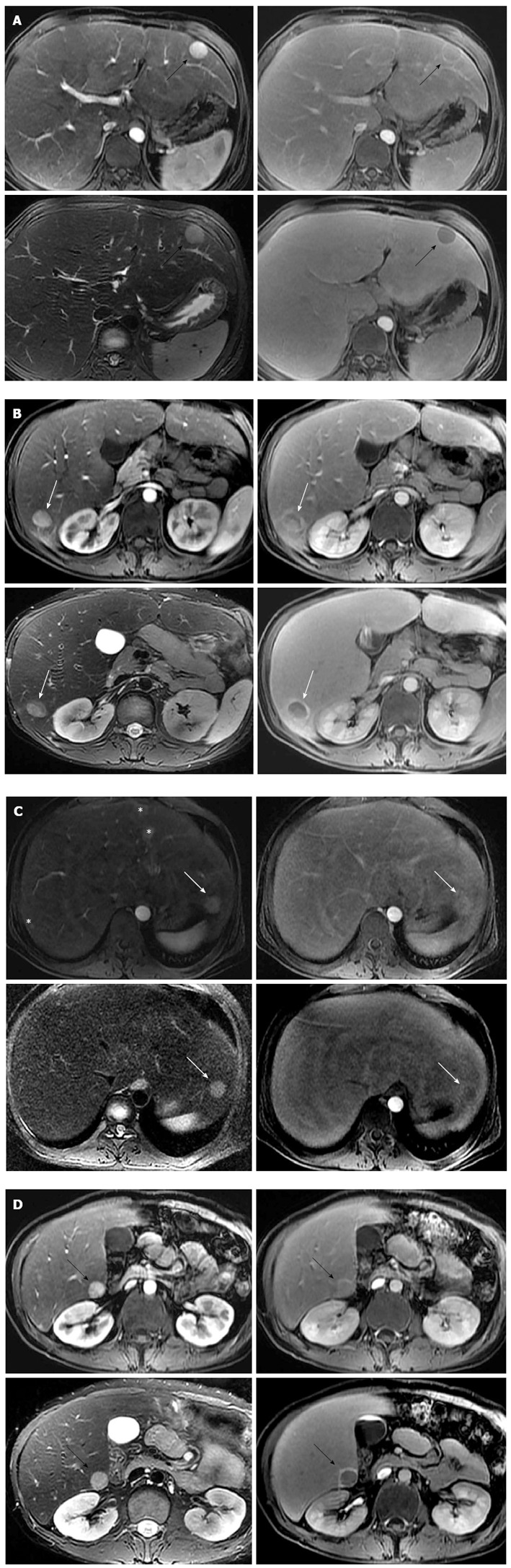
Hepatocellular adenoma (HCA) is one of the important complications of glycogen storage disease type Ia (GSD-Ia) because it can be transformed into hepatocellular carcinoma. Although surgical resection is a standard treatment of choice for solitary HCA, multiple HCAs in GSD-Ia patients present as therapeutic challenges for curative treatment. Therefore, treatment strategy according to malignant potential is important in management of HCAs in GSD-Ia. The authors present a case of histologically proven multiple HCAs without β-catenin mutations occurred in a GSD-Ia patient treated successfully with percutaneous radiofrequency ablation as a minimally invasive therapy.

Keywords: Glycogen storage disease; Hepatocellular adenoma; Radiofrequency ablation; β-catenin activation.


Figures

Figure 1
Figure 1
Hepatic adenomas associated with glycogen storage disease type Ia. A: Gd-EOB-DTPA enhanced axial T1-weighted image on arterial phase (left upper) reveals approximately 2 cm intense enhancing mass in the segment 3 of liver (black arrows). The mass shows nearly iso-signal intensity with capsular enhancement on 3 min delayed phase (right upper), intermediate high signal intensity on axial T2-weighted image (left lower), and clear defect on 20 min hepatobiliary phase (right lower); B: Approximately 1.8 cm sized intense enhancing mass on arterial phase (left upper) is noted in the segment 6 of liver. This mass shows suspicious focal eccentric wash-out enhancement (white arrows) on 3 min delayed phase (right upper). This portion reveals relatively low signal intensity on axial T2-weighted image (left lower) and 20 min hepatobiliary phase (right lower), compared to other tumor area. Surrounding rim enhancement in the tumor is seen on 20 min hepatobiliary phase; C: Approximately 1.8 cm arterial enhancing mass (white arrows) is seen in the segment 2 of liver. The mass reveals slightly high signal intensity on 3 min delayed phase (right upper), intermediate high signal intensity on axial T2-weighted image (left lower), and fuzzy defect on 20 min hepatobiliary phase (right lower). Multifocal arterioportal shunts (asterisks) are noted in the segments 3 and 7 of liver; D: Approximately 1.8 cm sized intense enhancing mass (black arrows) on arterial phase (left upper) is noted in the segment 6 of liver. This mass shows slightly high signal intensity with capsular enhancement on 3min delayed phase (right upper), and intermediate high signal intensity on axial T2-weighted image (left lower). On 20 min hepatobiliary phase (right lower), the tumor reveals clear defect with surrounding rim enhancement. Gd-EOB-DTPA: Gadolinium ethoxybenzyl diethylene triamine pentaacetic acid.
Figure 2
Figure 2
The histological features are consistent with the inflammatory type of hepatocellular adenoma. A: A hepatocellular adenoma in GSD-Ia has several unpaired arteries (thick black arrows), sinusoidal dilation (thin black arrows), and steatosis (white arrows), but the atypia of hepatocytes are low without a nuclear pleomorphism; B: A reticulin staining does not show thick hepatic cords; C: The lymphocytic infiltrates are found in a hepatic lobule; D: The lymphocytes infiltrate in the area surrounded a portal and a periportal tracts; E: Staining for glutamine synthetase is diffuse cytoplasmic with focal nuclear expression; F: β-catenin staining does not show abnormal expression. GSD-Ia: Glycogen storage disease type Ia.
Figure 3
Figure 3
Computed tomogram follow-up after 3 mo showed gradual reduction of radiofrequency ablation zone. A: Post-RFA follow-up CT revealed gradual contraction of ablated lesion in segment 3 to 1 cm; B: Complete ablation of enhancing lesion in segment 6 was observed in Post-RFA follow-up CT; C: Post-RFA follow-up CT showed complete ablation of 1.8 cm sized tumor in segment 2; D: Post-RFA CT revealed no residual lesion without adjacent organ damage. CT: Computed tomogram; RFA: Radiofrequency ablation.

Similar articles

Cited by

References

    1. Franco LM, Krishnamurthy V, Bali D, Weinstein DA, Arn P, Clary B, Boney A, Sullivan J, Frush DP, Chen YT, et al. Hepatocellular carcinoma in glycogen storage disease type Ia: a case series. J Inherit Metab Dis. 2005;28:153–162. - PubMed
    1. Wang DQ, Fiske LM, Carreras CT, Weinstein DA. Natural history of hepatocellular adenoma formation in glycogen storage disease type I. J Pediatr. 2011;159:442–446. - PMC - PubMed
    1. Sakellariou S, Al-Hussaini H, Scalori A, Samyn M, Heaton N, Portmann B, Tobal K, Quaglia A. Hepatocellular adenoma in glycogen storage disorder type I: a clinicopathological and molecular study. Histopathology. 2012;60:E58–E65. - PubMed
    1. Lee PJ. Glycogen storage disease type I: pathophysiology of liver adenomas. Eur J Pediatr. 2002;161 Suppl 1:S46–S49. - PubMed
    1. Bioulac-Sage P, Rebouissou S, Thomas C, Blanc JF, Saric J, Sa Cunha A, Rullier A, Cubel G, Couchy G, Imbeaud S, et al. Hepatocellular adenoma subtype classification using molecular markers and immunohistochemistry. Hepatology. 2007;46:740–748. - PubMed

Publication types

Supplementary concepts