. 2013 Nov 8;8(11):e78388.
doi: 10.1371/journal.pone.0078388. eCollection 2013.

Plakoglobin represses SATB1 expression and decreases in vitro proliferation, migration and invasion

Affiliations

Plakoglobin represses SATB1 expression and decreases in vitro proliferation, migration and invasion

Zackie Aktary et al. PLoS One. .

Abstract

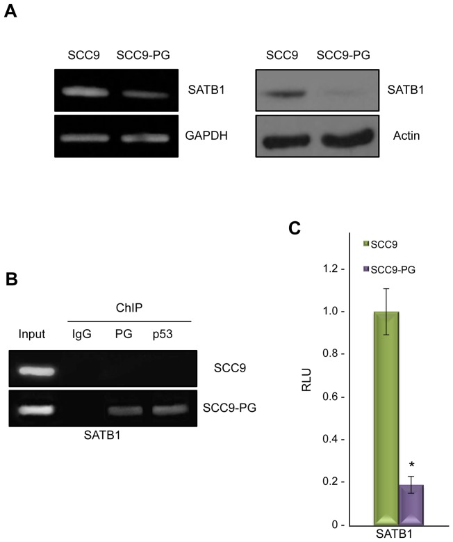
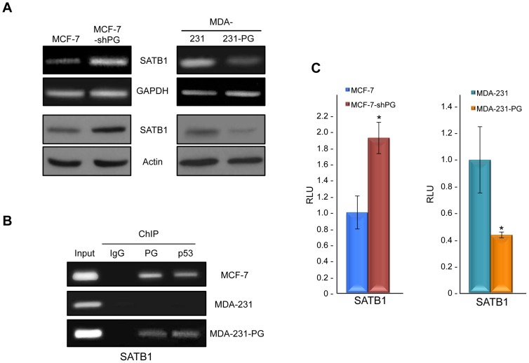
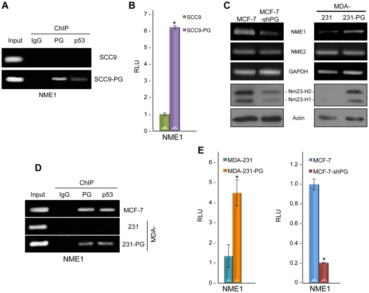
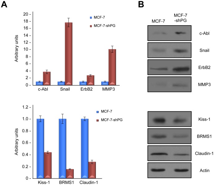
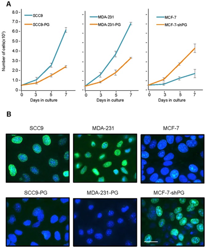
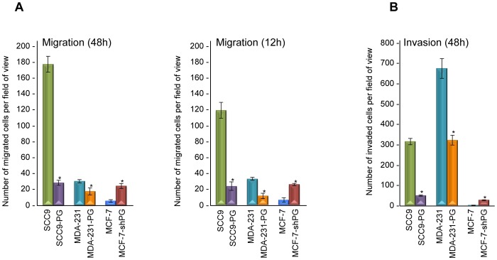
Plakoglobin (γ-catenin) is a homolog of β-catenin with dual adhesive and signaling functions. Plakoglobin participates in cell-cell adhesion as a component of the adherens junction and desmosomes whereas its signaling function is mediated by its interactions with various intracellular protein partners. To determine the role of plakoglobin during tumorigenesis and metastasis, we expressed plakoglobin in the human tongue squamous cell carcinoma (SCC9) cells and compared the mRNA profiles of parental SCC9 cells and their plakoglobin-expressing transfectants (SCC9-PG). We observed that the mRNA levels of SATB1, the oncogenic chromatin remodeling factor, were decreased approximately 3-fold in SCC9-PG cells compared to parental SCC9 cells. Here, we showed that plakoglobin decreased levels of SATB1 mRNA and protein in SCC9-PG cells and that plakoglobin and p53 associated with the SATB1 promoter. Plakoglobin expression also resulted in decreased SATB1 promoter activity. These results were confirmed following plakoglobin expression in the very low plakoglobin expressing and invasive mammary carcinoma cell line MDA-MB-231 cells (MDA-231-PG). In addition, knockdown of endogenous plakoglobin in the non-invasive mammary carcinoma MCF-7 cells (MCF-7-shPG) resulted in increased SATB1 mRNA and protein. Plakoglobin expression also resulted in increased mRNA and protein levels of the metastasis suppressor Nm23-H1, a SATB1 target gene. Furthermore, the levels of various SATB1 target genes involved in tumorigenesis and metastasis were altered in MCF-7-shPG cells relative to parental MCF-7 cells. Finally, plakoglobin expression resulted in decreased in vitro proliferation, migration and invasion in different carcinoma cell lines. Together with the results of our previous studies, the data suggests that plakoglobin suppresses tumorigenesis and metastasis through the regulation of genes involved in these processes.


Conflict of interest statement

Competing Interests: The authors have declared that no competing interests exist.

Figures

Figure 1
Figure 1. Plakoglobin associates with and suppresses the SATB1 promoter in SCC9-PG cells.
A. (Left) Total cellular RNA was isolated from SCC9 and SCC9-PG cells, reverse transcribed and processed for PCR using primers specific to SATB1 and GAPDH. (Right) Equal amounts of total cellular proteins from SCC9 and SCC9-PG cells were resolved by SDS-PAGE and processed for immunoblotting with antibodies to SATB1 and Actin. B. SCC9 and SCC9-PG cells were formaldehyde fixed and processed for chromatin immunoprecipitation. Following sonication, extracts were immunoprecipitated using control IgG, plakoglobin and p53 antibodies. Following extensive washes, immunoprecipitated DNA was separated from the immune complexes and purified using standard DNA purification protocols. The purified DNA was then processed for PCR using SATB1 primers. As a positive control, total cellular DNA (Input) was amplified using the same primers. C. SCC9 and SCC9-PG cells were transfected with luciferase reporter constructs under the control of a 1.2 kb sequence of the SATB1 promoter. Luciferase activities were measured 48 hours post-transfection. The levels of luciferase activities from the vector and SATB1 reporter constructs were determined from a minimum of three independent transfections and normalized for transfection efficiency by co-transfection with a β-galactosidase expression vector. The SATB1 promoter activity was normalized to the corresponding vector activity for each cell line and then normalized to SCC9 (*p<0.01). PG, plakoglobin; RLU, Relative Light Units.
Figure 2
Figure 2. Plakoglobin suppresses SATB1 in mammary epithelial cell lines.
A. (Top) Total cellular RNA was isolated from MCF-7, MCF-7-shPG, MDA-231 and MDA-231-PG cells, reverse transcribed and processed for PCR using primers specific to SATB1 and GAPDH. (Bottom) Equal amounts of total cellular proteins from these cells were resolved by SDS-PAGE and processed for immunoblotting with antibodies to SATB1 and Actin.B. MCF-7, MDA-231 and MDA-231-PG cells were formaldehyde fixed and processed for chromatin immunoprecipitation as described in Fig. 1B. The purified DNA was then processed for PCR using SATB1 primers. As a positive control, total cellular DNA (Input) was amplified using the same primers. C. MCF-7, MCF-7-shPG, MDA-231 and MDA-231-PG cells were transfected with luciferase reporter constructs and processed as described in Fig. 1C. The SATB1 promoter activity was normalized to the corresponding vector activity for each cell line and then normalized to MDA-231 or MCF-7, respectively (*p<0.01). PG, plakoglobin; RLU, Relative Light Units.
Figure 3
Figure 3. Plakoglobin associates with and activates NME1.
A. SCC9 and SCC9-PG cells were processed for chromatin immunoprecipitation using control IgG, plakoglobin and p53 antibodies as described in Fig. 1B. The purified DNA was then processed for PCR using NME1 primers. As a positive control, total cellular DNA (Input) was amplified using the same primers. B. SCC9 and SCC9-PG cells were transfected with luciferase reporter constructs under the control of a 2 kb sequence of the NME1 promoter. Luciferase activities were measured 48 hours post-transfection. The levels of luciferase activities from the vector and NME1 reporter constructs were determined from a minimum of three independent transfections and normalized for transfection efficiency by co-transfection with a β-galactosidase expression vector. The NME1 promoter activity was normalized to the corresponding vector activity for each cell line and then normalized to SCC9 (*p<0.01). PG, plakoglobin; RLU, Relative Light Units. C. (Top) Total cellular RNA was isolated from MCF-7, MCF-7-shPG, MDA-231 and MDA-231-PG cells, reverse transcribed and processed for PCR using primers specific to NME1, NME2 and GAPDH. (Bottom) Equal amounts of total cellular proteins from these cells were resolved by SDS-PAGE and processed for immunoblotting with antibodies to Nm23-H1, -H2 and Actin. D. MCF-7, MDA-231 and MDA-231-PG cells were processed for chromatin immunoprecipitation using control IgG, plakoglobin and p53 antibodies and the purified DNA processed for PCR using NME1 primers. As a positive control, total cellular DNA (Input) was amplified using the same primers. E. MCF-7, MCF-7-shPG, MDA-231 and MDA-231-PG cells were transfected with luciferase reporter constructs as described in Fig. 3B. The NME1 promoter activity was normalized to the corresponding vector activity for each cell line and then normalized to MDA-231 or MCF-7, respectively (*p<0.01). PG, plakoglobin; RLU, Relative Light Units.
Figure 4
Figure 4. Plakoglobin knockdown changes the levels of SATB1 target genes.
A. Total cellular RNA was isolated from MCF-7 and MCF-7-shPG cells, reverse transcribed and processed for PCR using primers specific to SATB1 target genes c-Abl, MMP3, ErbB2, Snail, BRMS1, Kiss1 and Claudin-1. B. Equal amounts of total cellular proteins from these cells were resolved by SDS-PAGE and processed for immunoblotting with antibodies to c-Abl, MMP3, ErbB2, Snail, BRMS1, Kiss1 and Claudin-1. PG, plakoglobin.
Figure 5
Figure 5. Plakoglobin decreases in vitro cell growth and proliferation.
A. Replicate cultures of SCC9, SCC9-PG, MDA-231, -231-PG, MCF-7 and MCF-7-shPG cells were established at single cell density and cells were counted at 3, 5 and 7 days. Each time point represents the average of three independent experiments. The absence of error bars at some time points is due to the small differences among the experiments. B. SCC9, SCC9-PG, MDA-231, -231-PG, MCF-7 and MCF-7-shPG cells were plated on glass coverslips and allowed to grow for 6 days at which time BrdU was added to the cell cultures for 24 hours. BrdU incorporation was then assessed by immunofluorescence staining using BrdU antibodies. Nuclei were countersatined with DRAQ5 and cells viewed using a 63X objective of an LSM510 META (Zeiss) laser scanning confocal microscope. Bar, 20 μm.
Figure 6
Figure 6. Plakoglobin decreases in vitro migration and invasion.
A. Forty-eight- and twelve-hour Transwell migration assays were performed in triplicates for SCC9, SCC9-PG, MDA-231, MDA-231-PG, MCF-7 and MCF-7-shPG cell lines. The membranes were fixed, stained, cut and mounted on slides and viewed under an inverted microscope. B. Forty-eight-hour matrigel invasion assays were performed as described in A using matrigel coated transwell membranes. The number of migrated/invaded cells in five random fields for each membrane were calculated using the ImageJ Cell Counter program and averaged. Histograms represent the average ± SD of the number of migrated/invaded cells for each cell line. *p<0.01). PG, plakoglobin.

References

    1. Jeanes A, Gottardi CJ, Yap AS (2008) Cadherins and cancer: how does cadherin dysfunction promote tumor progression? Oncogene 27: 6920–6929. - PMC - PubMed
    1. Makrilia N, Kollias A, Manolopoulos L, Syrigos K (2009) Cell adhesion molecules: role and clinical significance in cancer. Cancer Invest 27: 1023–1037. - PubMed
    1. Yilmaz M, Christofori G (2010) Mechanisms of motility in metastasizing cells. Mol Cancer Res 8: 629–642. - PubMed
    1. Saito M, Tucker DK, Kohlhorst D, Niessen CM, Kowalczyk AP (2012) Classical and desmosomal cadherins at a glance. J Cell Sci 125: 2547–2552. - PMC - PubMed
    1. Nelson WJ, Dickinson DJ, Weis WI (2013) Roles of cadherins and catenins in cell-cell adhesion and epithelial cell polarity. Prog Mol Biol Transl Sci 116: 3–23. - PubMed

Publication types

MeSH terms