. 2013 Nov 18;8(11):e79845.
doi: 10.1371/journal.pone.0079845. eCollection 2013.

Gli activity is critical at multiple stages of embryonic mammary and nipple development

Affiliations

Gli activity is critical at multiple stages of embryonic mammary and nipple development

Anupama Chandramouli et al. PLoS One. .

Abstract

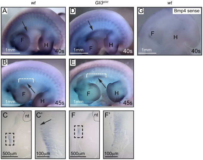
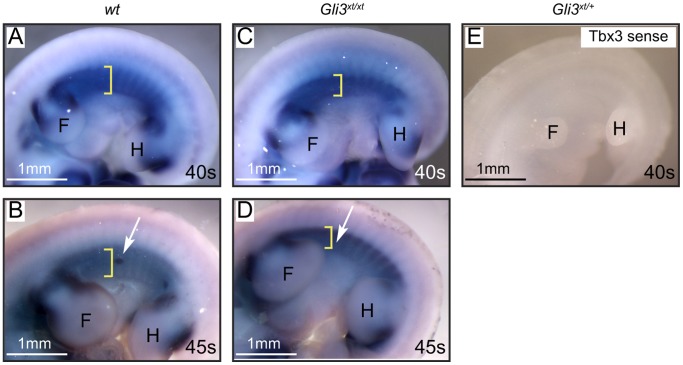
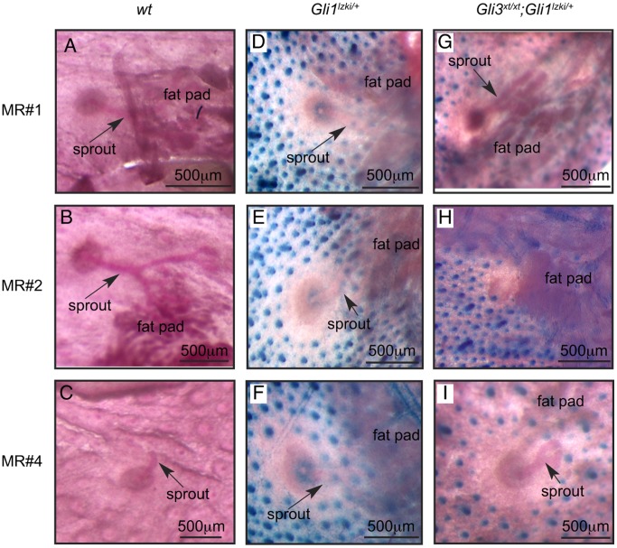
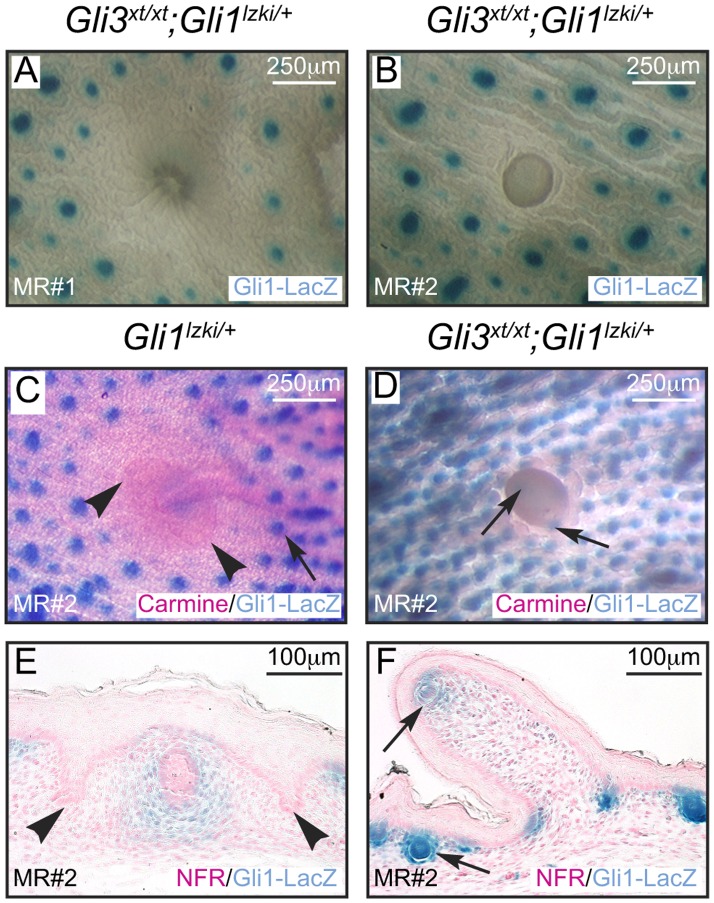
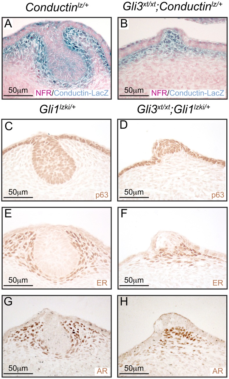
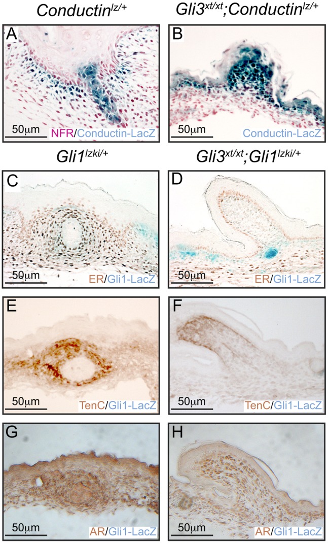
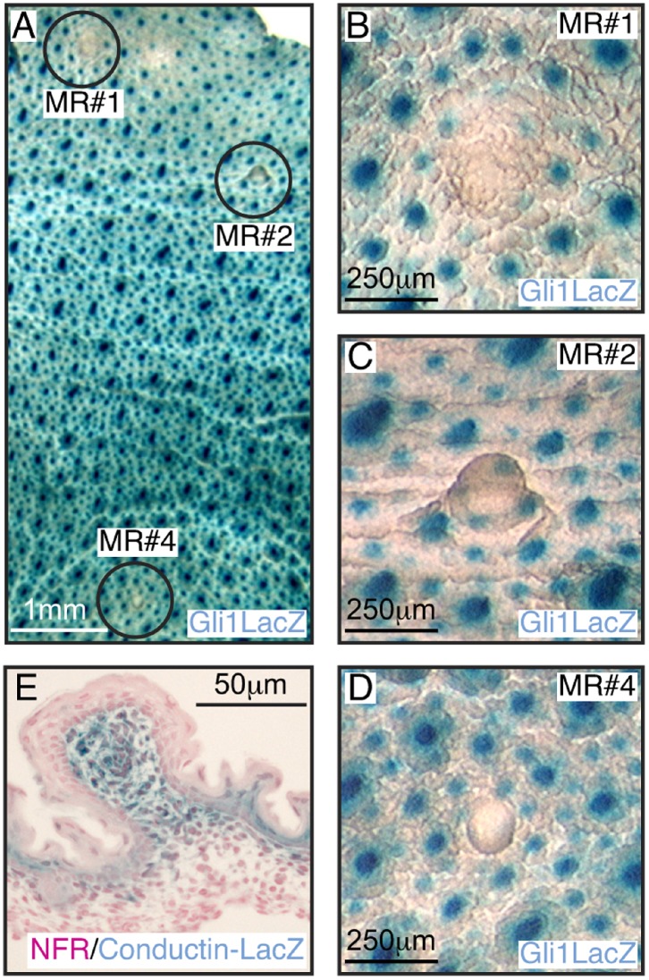
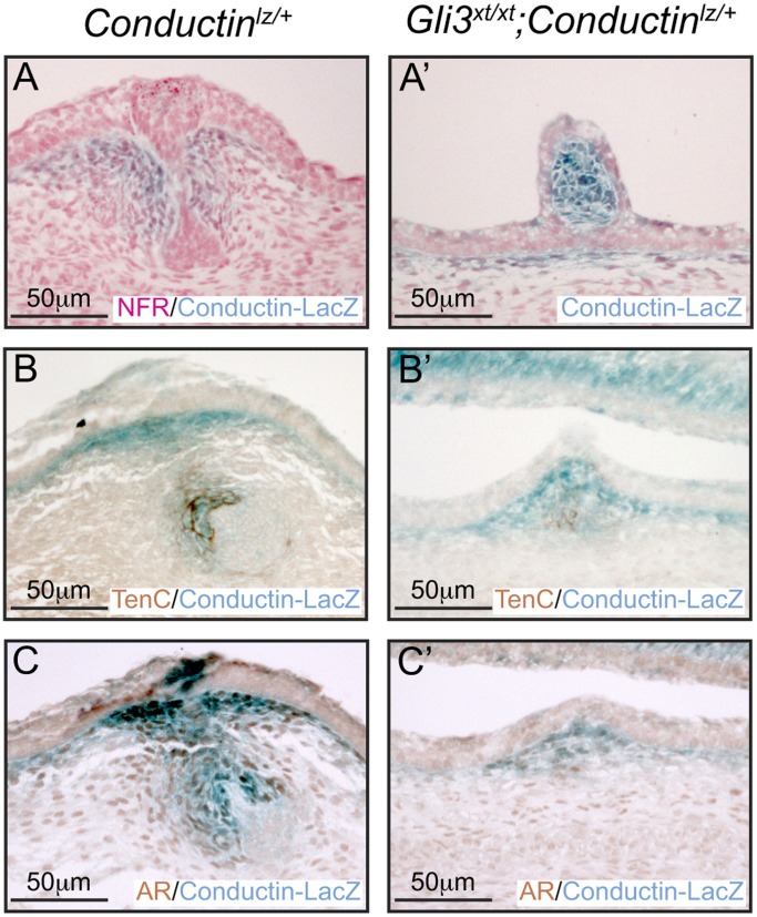
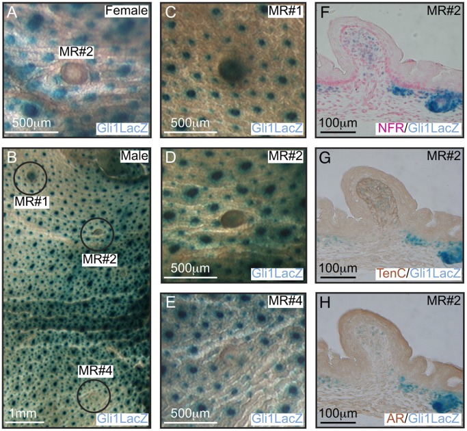
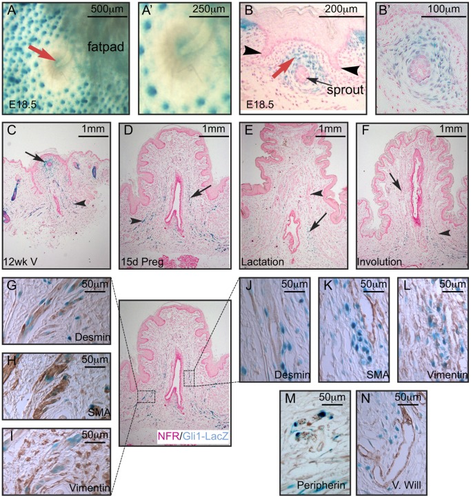
Gli3 is a transcriptional regulator of Hedgehog (Hh) signaling that functions as a repressor (Gli3(R)) or activator (Gli3(A)) depending upon cellular context. Previously, we have shown that Gli3(R) is required for the formation of mammary placodes #3 and #5. Here, we report that this early loss of Gli3 results in abnormal patterning of two critical regulators: Bmp4 and Tbx3, within the presumptive mammary rudiment (MR) #3 zone. We also show that Gli3 loss leads to failure to maintain mammary mesenchyme specification and loss of epithelial Wnt signaling, which impairs the later development of remaining MRs: MR#2 showed profound evagination and ectopic hairs formed within the presumptive areola; MR#4 showed mild invagination defects and males showed inappropriate retention of mammary buds in Gli3(xt/xt) mice. Importantly, mice genetically manipulated to misactivate Hh signaling displayed the same phenotypic spectrum demonstrating that the repressor function of Gli3(R) is essential during multiple stages of mammary development. In contrast, positive Hh signaling occurs during nipple development in a mesenchymal cuff around the lactiferous duct and in muscle cells of the nipple sphincter. Collectively, these data show that repression of Hh signaling by Gli3(R) is critical for early placodal patterning and later mammary mesenchyme specification whereas positive Hh signaling occurs during nipple development.


Conflict of interest statement

Competing Interests: The authors have declared that no competing interests exist.

Figures

Figure 1
Figure 1. Bmp4 mRNA expression is displaced in Gli3xt/xt embryos.
Whole-mount in situ hybridization for Bmp4 in wt (A, B) and Gli3xt/xt (D, E) embryos. In 40-somite stage wt embryos (A) Bmp4 is expressed ventrally. This expression is more intense in Gli3xt/xt embryos at this stage (D). At the 45-somite stage, expression is reduced in wt embryos (B arrow), but expression is displaced dorsally and centrally into the locale of the developing mammary placode #3 in Gli3xt/xt embryos (E arrow). Distance from the axilla to the most distal point of Bmp4 expression (B, E white dotted lines) was measured in mm. Sections through a 45-somite wt embryo (C) and higher power (C’) show mesenchymal expression ventral to the developing placode #3 (arrow). Sections through a 45-somite Gli3xt/xt embryo (F) and higher power (F’) show the displaced Bmp4 expression. Control in situ hybridization with Bmp4 sense probe is shown on a wt embryo in (G). Abbreviations: F – fore-limb, H – hind-limb, nt – neural tube, s – somite.
Figure 2
Figure 2. Tbx3 mRNA expression in Gli3xt/xt embryos.
Whole-mount in situ hybridization for Tbx3 in wt (A, B) and Gli3xt/xt (C, D) embryos. At the 40-somite stage, wt embryos (A) express Tbx3 mRNA in the mesenchyme between the fore- and hind-limb. This band of expression is narrower in Gli3xt/xt embryos at this stage (yellow bracket). At the 45-somite stage Tbx3 is reduced within the mesenchyme but is induced within the epithelium of developing mammary placodes #3 (white arrow) in wt embryos (B). Gli3xt/xt embryos (D), show a narrower band of Tbx3 expression between the fore- and hind-limb and fail to concentrate epithelial expression within placode #3 (white arrow). The control in situ hybridization for Tbx3 sense probe is shown in (E). Abbreviations: F – fore-limb, H – hind-limb, s – somite.
Figure 3
Figure 3. Failure of MR#2 invagination in Gli3xt/xt embryos.
Analysis of the inner surface of skin whole-mounts from E18.5 wt (A, B, C) and Gli1lzki/+ (D, E, F) and Gli3xt/xt; Gli1lzki/+ (G, H, I) embryos stained with X-Gal to detect Gli1-LacZ reporter expression (blue) within hair follicles and counterstained with carmine alum (pink). MRs#1 (A, D, G) and #4 (C, F, I) show comparable development at E18.5 in all three genotypes: sprouts are clearly visible (arrow). In control wt (B) and Gli1lzki/+ (E) embryos sprout #2 has elongated and branched several times but in Gli3xt/xt; Gli1lzki/+ embryos (H), MR#2 shows no evidence of sprouting towards the fatpad.
Figure 4
Figure 4. Evagination of MR#2 and encroachment of hair follicles in Gli3 xt/xt embryos.
Analysis of outer surface of E18.5 skin whole-mounts (A–D) stained with carmine (C, D) and sections stained with nuclear fast red (E, F) and X-Gal (blue) to detect hair follicles expressing the Gli1-LacZ reporter (A–F). MR#1 (A) from Gli3xt/xt; Gli1lzki/+ mutants and MR#2 from control Gli1lzki/+ (C) embryos show normal invagination and appropriate exclusion of hair follicles. In contrast MR#2 from Gli3xt/xt; Gli1lzki/+ mutants (B, D) showed prominent evagination and encroachment of hair follicles.
Figure 5
Figure 5. Mammary mesenchyme specification in E14.5 Gli3xt/xt embryos.
(A, B) Analysis of sections of MR#2 stained with X-Gal (blue) for expression of Conductin-LacZ reporter and counterstained with NFR. Control Conductinlz/+ embryos (A) show Wnt/β-catenin signaling pathway activity in the mammary mesenchyme and central epithelial cells whereas Gli3xt/xt; Conductinlz/+ embryos (B) show activation only within the mesenchymal compartment. Immunohistochemical analysis of serial sections from control E13 Gli1lzki/+ (C, E, G) and mutant Gli3xt/xt;Gli1lzki/+ (D, F, H) embryos for expression of (C, D) p63, (E, F) ER and (G, H)AR. Note the epithelium of Gli3xt/xt;Gli1lzki/+ mutant embryos fails to invaginate (D), the mammary mesenchyme shows no histological evidence of condensation and few cells express ER (F) and AR (H).
Figure 6
Figure 6. Gain of epithelial Wnt signaling and maintenance of mammary mesenchyme markers is compromised in MR#2 of Gli3xt/xt embryos.
Analysis of X-Gal and NFR stained sections from MR#2 at E18.5. (A) Conductin-LacZ is robustly expressed within the epithelial mammary sprout of control Conductinlz/+ embryos whereas (B) mutant Gli3xt/xt; Conductinlz/+ embryos lack expression within the epithelium and maintain robust mesenchymal expression. (C–J) Analysis of serial sections for mammary mesenchyme markers by immunohistochemistry revealed that ER (C), Tenascin C (E) and AR (G) are maintained in control Gli1lzki/+ embryos. In contrast, ER was lost (D) and Tenascin C and AR expression were weakened (F, H) in mutant Gli3xtxt;Gli1lzki/+ embryos.
Figure 7
Figure 7. Sexual dimorphism is lost in Gli3xt/xt embryos.
X-Gal stained skin whole-mounts from E18.5 male Gli3xt/xt;Gli1lzki/+ embryos show retention of MR#1, #2 and #4 (A). Examination of skins at high power revealed that MR#1 does not protruded from the surface of the skin (B), whereas MR#2 and #4 clearly evaginate (C, D). Elevated Wnt signaling activity can be seen in mesenchymal cells of protruding MR#2 from Gli3xt/xt;Conductinlz/+ embryos at this stage (E).
Figure 8
Figure 8. Mammary mesenchyme specification is impaired in male Gli3xt/xt embryos.
X-Gal stained sections from control Conductinlz/+ embryos showed appropriate constriction of mesenchymal cells coincident with the expression of Conductin-LacZ reporter (A), Tenascin C (B) and AR (C). However, mesenchymal cells surrounding the protruding MR#2 of Gli3xt/xt;Conductinlz/+ embryos retained mesenchymal Conductin-LacZ expression (A’) and showed weak Tenascin C (B’) and AR (C’) expression.
Figure 9
Figure 9. Misactivation of Hh signaling detrimentally affects MR invagination and hair follicle suppression in females and eradication of MRs in males.
X-Gal stained whole-mounts (A–E) and sections (F-H) of Gli21ki/1ki;Gli3xt/+;Gli1lzki/+ embryos were examined at E18.5. In whole-mounts of female skins, MR#2 protruded prominently and showed encroachment of hair follicles inappropriately close to the evaginated MR (A). Examination of male skin whole-mounts revealed retention of MR#1, #2 and #4 at low (B) and high power (C, D, E respectively), similar to that seen in Gli3xt/xt;Gli1lzki/+ embryos. Serial sections through a male MR#2 from Gli21ki/1ki;Gli3xt/+;Gli1lzki/+ embryos showed Gli1-LacZ-positive hair follicles close to the protruding bud (F; NFR counterstain) and weak expression of Tenascin C (G) and loss of AR (H) by immunohistochemistry.
Figure 10
Figure 10. Expression and modulation of Gli1-LacZ in the adult nipple during the pregnancy cycle.
Gli1-LacZ expression is visible at the neck of the mammary sprout (red arrow) in skin whole-mounts of E18.5 Gli1lzki/+ embryos at low (A) and higher power (A’). Histological section through the sprout shows expression of Gli1-LacZ within the stroma (red arrow) surrounding the sprout (black arrow) underneath the nipple sheath (black arrowheads) at low (B) and higher power (B’). Gli1-lacZ is expressed within the dermal component but not the epithelium in virgin (C), mid pregnant (D), lactating (E) and involuting (F) nipples. Immunohistochemistry for desmin (G, J), SMA (H, K) and vimentin (I, L) on serial sections of a 15.5 day pregnant nipple demonstrated that Gli1-lacZ was expressed in both smooth muscle cells and fibroblasts but not myofibroblasts. Gli1-lacZ was also found near and surrounding peripherin positive nerve tracts (M) and both Von Willebrand positive vessels (N).

Similar articles

Cited by

References

    1. Veltmaat JM, Mailleux AA, Thiery JP, Bellusci S (2003) Mouse embryonic mammogenesis as a model for the molecular regulation of pattern formation. Differentiation 71: 1–17. - PubMed
    1. Veltmaat JM, Van Veelen W, Thiery JP, Bellusci S (2004) Identification of the mammary line in mouse by Wnt10b expression. Dev Dyn 229: 349–356. - PubMed
    1. Cowin P, Wysolmerski J (2010) Molecular mechanisms guiding embryonic mammary gland development. Cold Spring Harbor perspectives in biology 2: a003251. - PMC - PubMed
    1. Lee MY, Racine V, Jagadpramana P, Sun L, Yu W, et al. (2011) Ectodermal influx and cell hypertrophy provide early growth for all murine mammary rudiments, and are differentially regulated among them by Gli3. PLoS One 6: e26242. - PMC - PubMed
    1. van Genderen C, Okamura RM, Farinas I, Quo R-G, Parslow TG, et al. (1994) Development of several organs that require inductive epithelial-mesenchymal interactions is impaired in Lef-1 deficient mice. Genes and Development 8: 2691–2704. - PubMed

Publication types

LinkOut - more resources