. 2013 Nov 18;8(11):e80526.
doi: 10.1371/journal.pone.0080526. eCollection 2013.

WNT5A encodes two isoforms with distinct functions in cancers

Affiliations

WNT5A encodes two isoforms with distinct functions in cancers

Matthieu Bauer et al. PLoS One. .

Abstract

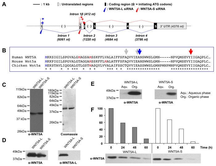
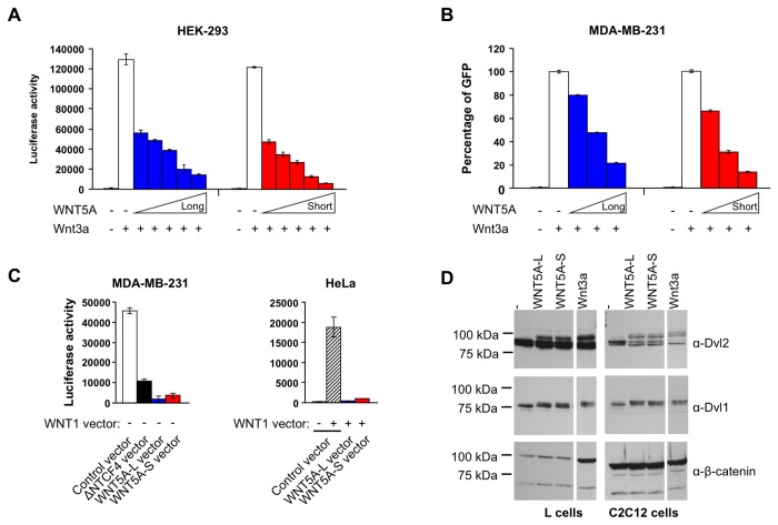
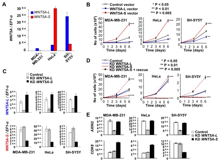
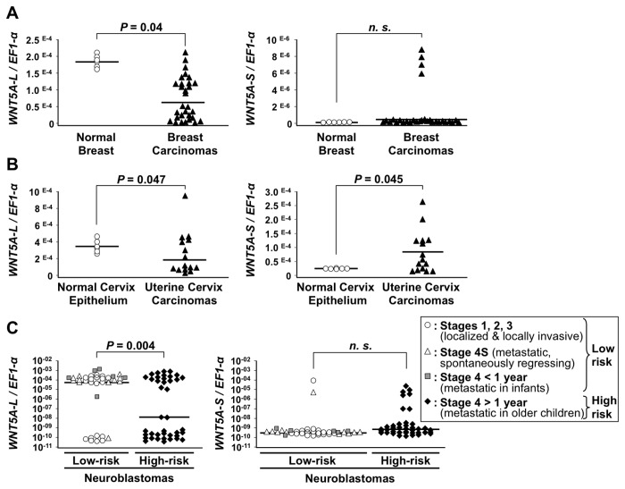
WNT5A, a member of the WNT family of secreted lipid-modified glycoproteins, is a critical regulator of a host of developmental processes, including limb formation, lung morphogenesis, intestinal elongation and mammary gland development. Altered WNT5A expression has been associated with a number of cancers. Interestingly, in certain types of cancers, such as hematological malignancies and colorectal carcinoma, WNT5A is inactivated and exerts a tumor suppressive function, while in other cancers, such as melanoma and gastric carcinoma, WNT5A is overexpressed and promotes tumor progression. The mechanism by which WNT5A achieves these distinct activities in cancers is poorly understood. Here, we provide evidence that the WNT5A gene produces two protein isoforms, WNT5A-long (WNT5A-L) and WNT5A-short (WNT5A-S). Amino-terminal sequencing and a WNT5A-L specific antibody demonstrate that the mature and secreted isoforms are distinct, with WNT5A-L carrying an additional 18 N-terminal amino acids. Biochemical analysis indicates that both purified proteins are similar with respect to their stability, hydrophobicity and WNT/β-catenin signaling activity. Nonetheless, modulation of these two WNT5A isoforms, either through ectopic expression or knockdown, demonstrates that they exert distinct activities in cancer cell lines: while WNT5A-L inhibits proliferation of tumor cell lines, WNT5A-S promotes their growth. Finally, we show that expression of these two WNT5A isoforms is altered in breast and cervix carcinomas, as well as in the most aggressive neuroblastoma tumors. In these cancers, WNT5A-L is frequently down-regulated, whereas WNT5A-S is found overexpressed in a significant fraction of tumors. Altogether, our study provides evidence that the distinct activities of WNT5A in cancer can be attributed to the production of two WNT5A isoforms.


Conflict of interest statement

Competing Interests: The authors have declared that no competing interests exist.

Figures

Figure 1
Figure 1. Description and biochemical chracterization of WNT5A isoforms.
A. Structure of the human WNT5A gene and alternative transcripts. Numbered boxes indicate the exons, with 5’ and 3’ untranslated (UTR) regions in grey and coding regions in black. nt: nucleotides, kb: kilo base. Blue and red arrows and dotted lines respectively indicate the positions of the different transcription initiation sites in exon 1 and exon 1β and splicing of exon 1- and exon 1β-initiated transcripts. Blue and red stars indicate the translation initiation codons for the WNT5A-L and WNT5A-S isoforms located in exon 1 and exon 2, respectively. Blue and red lightning bolt arrows indicate the position of the target sequences of WNT5A-L and WNT5A-S specific short interfering RNA (siRNA). B. Multiple amino acid sequence alignment of the N-terminus of the WNT5A precursor proteins. The blue M denotes the most likely start codon of WNT5A-L while the red Ms denote the most likely start codons of WNT5A-S. The grey arrows indicate the positions of signal peptide cleavage sites for both isoforms as predicted by SignalP 4.1 (http://www.cbs.dtu.dk/services/SignalP/). Blue and red arrows indicate the position of the observed first amino-acids of the mature WNT5A-L and WNT5A-S proteins, respectively, and thus the position of the observed signal peptide cleavage site for each isoform. C. Detection of purified WNT5A isoforms. Left panel: Immuno-blot for WNT5A demonstrates that both isoforms are of similar molecular weight (43kDa). Right panel: Coomassie stained gel shows that the purified WNT5A preparations are pure with largely undetectable contaminant proteins. D. An anti-mouse Wnt5a antibody detects both isoforms (left panel) while an anti-rabbit WNT5A-L specific antibody (right panel) detects the WNT5A-L but not the WNT5A-S isoform. E. Triton X-114 phase separation demonstrates that both WNT5A isoforms partition to the hydrophobic/organic phase. WNT5A proteins were detected by immuno-blot analysis with an anti-Wnt5a antibody. F. Incubation of purified WNT5A isoforms at 37°C for the indicated times indicates that their in vitro stability is similar. WNT5A proteins were detected as in E and band intensities were quantified using ImageJ software.
Figure 2
Figure 2. Effects of WNT5A isoforms on the β-catenin/TCF pathway and Disheveled proteins phosphorylation.
Both WNT5A proteins block Wnt3a-activation of β-catenin/TCF-driven transcriptional activity in HEK 293 (TOP-Luciferase, A.) and MDA-MB-231 (TOP-GFP reporter, B.). Cells were treated with purified Wnt3a and an increasing concentration of WNT5A for 24 hours (A) or 48 hours (B). C. Upon transfection, both WNT5A isoforms interfere with endogenous (MDA-MB-231, left panel) and WNT1-induced (HeLa, right panel) β-catenin/TCF-driven transcription in MDA-MB-231 (left panel) and HeLa cells (right panel). Cells were transfected with control, WNT5A-L or WNT5A-S expression vectors alone, or together with a WNT1 expression vector and assayed for β-catenin/TCF-driven TOP-Luciferase reporter activity. An expression vector encoding a dominant-negative form of TCF4 (ΔNTCF4) was used to interfere with the endogenous reporter activity in MDA-MB-231 cells. D. Both WNT5A isoforms promote DVL phosphorylation. L cells (left panel) and C2C12 cells (right panel) were treated with WNT5A isoforms (10 nM, 2 hours) and whole cell lysates were immuno-blotted with the indicated antibodies. Both WNT5A isoforms, as well as Wnt3a (10 nM, 2 hours), led to a mobility shift of Dvl1 and Dvl2 proteins, suggesting that Dvl proteins are post-translationally modified by phosphorylation in response to both canonical (Wnt3a) and non-canonical Wnt (WNT5A) signaling. Only Wnt3a induced accumulation of the β-catenin protein. Note: β-catenin accumulation in response to Wnt3a in C2C12 cells is not detectable in these whole cell lysates because these cells contain large amounts of membrane/adherens junction associated β-catenin.
Figure 3
Figure 3. Effects of WNT5A isoform expression on cancer cell line proliferation.
A. WNT5A isoform expression in three cancer cell lines. Transcript levels of WNT5A isoforms were determined in MDA-MB-231 (breast carcinoma), HeLa (cervix carcinoma), and SH-SY5Y (neuroblastoma) by qRT-PCR and normalized by EF1-α mRNA (normalization by GAPDH mRNA and 18S rRNA produced similar results). Mean ratios (WNT5A isoforms/ EF1-α) ± SEM from independent measurements are shown. B. Effects of ectopic expression of WNT5A-L and WNT5A-S isoforms on proliferation. The indicated cell lines were transduced with expression vectors encoding WNT5A-L (blue line), WNT5A-S (red line) or no WNT (Black line) and cell numbers were determined at 3 and 6 days. WNT5A-S increases while WNT5A-L decreases proliferative rates. Data (mean ± SEM from triplicate determinations) from a representative experiment are shown. Each experiment was performed at least three independent times. C. Isoform specific siRNAs reduce expression of each WNT5A isoform. The efficiency of WNT5A-L and WNT5A-S isoform knockdown (KD) was evaluated in MDA-MB-231, HeLa and SH-SY5Y transfected with control siRNA and WNT5A isoform-specific siRNA (KD). WNT5A isoforms transcript levels were measured by quantitative RT-PCR (qRT-PCR) and normalized by EF1-α mRNA (normalization by GAPDH mRNA and 18S rRNA produced similar results). D. Effects of siRNA-mediated knockdown (KD) of WNT5A-L and WNT5A-S isoforms on proliferation. Cells were transfected with siRNAs specific to WNT5A-L (blue line) or WNT5A-S (red line) and cell numbers were determined at 3 and 6 days. A scrambled siRNA served as control (black line). Co-transduction of cells with a WNT5A-S expression vector rescues the effect of the WNT5A-S-specific siRNA (dashed red line). Data (mean ± SEM from triplicate determinations) from a representative experiment are shown. Each experiment was performed at least three independent times. * P < 0.05; ** P < 0.01; *** P < 0.005; **** P < 0.001. E. Differential regulation of AXIN2 and CDK8 expression by isoform-specific knockdown of WNT5A. Transcript levels of AXIN2 and CDK8 were determined by quantitative RT-PCR analysis (normalized by EF1-α mRNA) in control MDA-MB-231 (breast carcinoma), HeLa (cervix carcinoma) and SH-SY5Y (neuroblastoma) cells, and in response to siRNA-mediated knock-down (KD) of WNT5A-L or WNT5A-S. Mean ratios ± SEM from independent measurements are shown.
Figure 4
Figure 4. Altered expression of WNT5A isoforms in cancers.
Transcript levels of WNT5A-L (left panels) and WNT5A-S (right panels) isoforms were measured in different types of cancers by qRT-PCR and normalized by EF1-α mRNA (normalization by GAPDH mRNA and 18S rRNA produced similar results). Symbols represent mean ratios (WNT5A isoforms/ EF1-α) from at least two independent measurements for each sample. Horizontal bars correspond to the mean expression value of WNT5A isoforms in each subgroup of samples. For breast (A) and uterine cervix (B), expression in carcinomas was compared to that in normal tissues. For neuroblastomas (NB, C), expression was compared between low risk tumors with good prognosis (Stages 1, 2, 3: localized or locally invasive NB; Stage 4S: spontaneously regressing metastatic NB; Stage 4 < 1 year, metastatic NB of infants) and high risk tumors with poor prognosis (Stage 4 > 1 year: metastatic NB of older children). n.s. = not significant.

References

    1. Bhanot P, Brink M, Samos CH, Hsieh JC, Wang Y et al. (1996) A new member of the frizzled family from Drosophila functions as a Wingless receptor. Nature 382: 225–230. doi:10.1038/382225a0. PubMed: 8717036. - DOI - PubMed
    1. Oishi I, Suzuki H, Onishi N, Takada R, Kani S et al. (2003) The receptor tyrosine kinase Ror2 is involved in non-canonical Wnt5a/JNK signalling pathway. Genes Cells 8: 645–654. doi:10.1046/j.1365-2443.2003.00662.x. PubMed: 12839624. - DOI - PubMed
    1. Mikels AJ, Nusse R (2006) Purified Wnt5a protein activates or inhibits beta-catenin-TCF signaling depending on receptor context. PLOS Biol 4: e115. doi:10.1371/journal.pbio.0040115. PubMed: 16602827. - DOI - PMC - PubMed
    1. Yoshikawa S, McKinnon RD, Kokel M, Thomas JB (2003) Wnt-mediated axon guidance via the Drosophila Derailed receptor. Nature 422: 583–588. doi:10.1038/nature01522. PubMed: 12660735. - DOI - PubMed
    1. Pinson KI, Brennan J, Monkley S, Avery BJ, Skarnes WC (2000) An LDL-receptor-related protein mediates Wnt signalling in mice. Nature 407: 535–538. doi:10.1038/35035124. PubMed: 11029008. - DOI - PubMed

Publication types

MeSH terms