. 2013 Nov 18;8(11):e80544.
doi: 10.1371/journal.pone.0080544. eCollection 2013.

The contributions of HIF-target genes to tumor growth in RCC

Affiliations

The contributions of HIF-target genes to tumor growth in RCC

Ting Zhang et al. PLoS One. .

Abstract

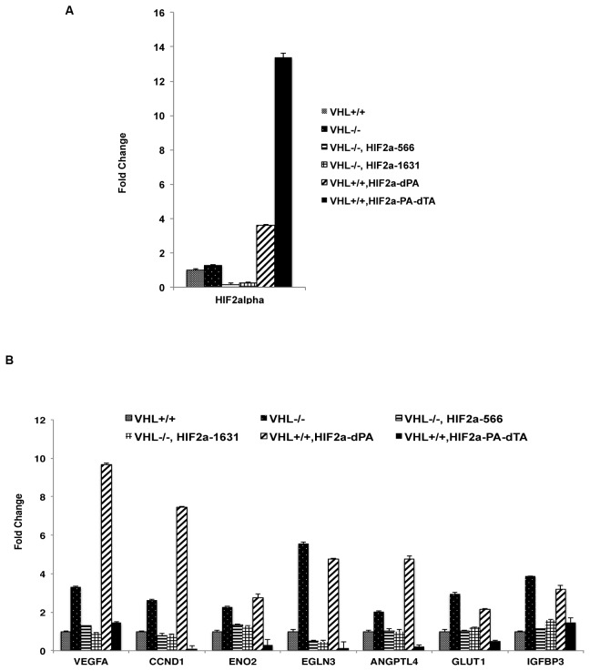
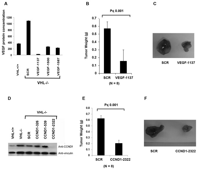
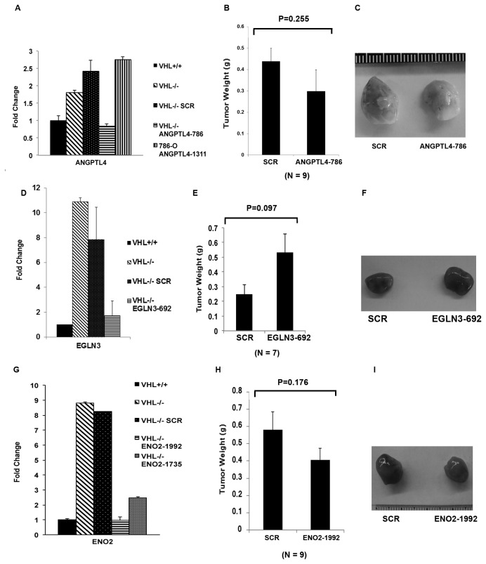
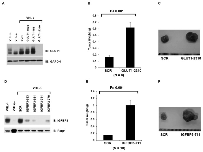
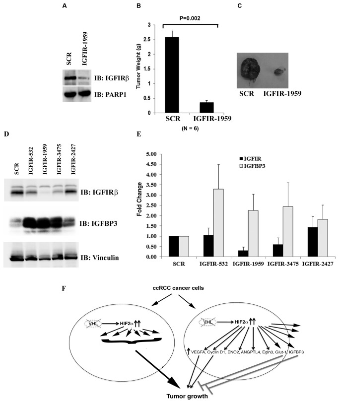
Somatic mutations or loss of expression of tumor suppressor VHL happen in the vast majority of clear cell Renal Cell Carcinoma, and it's causal for kidney cancer development. Without VHL, constitutively active transcription factor HIF is strongly oncogenic and is essential for tumor growth. However, the contribution of individual HIF-responsive genes to tumor growth is not well understood. In this study we examined the contribution of important HIF-responsive genes such as VEGF, CCND1, ANGPTL4, EGLN3, ENO2, GLUT1 and IGFBP3 to tumor growth in a xenograft model using immune-compromised nude mice. We found that the suppression of VEGF or CCND1 impaired tumor growth, suggesting that they are tumor-promoting genes. We further discovered that the lack of ANGPTL4, EGLN3 or ENO2 expression did not change tumor growth. Surprisingly, depletion of GLUT1 or IGFBP3 significantly increased tumor growth, suggesting that they have tumor-inhibitory functions. Depletion of IGFBP3 did not lead to obvious activation of IGFIR. Unexpectedly, the depletion of IGFIR protein led to significant increase of IGFBP3 at both the protein and mRNA levels. Concomitantly, the tumor growth was greatly impaired, suggesting that IGFBP3 might suppress tumor growth in an IGFIR-independent manner. In summary, although the overall transcriptional activity of HIF is strongly tumor-promoting, the expression of each individual HIF-responsive gene could either enhance, reduce or do nothing to the kidney cancer tumor growth.


Conflict of interest statement

Competing Interests: The authors have declared that no competing interests exist.

Figures

Figure 1
Figure 1. HIF is essential to tumor growth by VHL-deficient RCC cells.
A. Total cell lysates of human renal carcinoma 786-O cells stably expressing the control shRNA (SCR) or the HIF1b-1770 were prepared and immunoblotted with the indicated antibodies. B. 786-O VHL-/- cells infected to produce SCR or HIF1b-1770 were injected subcutaneously into the flanks of nude mice. Approximately 8-10 weeks later, tumors were excised and weighed. Thirteen tumors per line were analyzed. Error bars = standard error of the mean. P<=0.001 according to a Mann-Whitney U statistic analysis. C. Representative photographs of tumors analyzed in Figure 1B. Left: Tumor from cells expressing SCR; Right: Tumor from cells expressing HIF1b-1770. D. Total cell lysates of 786-O cells stably expressing the SCR or the HIF2a-566 were immunoblotted with the indicated antibodies. E. 786-O VHL-/- cells infected to produce SCR or HIF2a-566 were injected subcutaneously into the flanks of nude mice. Approximately 8-10 weeks later, tumors were excised and weighed. Ten tumors per line were analyzed. Error bars = standard error of the mean. P<=0.001 according to a Mann-Whitney U statistic analysis. F. Representative photographs of tumors analyzed in Figure 1E. Left: Tumor from cells expressing SCR; Right: Tumor from cells expressing HIF2a-566.
Figure 2
Figure 2. Validation of HIF target genes in VHL-deficient RCC cells.
A. The mRNA levels of HIF2α in 786-O VHL+/+ and VHL-/- cells expressing indicated plasmids or shRNA constructs were measured with real-time PCR. B. The mRNA levels of HIF target genes in 786-O VHL+/+ and VHL-/- cells expressing indicated plasmids or shRNA constructs were measured with real-time PCR.
Figure 3
Figure 3. VEGF and Cyclin D1 positively contribute to tumor growth by VHL-deficient RCC cells.
A. Lysates from 786-O cells stably expressing VHL, the SCR or the VEGF shRNAs were prepared and assayed with VEGF ELISA. B. 786-O VHL-/- cells infected to produce SCR or VEGF-1137 were injected subcutaneously into the flanks of nude mice. Approximately 8-10 weeks later, tumors were excised and weighed. Eight tumors per line were analyzed. Error bars = standard error of the mean. P<=0.001 according to a Mann-Whitney U statistic analysis. C. Representative photographs of tumors analyzed in Figure 3B. Left: Tumor from cells expressing SCR; Right: Tumor from cells expressing VEGF-1137. D. Total cell lysates of 786-O cells stably expressing VHL, the SCR or the Cyclin D1 shRNAs were immunoblotted with the indicated antibodies. E. 786-O VHL-/- cells infected to produce SCR or CCND1-2322 were injected subcutaneously into the flanks of nude mice. Approximately 8-10 weeks later, tumors were excised and weighed. Eight tumors per line were analyzed. Error bars = standard error of the mean. P<=0.001 according to a t-test. F. Representative photographs of tumors analyzed in Figure 3E. Left: Tumor from cells expressing SCR; Right: Tumor from cells expressing CCND1-2322.
Figure 4
Figure 4. ENO2, EGLN3, and ANGPTL4 make no significant contribution to tumor growth by VHL-deficient RCC cells.
A. The mRNA levels of ANGPTL4 in 786-O cells stably expressing VHL, the SCR or the ANGPTL4 shRNAs were analyzed with real-time PCR. B. 786-O VHL-/- cells infected to produce SCR or ANGPTL4-786 were injected subcutaneously into the flanks of nude mice. Approximately 8-10 weeks later, tumors were excised and weighed. Nine tumors per line were analyzed. Error bars = standard error of the mean. P=0.255 according to a t-test. C. Representative photographs of tumors analyzed in Figure 4B. Left: Tumor from cells expressing SCR; Right: Tumor from cells expressing ANGPTL4-786. D. The mRNA levels of EGLN3 in 786-O cells stably expressing VHL, the SCR or the EGLN3 shRNA were analyzed with real-time PCR. E. 786-O VHL-/- cells infected to produce SCR or EGLN3-692 were injected subcutaneously into the flanks of nude mice. Approximately 8-10 weeks later, tumors were excised and weighed. Seven tumors per line were analyzed. Error bars = standard error of the mean. P=0.097 according to a Mann-Whitney U statistic analysis. F. Representative photographs of tumors analyzed in Figure 4E. Left: Tumor from cells expressing SCR; Right: Tumor from cells expressing EGLN3-692. G. The mRNA levels of ENO2 in 786-O cells stably expressing VHL, the SCR or the ENO2 shRNAs were analyzed with real-time PCR. H. 786-O VHL-/- cells infected to produce SCR or ENO2-1992 were injected subcutaneously into the flanks of nude mice. Approximately 8-10 weeks later, tumors were excised and weighed. Nine tumors per line were analyzed. Error bars = standard error of the mean. P=0.176 according to a t-test. I. Representative photographs of tumors analyzed in Figure 4H. Left: Tumor from cells expressing SCR; Right: Tumor from cells expressing ENO2-1992.
Figure 5
Figure 5. GLUT1 and IGFBP3 are tumor suppressive HRGs in 786-O cells.
A. Lysates from 786-O cells stably expressing VHL, the SCR or the GLUT1 shRNAs were prepared and immunoblotted with indicated antibodies. B. 786-O VHL-/- cells infected to produce SCR or GLUT1-2310 were injected subcutaneously into the flanks of nude mice. Approximately 8-10 weeks later, tumors were excised and weighed. Eight tumors per line were analyzed. Error bars = standard error of the mean. P<=0.001 according to a Mann-Whitney U statistic analysis. C. Representative photographs of tumors analyzed in Figure 5B. Left: Tumor from cells expressing SCR; Right: Tumor from cells expressing GLUT1-2310. D. Total cell lysates of 786-O cells stably expressing VHL, the SCR or the IGFBP3 shRNAs were immunoblotted with the indicated antibodies. E. 786-O VHL-/- cells infected to produce SCR or IGFBP3-711 were injected subcutaneously into the flanks of nude mice. Approximately 8-10 weeks later, tumors were excised and weighed. Ten tumors per line were analyzed. Error bars = standard error of the mean. P<=0.001 according to a Mann-Whitney U statistic analysis. F. Representative photographs of tumors analyzed in Figure 5E. Left: Tumor from cells expressing SCR; Right: Tumor from cells expressing IGFBP3-711.
Figure 6
Figure 6. IGFIR loss suppresses tumor growth by VHL-defective RCC cells.
A. Lysates from 786-O VHL-/- cells expressing SCR or an IGFIR shRNA were immunoblotted with indicated antibodies. B. 786-O VHL-/- cells infected to produce the SCR or IGFIR-1959 were injected subcutaneously into the flanks of nude mice. Approximately 9 weeks later, tumors were excised and weighed. Six tumors per line were analyzed. Error bars = standard error of the mean. P=0.002 according to a Mann-Whitney U statistic analysis. C. Representative photographs of nude mice and tumors analyzed in Figure 6B. Left: Tumor from cells expressing SCR; Right: Tumor from cells expressing IGFIR-1959. D. Lysates from 786-O VHL-/- cells expressing SCR or IGFIR shRNAs were immunoblotted with indicated antibodies. E. The mRNA levels of IGFIR and IGFBP3 in 786-O VHL-/- cells expressing SCR or IGFIR shRNAs were analyzed with real-time PCR. F. A model depicting how HIF and HRGs regulate tumor growth in ccRCC.

Similar articles

Cited by

References

    1. Linehan WM, Vasselli J, Srinivasan R, Walther MM, Merino M et al. (2004) Genetic basis of cancer of the kidney: disease-specific approaches to therapy. Clin Cancer Res 10: 6282S-6289S. doi:10.1158/1078-0432.CCR-050013. PubMed: 15448018. - DOI - PubMed
    1. Kaelin WG Jr. (2002) Molecular basis of the VHL hereditary cancer syndrome. Nat Rev Cancer 2: 673-682. doi:10.1038/nrc885. PubMed: 12209156. - DOI - PubMed
    1. Ivan M, Kondo K, Yang H, Kim W, Valiando J et al. (2001) HIFalpha targeted for VHL-mediated destruction by proline hydroxylation: implications for O2 sensing. Science 292: 464-468. doi:10.1126/science.1059817. PubMed: 11292862. - DOI - PubMed
    1. Jaakkola P, Mole DR, Tian YM, Wilson MI, Gielbert J et al. (2001) Targeting of HIF-alpha to the von Hippel-Lindau ubiquitylation complex by O2-regulated prolyl hydroxylation. Science 292: 468-472. doi:10.1126/science.1059796. PubMed: 11292861. - DOI - PubMed
    1. Yu F, White SB, Zhao Q, Lee FS (2001) HIF-1alpha binding to VHL is regulated by stimulus-sensitive proline hydroxylation. Proc Natl Acad Sci U S A 98: 9630-9635. doi:10.1073/pnas.181341498. PubMed: 11504942. - DOI - PMC - PubMed

Publication types

MeSH terms

Substances