. 2013 Nov 15;8(11):e80915.
doi: 10.1371/journal.pone.0080915. eCollection 2013.

Neurosteroid 3α-androstanediol efficiently counteracts paclitaxel-induced peripheral neuropathy and painful symptoms

Affiliations

Neurosteroid 3α-androstanediol efficiently counteracts paclitaxel-induced peripheral neuropathy and painful symptoms

Laurence Meyer et al. PLoS One. .

Abstract

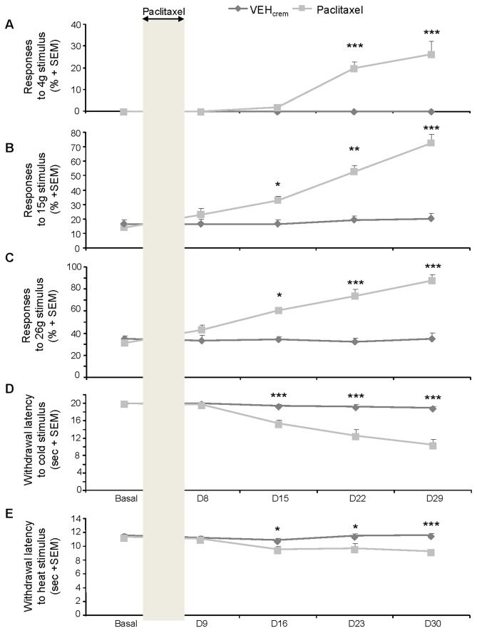
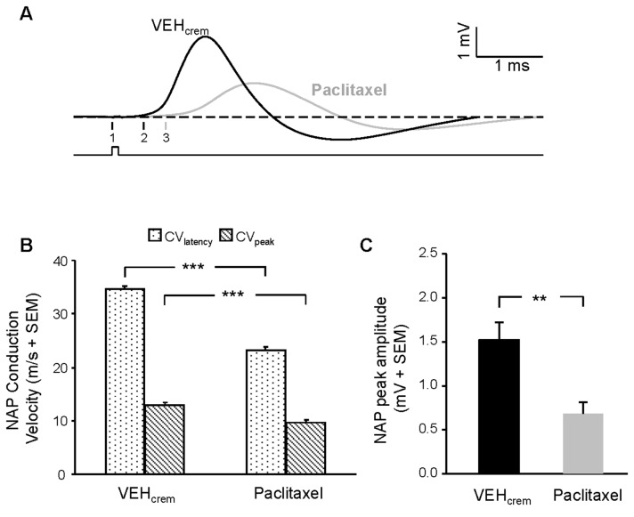
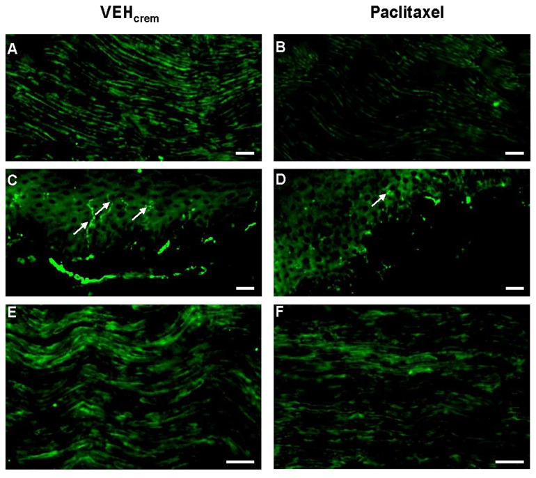
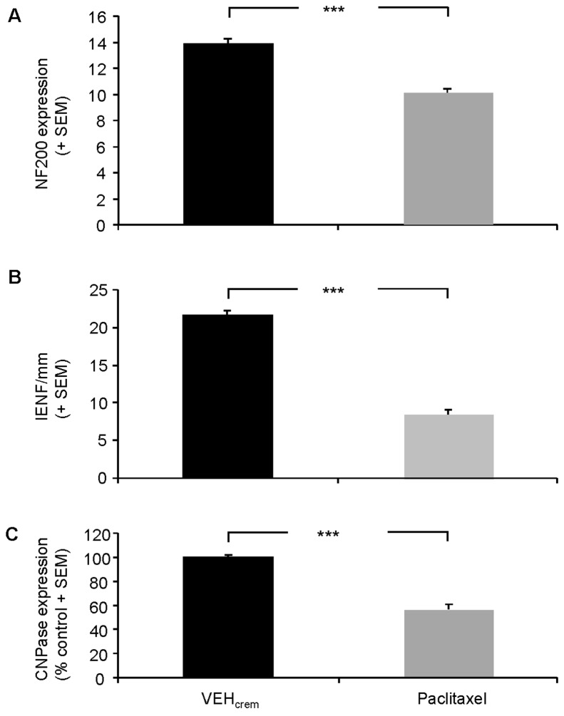
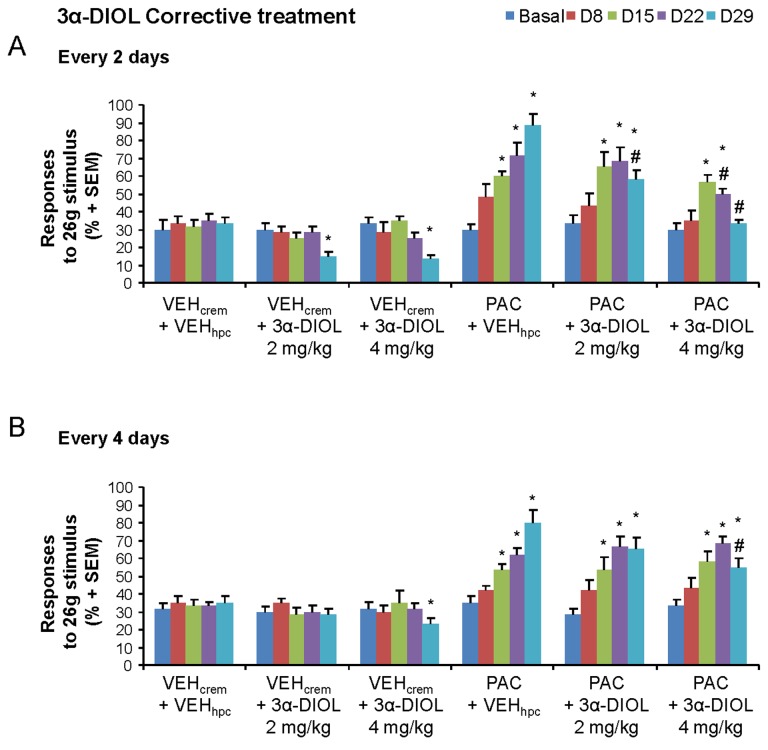
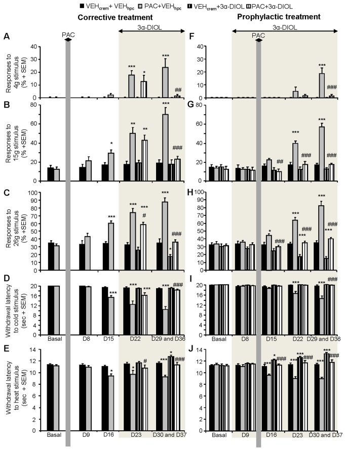
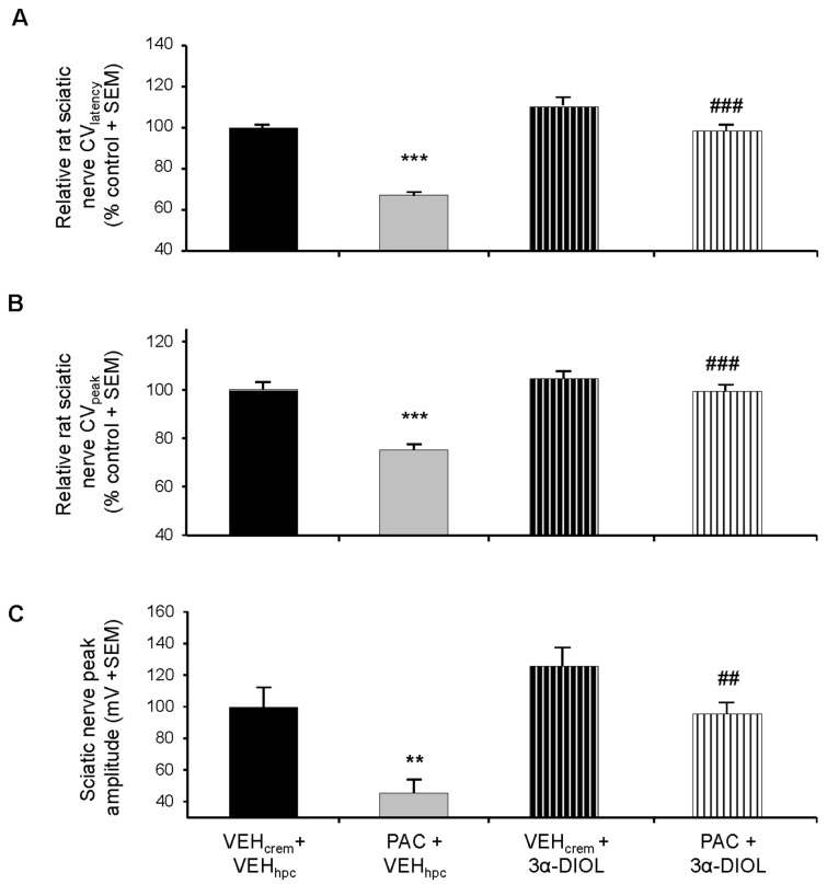
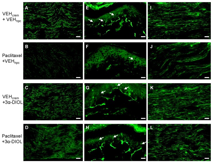
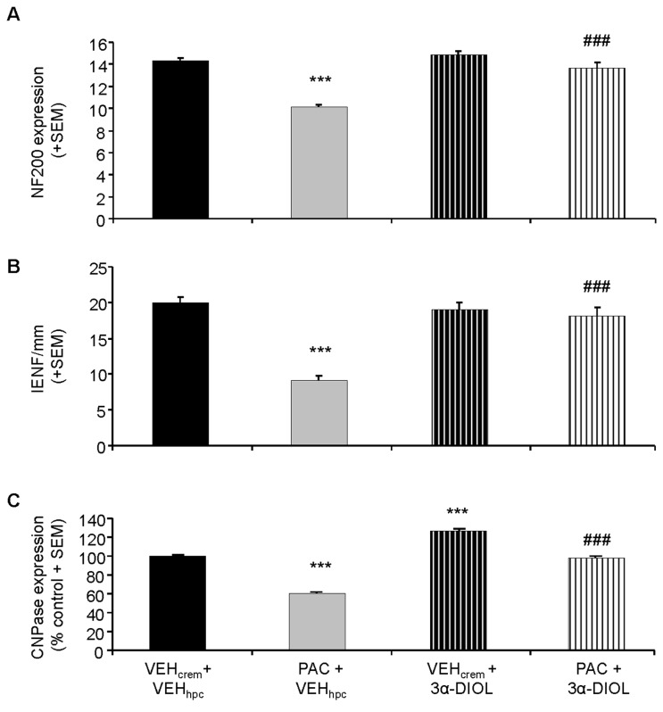
Painful peripheral neuropathy belongs to major side-effects limiting cancer chemotherapy. Paclitaxel, widely used to treat several cancers, induces neurological symptoms including burning pain, allodynia, hyperalgesia and numbness. Therefore, identification of drugs that may effectively counteract paclitaxel-induced neuropathic symptoms is crucial. Here, we combined histopathological, neurochemical, behavioral and electrophysiological methods to investigate the natural neurosteroid 3α-androstanediol (3α-DIOL) ability to counteract paclitaxel-evoked peripheral nerve tissue damages and neurological symptoms. Prophylactic or corrective 3α-DIOL treatment (4 mg/kg/2 days) prevented or suppressed PAC-evoked heat-thermal hyperalgesia, cold-allodynia and mechanical allodynia/hyperalgesia, by reversing to normal, decreased thermal and mechanical pain thresholds of PAC-treated rats. Electrophysiological studies demonstrated that 3α-DIOL restored control values of nerve conduction velocity and action potential peak amplitude significantly altered by PAC-treatment. 3α-DIOL also repaired PAC-induced nerve damages by restoring normal neurofilament-200 level in peripheral axons and control amount of 2',3'-cyclic-nucleotide-3'-phosphodiesterase in myelin sheaths. Decreased density of intraepidermal nerve fibers evoked by PAC-therapy was also counteracted by 3α-DIOL treatment. More importantly, 3α-DIOL beneficial effects were not sedation-dependent but resulted from its neuroprotective ability, nerve tissue repairing capacity and long-term analgesic action. Altogether, our results showing that 3α-DIOL efficiently counteracted PAC-evoked painful symptoms, also offer interesting possibilities to develop neurosteroid-based strategies against chemotherapy-induced peripheral neuropathy. This article shows that the prophylactic or corrective treatment with 3α-androstanediol prevents or suppresses PAC-evoked painful symptoms and peripheral nerve dysfunctions in rats. The data suggest that 3α-androstanediol-based therapy may constitute an efficient strategy to explore in humans for the eradication of chemotherapy-induced peripheral neuropathy.


Conflict of interest statement

Competing Interests: The authors have declared that no competing interests exist.

Figures

Figure 1
Figure 1. Effect of PAC treatment on the rat mechanical (A-C) and thermal (D, E) nociceptive threshold.
(A-C) Time-course of mechanical allodynia (A) and hyperalgesia (B,C) induced by PAC treatment. Graphs show the mean + SEM of the percentages of paw withdrawal responses to mechanical stimulation by von Frey filament 4 g (A), 15 g (B) or 26 g (C) (n=8 per group). (D,E) Effects of PAC treatment on rat cold (D) or heat (E) thermal nociceptive threshold assessed by acetone (D) or Plantar (E) tests. Each point represents the mean + SEM of 6 observations in each of 8 rats. Non-parametric Mann-Whitney U test was used for the analysis of the von Frey test results and one-way repeated measures ANOVAs followed by Newman-Keuls post hoc comparisons were used for acetone and Plantar tests. Statistical differences between control and paclitaxel group on each testing day are shown. * p<0.05, *** p<0.005.
Figure 2
Figure 2. PAC effects on rat sciatic nerve action potential (NPA) peak amplitude and conduction velocity.
(A) Mean traces of biphasic NAPs recorded from animals treated with vehicle (black curve) or PAC (grey curve). The nerve was stimulated with a supra-maximal pulse potential (1 V) as indicated by the protocol trace (lower trace). The arrows 1-3 indicate the beginning of the stimulation artifact (1) and the onset of NAP in control (2) and PAC (3) conditions giving a calculated NAP CVlatency in this case of 39.6 and 23.2 m/s for control and PAC, respectively. Note the reduction of NAP peak amplitude in PAC condition (1.2 mV versus 2.9 mV in controls). (B,C) NAP conduction velocity (B) and peak amplitude (C) histograms of statistical data obtained for vehicle- and PAC-treated rats (n=8 for each condition). ** p<0.01, *** p<0.005.
Figure 3
Figure 3. Photomicrographs of sagittal sections of sciatic nerves (A,B,E,F) or hind paw intraplantar skins (C,D) dissected from vehicle (A,C,E)- and PAC (B,D,F)-treated rats.
Nerve sections were labeled with the monoclonal NF200 antibody (A,B) or with the monoclonal anti-CNPase (E,F) revealed with Alexa-488-conjugated donkey anti-mouse. (C,D) Intraplantar skin sections were labeled with the polyclonal anti-PGP9.5 revealed with FITC-conjugated goat anti-rabbit. White arrows indicated intraepidermal nerve fibers. Scale bar=50µm.
Figure 4
Figure 4. Comparison of IENF density, NF-200 or CNPase expression in vehicle- and PAC-treated rat nerves.
(A) Comparative analysis of NF200-immunostaining density (actual counts for NF200-positive fibers) detected in sciatic nerve sections dissected from vehicle- and PAC-treated rats. (B) Comparative analysis of IENF density (counts for PGP9.5-positive terminals) measured in vehicle- and PAC-treated rat intraplantar skin sections. (C) Comparative analysis of the numbers of CNPase-positive Schwann cell bodies detected in sciatic nerve sections dissected from vehicle- and PAC-treated rats. Each value is expressed as percent (+ SEM) of CNPase-positive cells bodies detected in sciatic nerve sections of control (vehicle-treated) rats. n=8 per group. *** p<0.001.
Figure 5
Figure 5. Dose- and injection frequency-dependent effects of corrective 3α-DIOL treatment on the mechanical nociceptive thresholds of control- and PAC-treated rats.
Corrective treatment every 2- (A) or 4-days (B) consisted in starting 3α-DIOL (2 or 4 mg/kg) or VEHhpc i.p. administrations 2 days after the end of PAC treatment. Threshold values represent responses to 26 g von Frey filament (% ± SEM). Non-parametric Mann-Whitney U test was used. Statistical differences between controls and each treatment group at each testing day are shown. n=6 per group; * p<0.05. * compared to VEHcrem+VEHhpc; # compared to PAC+VEHhpc.
Figure 6
Figure 6. Effects of 3α-DIOL (4 mg/kg/2 days) corrective (A-E) or prophylactic (F-J) treatment on PAC-induced neuropathic pain symptoms.
(A-C,F-H) Action of 3α-DIOL against PAC-induced mechanical allodynia (A,F) and hyperalgesia (B,C,G,H). Chartbars show the mean + SEM of the percentages of paw withdrawal responses to mechanical stimulation by von Frey filament 4 g (A,F), 15 g (B,G) or 26 g (C,H) (n=8 per group). (D,I) Effect of 3α-DIOL against PAC-evoked cold-allodynia. (E,J) 3α-DIOL effects on the heat thermal nociceptive thresholds of vehicle- and PAC-treated rats. Each bar represents the mean + SEM of 6 observations in each of 8 rats. Non-parametric Mann-Whitney U test was used for the analysis of the von Frey test results and two-way repeated measures ANOVAs followed by Newman-Keuls post hoc comparisons were used for acetone and Plantar Tests. Statistical differences between controls and each treatment group at each testing day are shown. * p<0.05, ** p<0.01, *** p<0.005. * vs (VEHcrem + VEHhpc), # vs (PAC + VEHhpc).
Figure 7
Figure 7. Curative effects of 3α-DIOL against PAC-induced reduction of sciatic nerve action potential parameters (CVlatency: A; CVpeak: B and peak amplitude: C).
Histograms of normalized CVlatency and CVpeak show their reduction by PAC and recovery by 3α-DIOL treatment (A,B). Mean CV values were calculated as % of the mean CV obtained from vehicle-treated rats. (C) Recovery from PAC-induced NAP peak amplitude decrease by 3α-DIOL treatment. Each value is the mean (+SEM) of NAP peak amplitude obtained from 8 rat sciatic nerves per each group investigated. ** p<0.01, *** p<0.005. * vs (VEHcrem + VEHhpc), # vs (PAC + VEHhpc).
Figure 8
Figure 8. Photomicrographs of sagittal sections of sciatic nerves (A-D,I-L) or intraplantar skins (E-H) dissected from (VEHcrem + VEHhpc)(A,E,I)-, (PAC + VEHhpc)(B,F,J)-, (VEHcrem + 3α-DIOL)(C,G,K)- or (PAC + 3α-DIOL)(D,H,L)-treated rats.
Nerve sections were labeled with the monoclonal NF200 antibody (A-D) or with the monoclonal anti-CNPase (I-L) revealed with Alexa-488-conjugated donkey anti-mouse. (E-H) Intraplantar skin sections were labeled with the polyclonal anti-PGP9.5 revealed with FITC-conjugated goat anti-rabbit. White arrows indicated intraepidermal nerve fibers. Scale bar, 50 µm.
Figure 9
Figure 9. Neuroprotective effects of 3α-DIOL corrective treatment against PAC-induced alterations in sciatic nerves and intraplantar skin.
(A-C) Chartbars show NF200 (A) or CNPase (C) expression level in sciatic nerve sections or IENF density in intraplantar skin sections (B) dissected from (VEHcrem + VEHhpc)-, (PAC + VEHhpc)-, (VEHcrem + 3α-DIOL)- and (PAC + 3α-DIOL)-treated rats. (A) Each value is expressed as mean (+ SEM) of actual counts for NF200-positive fibers detected in sciatic nerve sections. (B) Each value is expressed as mean (+ SEM) of IENF density (counts for PGP9.5-positive terminals) detected in intraplantar skin sections. (C) Each value is expressed as percent (+ SEM) of CNPase-positive cells bodies detected in sciatic nerve sections of control (VEHcrem + VEHhpc)-treated rats. n=8 per group. *** p<0.001 vs (VEHcrem + VEHhpc), ### p<0.001 vs (PAC + VEHhpc).

Similar articles

Cited by

References

    1. Balayssac D, Ferrier J, Descoeur J, Ling B, Pezet D, Eschalier A, Authier N (2011) Chemotherapy-induced peripheral neuropathies: from clinical relevance to preclinical evidence. Expert Opin Drug Saf 10: 407-417. doi:10.1517/14740338.2011.543417. PubMed: 21210753. - DOI - PubMed
    1. Quasthoff S, Hartung HP (2002) Chemotherapy-induced peripheral neuropathy. J Neurol 249: 9-17. doi:10.1007/PL00007853. PubMed: 11954874. - DOI - PubMed
    1. Authier N, Balayssac D, Marchand F, Ling B, Zangarelli A, Descoeur J, Coudore F, Bourinet E, Eschalier A (2009) Animal models of chemotherapy-evoked painful peripheral neuropathies. Neurotherapeutics 6: 620-629. doi:10.1016/j.nurt.2009.07.003. PubMed: 19789067. - DOI - PMC - PubMed
    1. Dougherty PM, Cata JP, Cordella JV, Burton A, Weng HR (2004) Taxol-induced sensory disturbance is characterized by preferential impairment of myelinated fiber function in cancer patients. Pain 109: 132-142. doi:10.1016/j.pain.2004.01.021. PubMed: 15082135. - DOI - PubMed
    1. Verstappen CC, Heimans JJ, Hoekman K, Postma TJ (2003) Neurotoxic complications of chemotherapy in patients with cancer: clinical signs and optimal management. Drugs 63: 1549-1563. doi:10.2165/00003495-200363150-00003. PubMed: 12887262. - DOI - PubMed

Publication types

MeSH terms