

Functionalized carbon nanotubes in the brain: cellular internalization and neuroinflammatory responses
- PMID: 24260521
- PMCID: PMC3832421
- DOI: 10.1371/journal.pone.0080964
Functionalized carbon nanotubes in the brain: cellular internalization and neuroinflammatory responses
Abstract
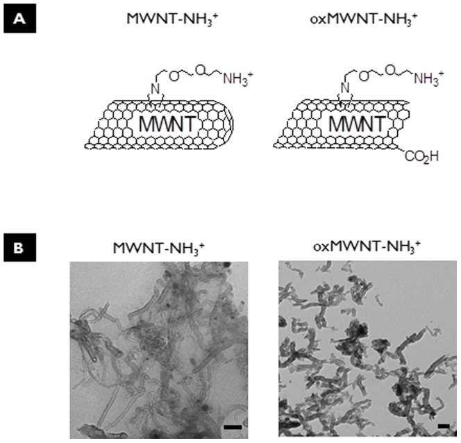
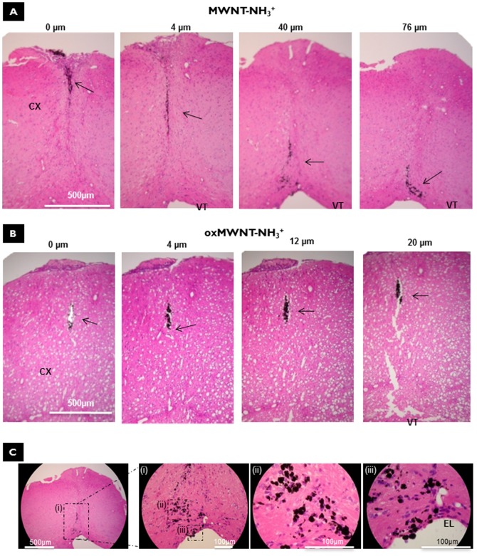
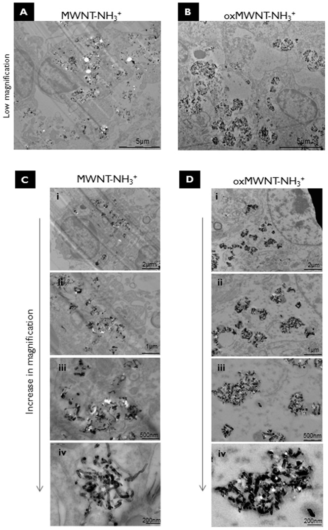
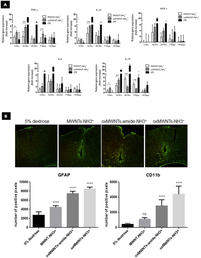
The potential use of functionalized carbon nanotubes (f-CNTs) for drug and gene delivery to the central nervous system (CNS) and as neural substrates makes the understanding of their in vivo interactions with the neural tissue essential. The aim of this study was to investigate the interactions between chemically functionalized multi-walled carbon nanotubes (f-MWNTs) and the neural tissue following cortical stereotactic administration. Two different f-MWNT constructs were used in these studies: shortened (by oxidation) amino-functionalized MWNT (oxMWNT-NH3(+)) and amino-functionalized MWNT (MWNT-NH3(+)). Parenchymal distribution of the stereotactically injected f-MWNTs was assessed by histological examination. Both f-MWNT were uptaken by different types of neural tissue cells (microglia, astrocytes and neurons), however different patterns of cellular internalization were observed between the nanotubes. Furthermore, immunohistochemical staining for specific markers of glial cell activation (GFAP and CD11b) was performed and secretion of inflammatory cytokines was investigated using real-time PCR (qRT-PCR). Injections of both f-MWNT constructs led to a local and transient induction of inflammatory cytokines at early time points. Oxidation of nanotubes seemed to induce significant levels of GFAP and CD11b over-expression in areas peripheral to the f-MWNT injection site. These results highlight the importance of nanotube functionalization on their interaction with brain tissue that is deemed critical for the development nanotube-based vector systems for CNS applications.
Conflict of interest statement
Figures
Similar articles
-
Microglia Determine Brain Region-Specific Neurotoxic Responses to Chemically Functionalized Carbon Nanotubes.ACS Nano. 2015 Aug 25;9(8):7815-30. doi: 10.1021/acsnano.5b02358. Epub 2015 Jun 26. ACS Nano. 2015. PMID: 26043308
-
Translocation of LRP1 targeted carbon nanotubes of different diameters across the blood-brain barrier in vitro and in vivo.J Control Release. 2016 Mar 10;225:217-29. doi: 10.1016/j.jconrel.2016.01.031. Epub 2016 Jan 23. J Control Release. 2016. PMID: 26809004 Free PMC article.
-
Binding and condensation of plasmid DNA onto functionalized carbon nanotubes: toward the construction of nanotube-based gene delivery vectors.J Am Chem Soc. 2005 Mar 30;127(12):4388-96. doi: 10.1021/ja0441561. J Am Chem Soc. 2005. PMID: 15783221
-
99mTc-Glucosamine-multiwall carbon nanotubes.2007 Oct 2 [updated 2007 Nov 5]. In: Molecular Imaging and Contrast Agent Database (MICAD) [Internet]. Bethesda (MD): National Center for Biotechnology Information (US); 2004–2013. 2007 Oct 2 [updated 2007 Nov 5]. In: Molecular Imaging and Contrast Agent Database (MICAD) [Internet]. Bethesda (MD): National Center for Biotechnology Information (US); 2004–2013. PMID: 20641677 Free Books & Documents. Review.
-
Significant roles of neuroinflammation in Parkinson's disease: therapeutic targets for PD prevention.Arch Pharm Res. 2019 May;42(5):416-425. doi: 10.1007/s12272-019-01133-0. Epub 2019 Mar 4. Arch Pharm Res. 2019. PMID: 30830660 Review.
Cited by
-
Carbon Nanomaterials Interfacing with Neurons: An In vivo Perspective.Front Neurosci. 2016 Jun 9;10:250. doi: 10.3389/fnins.2016.00250. eCollection 2016. Front Neurosci. 2016. PMID: 27375413 Free PMC article. Review.
-
Nanotechnological advances in cancer: therapy a comprehensive review of carbon nanotube applications.Front Bioeng Biotechnol. 2024 Mar 6;12:1351787. doi: 10.3389/fbioe.2024.1351787. eCollection 2024. Front Bioeng Biotechnol. 2024. PMID: 38562672 Free PMC article. Review.
-
Nanoparticles: A Hope for the Treatment of Inflammation in CNS.Front Pharmacol. 2021 May 26;12:683935. doi: 10.3389/fphar.2021.683935. eCollection 2021. Front Pharmacol. 2021. PMID: 34122112 Free PMC article. Review.
-
Targeting specific cells in the brain with nanomedicines for CNS therapies.J Control Release. 2016 Oct 28;240:212-226. doi: 10.1016/j.jconrel.2015.12.013. Epub 2015 Dec 11. J Control Release. 2016. PMID: 26686078 Free PMC article. Review.
-
Current and novel polymeric biomaterials for neural tissue engineering.J Biomed Sci. 2018 Dec 20;25(1):90. doi: 10.1186/s12929-018-0491-8. J Biomed Sci. 2018. PMID: 30572957 Free PMC article. Review.
References
-
- Bjartmarz H, Rehncrona S (2007) Comparison of accuracy and precision between frame-based and frameless stereotactic navigation for deep brain stimulation electrode implantation. Stereot Funct Neuros 85: 235–242. - PubMed
-
- Fasano A, Daniele A, Albanese A (2012) Treatment of motor and non-motor features of Parkinson's disease with deep brain stimulation. The Lancet Neurology 11: 429–442. - PubMed
-
- Gallina P, Paganini M, Di Rita A, Lombardini L, Moretti M, et al. (2008) Human fetal striatal transplantation in huntington's disease: a refinement of the stereotactic procedure. Stereotact Funct Neurosurg 86: 308–313. - PubMed
-
- Kang GA, Heath S, Rothlind J, Starr PA (2011) Long-term follow-up of pallidal deep brain stimulation in two cases of Huntington's disease. J Neurol Neurosurg Psychiatry 82: 272–277. - PubMed
-
- Malarkey EB, Parpura V (2007) Applications of carbon nanotubes in neurobiology. Neurodegener Dis 4: 292–299. - PubMed
Publication types
MeSH terms
Substances
LinkOut - more resources
Full Text Sources
Other Literature Sources
Research Materials
Miscellaneous
