. 2013 Nov 18;8(11):e81356.
doi: 10.1371/journal.pone.0081356. eCollection 2013.

The LIM-only protein FHL2 attenuates lung inflammation during bleomycin-induced fibrosis

Affiliations

The LIM-only protein FHL2 attenuates lung inflammation during bleomycin-induced fibrosis

Abdulaleem Alnajar et al. PLoS One. .

Abstract

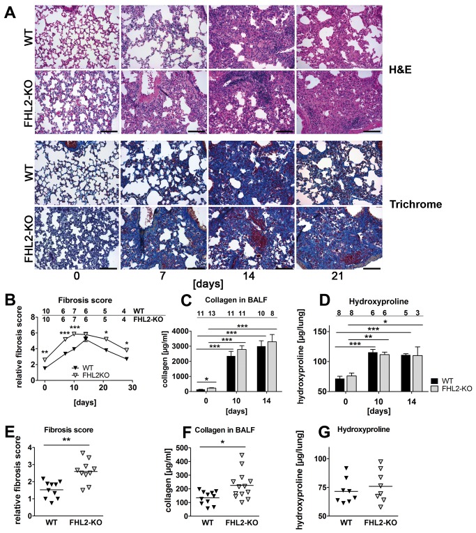
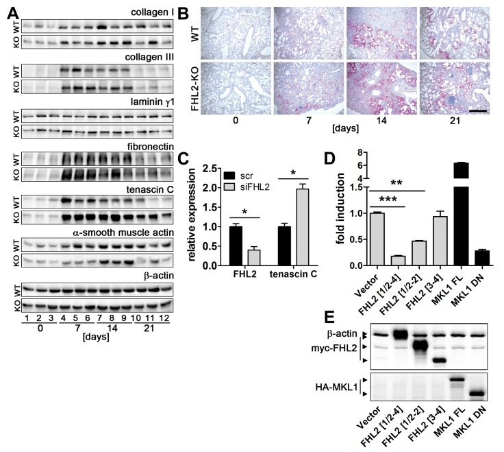
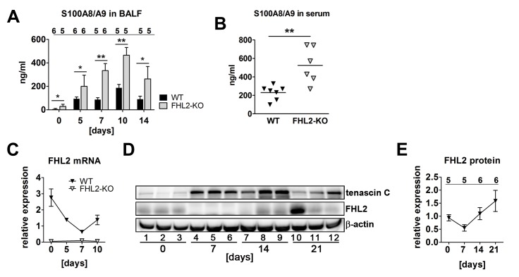
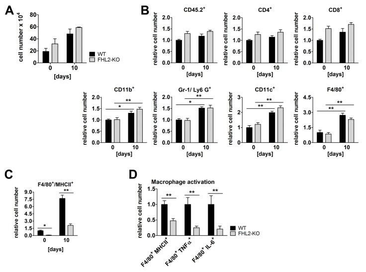
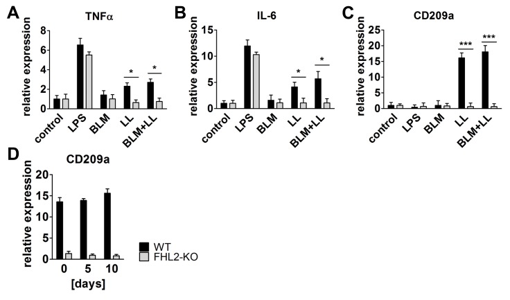
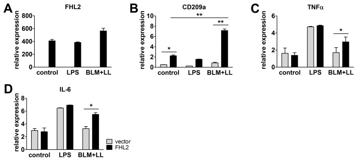
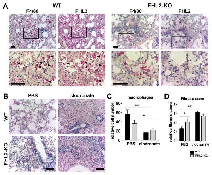
Fibrogenesis is usually initiated when regenerative processes have failed and/or chronic inflammation occurs. It is characterised by the activation of tissue fibroblasts and dysregulated synthesis of extracellular matrix proteins. FHL2 (four-and-a-half LIM domain protein 2) is a scaffolding protein that interacts with numerous cellular proteins, regulating signalling cascades and gene transcription. It is involved in tissue remodelling and tumour progression. Recent data suggest that FHL2 might support fibrogenesis by maintaining the transcriptional expression of alpha smooth muscle actin and the excessive synthesis and assembly of matrix proteins in activated fibroblasts. Here, we present evidence that FHL2 does not promote bleomycin-induced lung fibrosis, but rather suppresses this process by attenuating lung inflammation. Loss of FHL2 results in increased expression of the pro-inflammatory matrix protein tenascin C and downregulation of the macrophage activating C-type lectin receptor DC-SIGN. Consequently, FHL2 knockout mice developed a severe and long-lasting lung pathology following bleomycin administration due to enhanced expression of tenascin C and impaired activation of inflammation-resolving macrophages.


Conflict of interest statement

Competing Interests: The authors have declared that no competing interests exist.

Figures

Figure 1
Figure 1. Development of BLM-induced lung fibrosis in FHL2 WT and knockout mice.
(A) FHL2 WT and knockout mice received a single intranasal application of BLM (2 U/kg) and the right lungs were fixed in 4% paraformaldehyde and embedded into paraffin. Four-micrometre-thick sections were stained with hematoxylin and eosin (upper panel) or with AZAN trichrome according to Heidenhain (lower panel). Bar=200 µm. Representative images from N = 2 and n = 5 to 10 (see B) are shown. (B) Lung sections shown in A were scored for fibrotic alterations according to Ashcroft criteria [30] by three observers in a blinded fashion. N = 2. Ciphers above the curves represent the numbers of analysed animals per time point, with upper and lower lines showing WT and FHL2-KO group of mice, respectively. (C and D) Total collagen amount in the bronchoalveolar spaces (C) and hydroxyproline content in lungs (D) of BLM-treated mice. N = 2 – 3, depending on the time point. Ciphers above the curves represent the numbers of analysed animals per group of mice. (E - G) Lung fibrosis score (E), collagen amount in BALF (F) and hydroxyproline content (G) in lungs of non-induced control mice.
Figure 2
Figure 2. FHL2 inhibits expression of tenascin C.
(A) FHL2 WT and knockout mice were administered BLM for the indicated times. The left lung was then lysed with RIPA buffer and 20 µg of total lysates were analysed by Western blotting for the expression of different proteins. N = 2, n = 5 to 6 per time point, see Figure S3 for details. Lung lysates from three animals per time point are shown. Equal protein loads were verified by β-actin immunoblotting. (B) Paraffin lung sections of control and BLM-induced mice were stained for tenascin C (red). Nuclei (blue) were counterstained with haematoxylin. Bar=500 µm. (C) Immortalised embryonal fibroblasts from C57Bl/6 mice were transfected with scrambled or FHL2-specific siRNA for 24 h and the expression of tenascin C examined by TaqMan qRT-PCR. N = 3. (D) HEK 293 cells were cotransfected for 24 h with a luciferase reporter gene construct containing a 2000-bp tenascin C promoter sequence and indicated plasmids and the luciferase activity was then measured. Relative scores are presented. Mean values of N = 4 shown. (E) The expression of transfected plasmids was verified by Western blotting. A mixture of anti-myc and anti-actin antibodies was used to visualize FHL2 protein and the loading control β-actin on the same blot (upper panel). FHL2[1/2-4] represents the full length protein containing aa 1-279. FHL2[1/2-2] and FHL2 [3,4] represent C-terminal and N-terminal truncations containing the LIM domains ½ to 2 and LIM domains 3 to 4 or aa 1-157 and aa 159-279, respectively, and are described in [28]. MKL1 FL represent the full-length MKL1 protein and MKL1 DN, the dominant-negative MKL1 protein truncated at its N-terminal PPEL motifs and C-terminal transactivation domain, described in [29]. The MKL1 proteins were used as positive and negative control, respectively, for TNC promoter activity.
Figure 3
Figure 3. FHL2-deficient mice have a higher inflammation status in the lungs.
(A) The content of the S100A8/A9 soluble proteins was measured in the BALF of mice by ELISA. The number of analysed animals per time point is shown above the columns. (B) Concentration of S100A8/A9 proteins in the serum of control mice measured by ELISA. (C) FHL2 transcripts in the lungs of BLM-treated mice were estimated by TaqMan qRT-PCR. The number of animals analysed per time point varied from 6 to 12. (D) Western blot analysis of FHL2 expression in the lungs of WT mice after BLM-treatment. Representative images of five to six analysed animals per each time point are shown. (E) Quantification of FHL2 WB images. The FHL2 amount was estimated densitometrically as the relative intensity of the FHL2 bands compared to those of the loading controls. Values at time point 0 were taken as unity. N = 2. The number of analysed animals per time point is shown above the curve.
Figure 4
Figure 4. Immune status of WT and FHL2-KO lungs.
(A) Cell count in BALF of control and 10-day BLM-treated mice. N = 4 and 2 for control and BLM-treated mice, respectively, with 3 to 4 mice per group in each experiment. (B) Single cell suspensions of lung tissue from control or 10-day BLM-treated mice were studied for the indicated surface receptors by flow cytometry. A total of 105 cells per lung were analysed. n = 8 and 9 for control and n = 5 and 6 for BLM-treated WT and FHL2-KO mice, respectively. The relative mean amount of immune cells presented in the lungs of wild type mice was always assigned a value of 1. (C) Lung cell suspensions from (B) were analysed for MHCII and F4/80 surface marker. (D) The F4/80-positive macrophages of BLM-induced mice were further analysed for expression of activation markers: the MHCII receptor and the intracellular TNFα and IL-6 proteins.
Figure 5
Figure 5. FHL2 deficiency abrogates CD209a/DC-SIGN-mediated activation of macrophages.
(A-C) FHL2 WT or knockout mice were injected i.p. with 0.8 ml of 4% starch in PBS and three days later, peritoneal macrophages isolated from three to four mice were combined and plated into 6-well plate dishes. After incubation for 2-3 h, the non-adherent cells were washed off with PBS and the remaining attached macrophages were stimulated with LPS (1µg/ml), BLM (15 U/ml), lung tissue lysate (LL) or BLM+LL for 24 h. After isolation of total RNA, the transcripts of the indicated genes were determined by TaqMan qRT-PCR. Mean values of three repeated experiments are shown. (D) The mRNA levels of D209a/DC-SIGN in the lungs of WT and FHL2-KO mice were determined by qRT-PCR (n=4-5 per group).
Figure 6
Figure 6. Rescue of FHL2 in FHL2-deficient macrophages restores their ability to upregulate the CD209a receptor and to respond to BLM and lung lysate.
Peritoneal macrophages from FHL2-KO mice were transfected with empty vector or human FHL2 cDNA, stimulated with LPS or BLM plus mouse lung lysate, and analysed for expression of (A) human FHL2 and (B) endogenous mouse CD209a, (C) TNFα and (D) IL-6 by TaqMan qRT-PCR. N = 3, with macrophage samples being pooled from three to four animals in each experiment.
Figure 7
Figure 7. Depletion of macrophages aggravates lung fibrosis.
(A) Immunostaining of 2 µm-thick serial paraffin lung sections of 10-days BLM-treated WT (left panel) and FHL2-KO (right panel) mice for macrophage marker F4/80 and FHL2. Samples were counterstained with hematoxylin to reveal nuclei (blue). Arrowheads mark several cells with prominent staining of both proteins. Lower images represent larger magnifications of marked areas of upper images. Bars = 50 µm. (B-D) FHL2 WT and knockout mice were injected i.v. with 100 µl/mouse of PBS- or clodronate-liposomes. The next day, they repeatedly received 75 µl/mouse of PBS- or clodronate-liposomes, but intranasally. On the following day, the mice received 3U/kg of BLM intranasally and 5 days later, they received another dose of PBS- or clodronate-liposomes intranasally. A higher dose of BLM was used to strengthen the acute inflammation and the impact of infiltrated macrophages. On day 10 after BLM application, the right lungs were fixed in 4% paraformaldehyde, embedded into paraffin and analysed for the presence of macrophages by immunohistochemistry using the anti-F4/80 antibody (B) and for fibrotic alterations according to the Ashcroft criteria (D) after H&E staining (Figure S4A). Bars = 100 µm. Representative images of F4/80 stained lung sections are shown in (B) and F4/80-positive cells that were counted in five microscopic fields with a size of 0.75 mm2 in each lung sample are presented in (C). Four animals per group were analysed.

References

    1. Gurtner GC, Werner S, Barrandon Y, Longaker MT (2008) Wound repair and regeneration. Nature 453: 314-321. doi:10.1038/nature07039. PubMed: 18480812. - DOI - PubMed
    1. Krieg T, Abraham D, Lafyatis R (2007) Fibrosis in connective tissue disease: the role of the myofibroblast and fibroblast-epithelial cell interactions. Arthritis Res Ther 9 (Suppl 2): S4. doi:10.1186/ar2168. PubMed: 17767742. - DOI - PMC - PubMed
    1. Shaw TJ, Martin P (2009) Wound repair at a glance. J Cell Sci 122: 3209-3213. doi:10.1242/jcs.031187. PubMed: 19726630. - DOI - PMC - PubMed
    1. Wynn TA (2011) Integrating mechanisms of pulmonary fibrosis. J Exp Med 208: 1339-1350. doi:10.1084/jem.20110551. PubMed: 21727191. - DOI - PMC - PubMed
    1. Fernandez IE, Eickelberg O (2012) The impact of TGF-beta on lung fibrosis: from targeting to biomarkers. Proc Am Thorac Soc 9: 111-116. doi:10.1513/pats.201203-023AW. PubMed: 22802283. - DOI - PubMed

Publication types

MeSH terms