. 2013 Nov 18;8(11):e81405.
doi: 10.1371/journal.pone.0081405. eCollection 2013.

Antagonist properties of Conus parius peptides on N-methyl-D-aspartate receptors and their effects on CREB signaling

Affiliations

Antagonist properties of Conus parius peptides on N-methyl-D-aspartate receptors and their effects on CREB signaling

Shailaja Kunda et al. PLoS One. .

Abstract

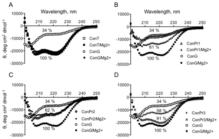
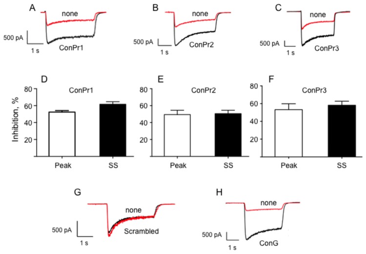
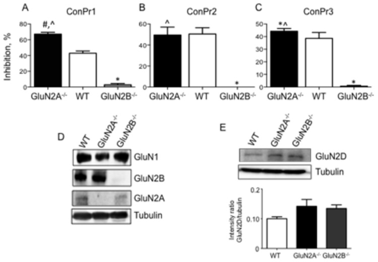
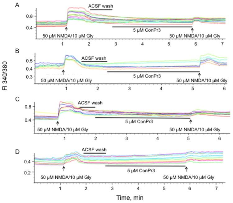
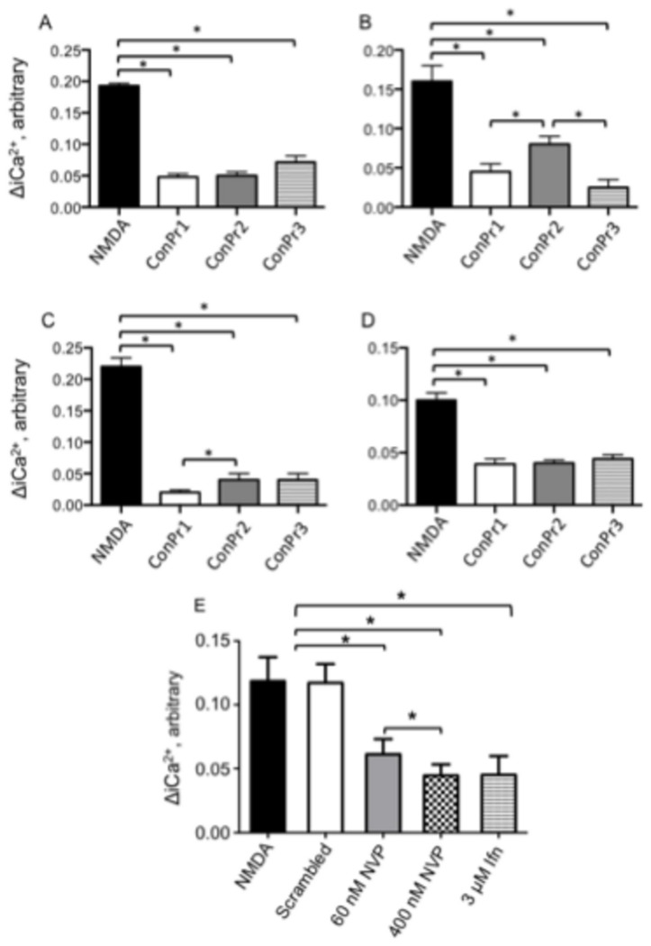
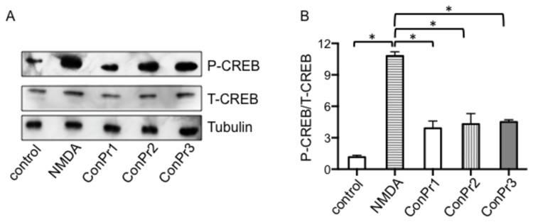
Three members of a family of small neurotoxic peptides from the venom of Conus parius, conantokins (Con) Pr1, Pr2, and Pr3, function as antagonists of N-methyl-D-aspartate receptors (NMDAR). We report structural characterizations of these synthetic peptides, and also demonstrate their antagonistic properties toward ion flow through NMDAR ion channels in primary neurons. ConPr1 and ConPr2 displayed moderate increases in α-helicity after addition of Mg(2+). Native apo-ConPr3 possessed an α-helical conformation, and the helicity increased only slightly on addition of Mg(2+). Additionally, these peptides diminished NMDA/Gly-mediated currents and intracellular Ca(2+) (iCa(2+)) influx in mature rat primary hippocampal neurons. Electrophysiological data showed that these peptides displayed slower antagonistic properties toward the NMDAR than conantokins from other species of cone snails, e.g., ConT and ConG. Furthermore, to demonstrate selectivity of the C. parius-derived conantokins towards specific NMDAR subunits, cortical neurons from GluN2A(-/-) and GluN2B(-/-) mice were utilized. Robust inhibition of NMDAR-mediated stimulation in GluN2A(-/-)-derived mouse neurons, as compared to those isolated from GluN2B(-/-)-mouse brains, was observed, suggesting a greater selectivity of these antagonists towards the GluN2B subunit. These C. parius conantokins mildly inhibited NMDAR-induced phosphorylation of CREB at Ser(133), suggesting that the peptides modulated iCa(2+) entry and, thereby, activation of CREB, a transcription factor that is required for maintaining long-term synaptic activity. Our data mechanistically show that while these peptides effectively antagonize NMDAR-directed current and iCa(2+) influx, receptor-coupled CREB signaling is maintained. The consequence of sustained CREB signaling is improved neuronal plasticity and survival during neuropathologies.


Conflict of interest statement

Competing Interests: The authors have declared that no competing interests exist.

Figures

Figure 1
Figure 1. Representative CD spectra of C. parius-derived conantokins.
Scans from 200-250 nm of: (A) ConT or ConG ± MgCl2; (B) ConG or ConPr1 ± MgCl2; (C) ConG or ConPr2 ± MgCl2; (D) ConG or ConPr3 ± MgCl2. The peptide concentrations were 90 μM and the MgCl2 concentration was 2 mM, when present. The buffer was 10 mM HEPES, pH 7, at 25° C. Each curve represents an average of three scans and the percent helicity is determined as a percent of molar ellipticity of -22,081 deg cm2 dmol-1, which is observed for ConG + 2 mM MgCl2 and which represented 100% α-helicity of these classes of peptides.
Figure 2
Figure 2. Inhibition of the NMDA/Gly-induced current by C. parius-derived conantokins in rat hippocampal neurons.
Representative traces of the NMDA-induced currents using DIV 13-20 primary rat hippocampal neurons in the presence or absence of C. parius conantokins. (A) ConPr1. (B) ConPr2. (C) ConPr3. In each case, 2 μM solutions of antagonist peptides were perfused for 5 min. NMDAR currents were recorded before (black) and after (red) the conantokin perfusions. (D-F) The bar graphs represent the percentage inhibition observed for both peak and steady state (SS) components of NMDAR current for C. parius conantokins, (D) ConPr1, (E) ConPr2, (F) ConPr3. For control experiments, (G) 5 μM scrambled peptide (based on the composition of ConPr1) was perfused for 5 min, or (H) 5 μM ConG was perfused for 3 min. The data are averages of N = 7 separate neurons for ConPr1, N = 10 for ConPr2, N = 10 for ConPr3, and N = 3 for ConG and for the scrambled peptide.
Figure 3
Figure 3. Lack of inhibition of NMDA-induced currents by C. parius-derived peptides in GluN2B-/- mouse cortical neurons.
DIV 13-20 primary cortical neurons from WT, GluN2A-/-, and GluN2B-/- mouse brains were utilized to observe inhibition of NMDA-induced currents. (A) ConPr1. (B) ConPr2. (C) ConPr3. The percent inhibition of NMDAR currents in the absence of conantokins is plotted. The antagonist conantokins (2 μM) were perfused for 5 min and NMDAR currents were recorded before and after the perfusion. Data were obtained from at least three independent recordings for each genotype. (D) Western blot of neuronal cell lysates probed with anti-GluN1, anti-GluN2B, or anti-GluN2A. The lysates were also probed for α-tubulin, shown in lower panel, as loading controls. (E) Representative Western blot and densitometric analysis of WT, GluN2A-/-, and GluN2B-/- cells probed with anti-GluN2D. α-tubulin is the loading control. The bar graph display the means ± SEM of 3 independent experiments. *p < 0.05 for comparison between GluN2B-/- and WT neurons, #p < 0.05 between GluN2A-/- and WT neurons, ^p < 0.05 between GluN2A-/- and GluN2B-/- neurons.
Figure 4
Figure 4. Representative traces of the effect of ConPr3 on NMDA/Gly-dependent Ca2+ influx in neurons.
(A) DIV 12-15 rat hippocampal neurons. (B) WT mouse cortical neurons. (C) GluN2A-/- mouse cortical neurons. (D) GluN2B-/- mouse cortical neurons. The cells were first stimulated with 50 μM NMDA/10 μM glycine, then freed of coagonists by washing with ACSF buffer. Lastly, the neurons were treated with 5 μM ConPr3 for 3 min. The neurons were then again stimulated with 50 μM NMDA/10 μM Gly to determine the extent of inhibition of Ca2+ influx. Changes in iCa2+ were monitored by changes in the fluorescence (FI) ratio at 340/380 nm.
Figure 5
Figure 5. ConPr1 inhibited Ca2+ influx into cultured neurons most robustly in GluN2A-/- cortical neurons.
(A) Inhibition of NMDA/Gly-dependent Ca2+ influx in DIV 12-15 rat hippocampal neurons. (B) DIV 12-15 WT mouse cortical neurons. (C) DIV 12-15 mouse GluN2A-/- cortical neurons. (D) DIV 12-15 mouse GluN2B-/- cortical neurons, after exposure to 5 μM ConPr1, ConPr2, or ConPr3 for 3 min and then re-stimulation with 50 μM NMDA/10 μM glycine. (E) Effect of 5 μM scrambled peptide (N = 8), 60 nM NVP-AAM077 (NVP) (N = 15), 400 nM NVP-AAM077 (NVP) (N = 15), and 3 μM ifenprodil (N = 8) on NMDA/Gly-dependent Ca2+ influx in DIV 12-15 rat hippocampal neurons. The bar graphs display mean ± S.E.M of 2-3 independent experiments performed using different batches of neurons and one batch of neurons (N = 8) for scrambled peptide. *p < 0.05 for comparison between NMDA treated neurons and neurons pre-incubated with ConPr1, ConPr2, and ConPr3. For panel E, no significant differences were observed in [iCa2+] changes between neurons treated with NMDA alone and with neurons treated with NMDA/scrambled peptide. In panel E, *p < 0.05 for pairwise comparisons between NMDA alone or NMDA/scrambled peptide and neurons treated with 60 nM NVP-AAM077, 400 nM NVP-AAM077, 3 μM ifenprodil.
Figure 6
Figure 6. Conus parius-derived conantokins diminished NMDA/Gly-stimulated P-CREB levels in neurons.
(A) Representative Western blot of cultured rat hippocampal neurons at DIV 16 that were not stimulated (control), stimulated with 50 μM NMDA, or pre-incubated with 5 μM ConPr1, ConPr2, or ConPr3 for 5 min, and then stimulated with 50 μM NMDA for 5 min. Total cell lysates were obtained and immunoblotted for P-CREB and total CREB (T-CREB). α-tubulin was probed as loading control. (B) Densitometric analysis of the blots representing the ratios of P-CREB/T-CREB in the samples.

Similar articles

Cited by

References

    1. Terlau H, Olivera BM (2004) Conus venoms: a rich source of novel ion channel-targeted peptides. Physiol Rev 84: 41-68. doi:10.1152/physrev.00020.2003. PubMed: 14715910. - DOI - PubMed
    1. McIntosh JM, Olivera BM, Cruz LJ, Gray WR (1984) γ-Carboxyglutamate in a neuroactive toxin. J Biol Chem 259: 14343-14346. PubMed: 6501296. - PubMed
    1. Haack JA, Rivier J, Parks TN, Mena EE, Cruz LJ et al. (1990) Conantokin-T. A gamma-carboxyglutamate containing peptide with N-methyl-d-aspartate antagonist activity. J Biol Chem 265: 6025-6029. PubMed: 2180939. - PubMed
    1. White HS, McCabe RY, Armstrong H, Donevan SD, Cruz LJ et al. (2000) In vitro and in vivo characterization of conantokin-R, a selective NMDA receptor antagonist isolated from the venom of the fish-hunting snail Conus radiatus. J Pharmacol Exp Ther 292: 425-432. PubMed: 10604979. - PubMed
    1. Dai Q, Sheng Z, Geiger JH, Castellino FJ, Prorok M (2007) Helix-helix interactions between homo- and heterodimeric gamma-carboxyglutamate-containing conantokin peptides and their derivatives. J Biol Chem 282: 12641-12649. doi:10.1074/jbc.M609087200. PubMed: 17347154. - DOI - PubMed

Publication types

MeSH terms

LinkOut - more resources