. 2013 Nov 22:10:321.
doi: 10.1186/1743-422X-10-321.

Influenza A penetrates host mucus by cleaving sialic acids with neuraminidase

Affiliations

Influenza A penetrates host mucus by cleaving sialic acids with neuraminidase

Miriam Cohen et al. Virol J. .

Abstract

Background: Influenza A virus (IAV) neuraminidase (NA) cleaves sialic acids (Sias) from glycans. Inhibiting NA with oseltamivir suppresses both viral infection, and viral release from cultured human airway epithelial cells. The role of NA in viral exit is well established: it releases budding virions by cleaving Sias from glycoconjugates on infected cells and progeny virions. The role of NA in viral entry remains unclear. Host respiratory epithelia secrete a mucus layer rich in heavily sialylated glycoproteins; these could inhibit viral entry by mimicking sialylated receptors on the cell surface. It has been suggested that NA allows influenza to penetrate the mucus by cleaving these sialylated decoys, but the exact mechanism is not yet established.

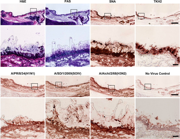
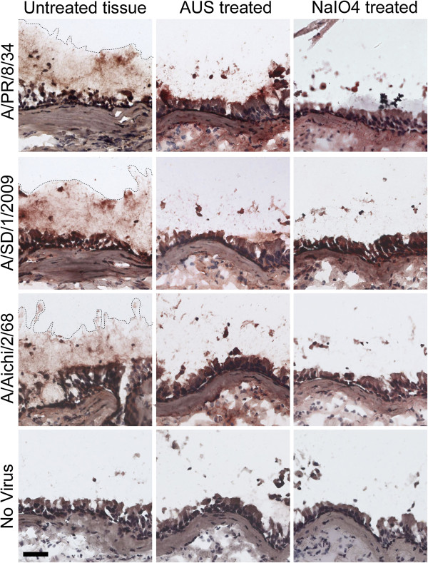
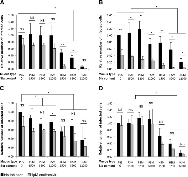
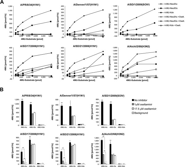
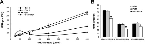
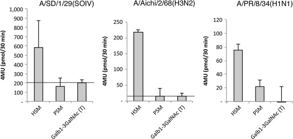
Methods: We tested IAV interaction with secreted mucus using frozen human trachea/bronchus tissue sections, and bead-bound purified human salivary mucins (HSM) and purified porcine submaxillary mucins (PSM). The protective effect of mucus was analyzed using MDCK cells coated with purified HSM and PSM with known Sia content. Oseltamivir was used to inhibit NA activity, and the fluorescent reporter substrate, 4MU-Neu5Ac, was used to quantify NA activity.

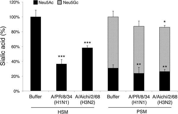
Results: IAV binds to the secreted mucus layer of frozen human trachea/bronchus tissues in a Sia dependent manner. HSM inhibition of IAV infection is Sia dose-dependent, but PSM cannot inhibit infection of underlying cells. HSM competitively inhibits NA cleavage of 4MU-Neu5Ac, reporter substrate. Human IAV effectively cleaves Sias from HSM but not from PSM, and binds to HSM but not to PSM.

Conclusion: IAV interacts with human mucus on frozen tissue sections and mucus-coated beads. Inhibition of IAV infection by sialylated human mucus is dose-dependent, and enhanced when NA is inhibited with oseltamivir. Thus NA cleaves sialylated decoys during initial stages of infection. Understanding IAV interactions with host mucins is a promising new avenue for drug development.


Figures

Figure 1
Figure 1
IAV binds to secreted mucus in human trachea tissues. Frozen human trachea tissue sections were stained with Hematoxylin and Eosin (H&E), periodic acid Schiff (PAS, mucin staining in pink), Sambucus nigra agglutinin (SNA, binds to Siaα2-6Gal/GalNAc) or TKH2 antibody (binds to Sialyl Tn: Siaα2-6GalNAc on O-linked glycans). Dashed lines specify the location of secreted mucus, which is preserved in the frozen tissues, as seen in H&E and PAS staining. Both SNA and TKH2 bound to the secreted mucus (dark brown staining), indicating abundant potential ligands for IAV binding. TKH2 binding was confined to the lining of the epithelium and the glands, further confirming that the secreted mucus layer is adequately preserved in these tissues. Binding of IAV to the tissues was tested by incubating 600 HAU of virus 1.5 h at room temperature. All three strains bound to the secreted mucus layer (dashed line), and to ciliated cells (cells stained in dark brown). Virus was detected by anti-NP antibodies. Boxed area is enlarged below each image. Scale bar indicates 500 μm, scale bar of enlarged area indicates 50 μm.
Figure 2
Figure 2
IAV binding to secreted mucus is Sia-dependent. Human trachea tissue sections were treated with Arthrobacter ureafaciens sialidase (AUS), which cleaves Sias, or with mild sodium periodate (NaIO4), which truncates the Sia side chain. Both treatments reduce IAV binding to the secreted mucus on human trachea tissues compared to untreated control tissues, confirming that IAV binding to the secreted mucus is Sia-dependent. Dashed lines specify location of virus binding to secreted mucus. Scale bar indicates 50 μm.
Figure 3
Figure 3
HSM and oseltamivir have additive inhibitory effects. MDCK cells were layered with PSM or HSM at 1,500, 3,200, 12,000 pmol Sia/well or with PBS buffer as control. The cells were challenged for 1h at 37°C with 109 TCID50 of (A) A/PR/8/34(H1N1), (B) A/SD/1/2009(H1N1), (C) A/SD/17/2008(H1N1), or (D) A/Aichi/2/68(H3N2) in the presence (gray bars) or absence (black bars) of 1 μM oseltamivir. Infected cells were identified by staining with anti-NP antibodies, and quantified in twenty randomly selected images from each sample. Experiments were repeated three times, for each experiment the number of infected cells in the PBS-coated sample was set to 1, and the relative number of infected cells for each treatment was calculated. Lower number of infected cells was observed in HSM coated monolayers compared to PBS-coated monolayers for all tested virus strains (A-D, P<0.05). Dose effects of Sia content in HSM-coated samples were observed for three IAV strains (A-B, D). Significant reduction in the number of infected cells in PSM-coated monolayers was observed only for one strain (C, P<0.05). With exception of the A/SD/1/2009(H1N1) strain, oseltamivir did not inhibit infection of cell coated with either PBS or PSM (A, C-D, gray bars). In contrast, addition of oseltamivir to HSM-coated samples further reduced infection of A/PR/8/34(H1N1), A/SD/1/2009(H1N1) strains (A-B). Both of the oseltamivir-insensitive strains, A/SD/17/2008(H1N1) and A/Aichi/2/68(H3N2), were not affected by addition of the drug (C-D). Data was analyzed by 3-way ANOVA, corrected for multiple comparisons using Tukey’s HSD (see Additional file 2 for complete statistical analysis data). Error bars represent standard deviation. *P<0.05, **P<0.005.
Figure 4
Figure 4
IAV effectively cleaves sialylated HSM. Magnetic beads-conjugated to HSM or PSM were incubated with A/PR/8/34(H1N1), A/Aichi/2/68(H3N2), or buffer at room temperature to allow cleavage of sialylated beads. After 1.5 h incubation the Sia content of the beads was analyzed by DMB-HPLC, and is expressed as percent of Sia content in buffer-incubated beads. Solid bars represent Neu5Ac content, and hatched bars represent Neu5Gc content. Both viruses reduce Neu5Ac content of HSM by 40%-60%, in contrast, only mild cleavage of PSM Sias was observed. ***P<0.001, **P<0.012 *P=0.03 values indicate the significance in difference between Sia content in the virus-treated samples and the corresponding buffer control (two-tailed T-Test, n=3).
Figure 5
Figure 5
Sia cleavage preference and susceptibility to oseltamivir inhibition. (A) The cleaving preference of IAV NA was tested by incubation of IAV with the fluorescent reporter, 4-methyl-umbelliferyl (4MU), linked to Neu5Ac, Neu5Gc, or 2-keto-3-deoxynononic acid (Kdn) as substrate. All virus strains cleaved Neu5Ac and Neu5Gc but not Kdn. Background from spontaneous degradation of the 4MU-Sia compounds was subtracted from the results. (B) Virus susceptibility to oseltamivir inhibition was tested by incubation of IAV with 2.5 nmol 4MU-Neu5Ac in the presence of 0, 1 or 17.5 μM oseltamivir in triplicates. Certain strains were only partially inhibited by 1 μM oseltamivir, however, all of the strains were inhibited by 17.5 μM oseltamivir. Substrate cleavage was quantified by measuring fluorescence from the released 4MU reporter compound. *P<0.05, **P<0.01 (two-tailed T-Test, n=3).
Figure 6
Figure 6
HSM inhibits IAV cleavage of the 4MU-Neu5Ac reporter substrate. (A) Cleavage of 4MU-Neu5Ac reporter substrate by A/Aichi/2/68(H3N2) was tested in the presence of HSM from two donors, PSM (10 nmol Sia), or PBS buffer. HSM from both donors inhibited the cleavage of 4MU-Ne5Ac. In contrast PSM did not inhibit cleavage of 4MU-Neu5Ac, similar to the buffer control. (B) Cleavage of 0.1 nmol 4MU-Neu5Ac by three IAV strains was tested in the presence of HSM, PSM (4.7 pmol Sia), or PBS buffer in triplicates. All virus strains were inhibited by HSM but not by PSM. Bars represent standard error, *P<0.05, **P<0.01 (two-tailed T-Test, n=3).
Figure 7
Figure 7
Direct binding of IAV to HSM. Direct binding of IAV to bead-bound HSM and PSM was tested. Virus was incubated with the mucus-coated beads in the presence of oseltamivir to inhibit NA activity. Following incubation, beads were washed to remove both non-bound virus and oseltamivir. The bound virus was quantified by measuring NA activity using 4MU-Neu5Ac-reporter method. Average of three independent experiments is shown. As control for non-specific binding, beads conjugated to non-sialylated polymer were used. Black line indicates the background. All virus strains bound to HSM, but only one strain bound to PSM.

References

    1. Andrewes CH. Nomenclature of viruses. Nature. 1954;10:620–621. - PubMed
    1. Burnet FM. Mucoproteins in relation to virus action. Physiol Rev. 1951;10:131–150. - PubMed
    1. Fahy JV, Dickey BF. Airway mucus function and dysfunction. N Engl J Med. 2010;10:2233–2247. doi: 10.1056/NEJMra0910061. - DOI - PMC - PubMed
    1. Cone RA. Barrier properties of mucus. Adv Drug Deliv Rev. 2009;10:75–85. doi: 10.1016/j.addr.2008.09.008. - DOI - PubMed
    1. Duez JM, Sixt N, Pechinot A. Influenza virus infection: don’t forget the role of the mucociliary system! J Antimicrob Chemother. 2009;10:421–422. - PubMed

Publication types