. 2013 Nov 22;10(1):33.
doi: 10.1186/2045-8118-10-33.

Comparative study of four immortalized human brain capillary endothelial cell lines, hCMEC/D3, hBMEC, TY10, and BB19, and optimization of culture conditions, for an in vitro blood-brain barrier model for drug permeability studies

Affiliations

Comparative study of four immortalized human brain capillary endothelial cell lines, hCMEC/D3, hBMEC, TY10, and BB19, and optimization of culture conditions, for an in vitro blood-brain barrier model for drug permeability studies

Daniela E Eigenmann et al. Fluids Barriers CNS. .

Abstract

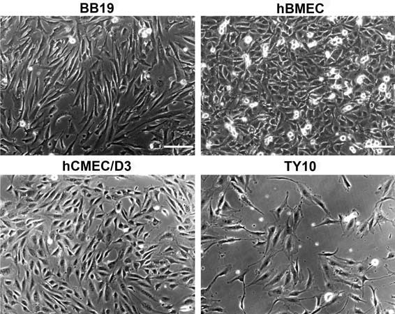
Background: Reliable human in vitro blood-brain barrier (BBB) models suitable for high-throughput screening are urgently needed in early drug discovery and development for assessing the ability of promising bioactive compounds to overcome the BBB. To establish an improved human in vitro BBB model, we compared four currently available and well characterized immortalized human brain capillary endothelial cell lines, hCMEC/D3, hBMEC, TY10, and BB19, with respect to barrier tightness and paracellular permeability. Co-culture systems using immortalized human astrocytes (SVG-A cell line) and immortalized human pericytes (HBPCT cell line) were designed with the aim of positively influencing barrier tightness.

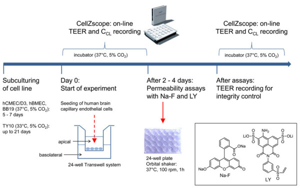
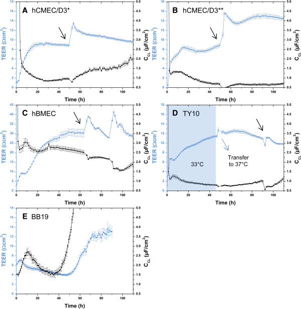
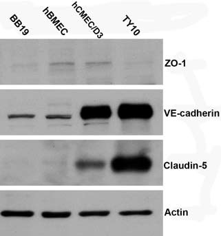
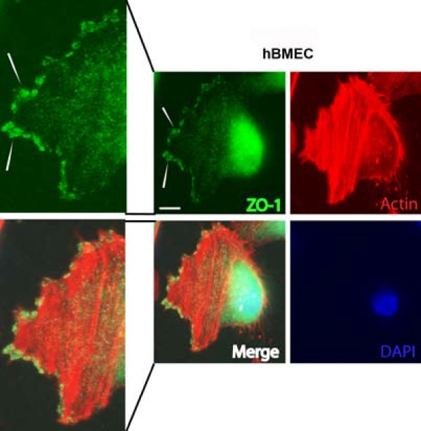
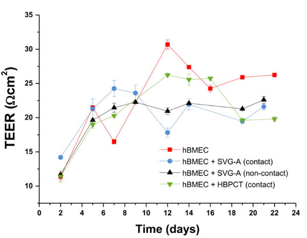
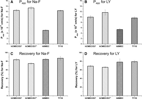
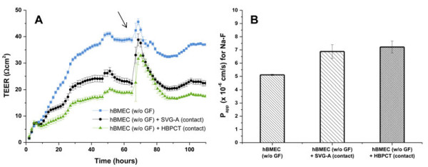
Methods: Tight junction (TJ) formation was assessed by transendothelial electrical resistance (TEER) measurements using a conventional epithelial voltohmmeter (EVOM) and an automated CellZscope system which records TEER and cell layer capacitance (CCL) in real-time.Paracellular permeability was assessed using two fluorescent marker compounds with low BBB penetration (sodium fluorescein (Na-F) and lucifer yellow (LY)). Conditions were optimized for each endothelial cell line by screening a series of 24-well tissue culture inserts from different providers. For hBMEC cells, further optimization was carried out by varying coating material, coating procedure, cell seeding density, and growth media composition. Biochemical characterization of cell type-specific transmembrane adherens junction protein VE-cadherin and of TJ proteins ZO-1 and claudin-5 were carried out for each endothelial cell line. In addition, immunostaining for ZO-1 in hBMEC cell line was performed.

Results: The four cell lines all expressed the endothelial cell type-specific adherens junction protein VE-cadherin. The TJ protein ZO-1 was expressed in hCMEC/D3 and in hBMEC cells. ZO-1 expression could be confirmed in hBMEC cells by immunocytochemical staining. Claudin-5 expression was detected in hCMEC/D3, TY10, and at a very low level in hBMEC cells. Highest TEER values and lowest paracellular permeability for Na-F and LY were obtained with mono-cultures of hBMEC cell line when cultivated on 24-well tissue culture inserts from Greiner Bio-one® (transparent PET membrane, 3.0 μm pore size). In co-culture models with SVG-A and HBPCT cells, no increase of TEER could be observed, suggesting that none of the investigated endothelial cell lines responded positively to stimuli from immortalized astrocytic or pericytic cells.

Conclusions: Under the conditions examined in our experiments, hBMEC proved to be the most suitable human cell line for an in vitro BBB model concerning barrier tightness in a 24-well mono-culture system intended for higher throughput. This BBB model is being validated with several compounds (known to cross or not to cross the BBB), and will potentially be selected for the assessment of BBB permeation of bioactive natural products.


Figures

Figure 1
Figure 1
Phase contrast microscopy of four immortalized human brain capillary endothelial cell lines. Four endothelial cell lines were cultured in growth medium containing 20% FBS for 2 days and the representative cell morphology was imaged with a phase contrast microscope. Scale bar: 150 μm.
Figure 2
Figure 2
Scheme of preparation of the in vitro human BBB model. After subculturing the human cell lines for several days, the brain capillary endothelial cells were seeded on microporous filter membranes of 24-well tissue culture inserts (Greiner Bio-one®, transparent PET membrane, 3.0 μm pore size). The inserts were immediately transferred to a 24-well cell module of a CellZscope system which was placed inside an incubator (37°C and 5% CO2). TEER and CCL values were recorded on-line every hour. After 2–4 days (at the maximal TEER), paracellular permeability of Na-F and LY was assessed. To monitor barrier integrity after the assay, TEER and CCL values were again recorded real-time using the CellZscope system.
Figure 3
Figure 3
Mean TEER values (blue curve) and CCL values (black curve) recorded real-time by the CellZscope system of four human brain capillary endothelial cell lines grown on 24-well tissue culture inserts. Black arrows indicate the time when the permeability assay with Na-F was performed (for resulting Papp values see Figures 7A and B). For hCMEC/D3 and TY10, the permeability assay was performed at TEER values in the range of 10 Ωcm2 (hCMEC/D3: *cultured with initial medium (A), **cultured with growth medium 20% FBS (B). For hBMEC, the assay was carried out at TEER values in the range of 30 Ωcm2. TEER values after the assay were in the same range or even higher as before, suggesting that cell layers were robust during the experiment and barrier integrity was maintained (A-D). For each of the three cell lines, CCL values were in the acceptable range of 0.5-5.0 μF/cm2, indicating that cells were confluent. BB19 cell line (E) yielded very low TEER values in the range of 5 Ωcm2. Since CCL values were drastically increasing after 55 h, the experiment was stopped. BB19 cells were not included into permeability studies with Na-F and LY due to their low TEER. All experiments were performed with Greiner Bio-one® inserts (transparent PET membrane, 3.0 μm pore size, 0.6 × 10⁶ pores/cm2, n = 2–5).
Figure 4
Figure 4
Western blot analysis of tight junction proteins ZO-1, claudin-5, and adherens junction protein VE-cadherin from cell lysates of four endothelial cell lines. The cells were lysed with RIPA buffer. 50 μg of total protein was subjected to SDS-PAGE and western blotting analysis for individual markers. Actin was used as loading control. The four cell lines all expressed the adherens junction protein VE-cadherin. The TJ protein ZO-1 was detected in hCMEC/D3, in hBMEC, and at much lower levels in BB19 and TY10 cell lines. Claudin-5 expression was detected in hCMEC/D3, TY10, and at a very low level in hBMEC cells.
Figure 5
Figure 5
Immunofluorescence staining of tight junction protein ZO-1 in hBMEC cell line. hBMEC cells were grown on collagen-coated glass coverslips for 48 h followed by paraformaldehyde fixation. Even with a high background noise level especially on nuclei, ZO-1 was detected at the leading edge of migrating hBMEC cells (see white arrows). Scale bar: 30 μm.
Figure 6
Figure 6
Mean TEER values for hBMEC cell line in mono-cultures and co-cultures with immortalized astrocytes (SVG-A) and immortalized pericytes (HBPCT). Experiments using 24-well tissue culture inserts from Corning® (transparent PES membrane, 0.4 μm pore size, 4.0 × 10⁶ pores/cm2, n = 2). Maximal TEER values (30.7 ± 0.660 Ωcm2 on day 12) were obtained with hBMEC mono-cultures. Co-culture models with SVG-A and HBPCT cells did not result in increased TEER values. Data were recorded with an EVOM coupled to an Endohm-6 measurement chamber.
Figure 7
Figure 7
Mean Papp values (A, B) and mean recoveries (C, D) for Na-F and LY through hCMEC/D3, hBMEC, and TY10 monolayers. 24-Well tissue culture inserts from Greiner Bio-one® (transparent PET membrane, 3.0 μm pore size, 0.6 × 10⁶ pores/cm2) were used (n = 3–5). A, B: Lowest paracellular permeability of Na-F and LY was obtained through hBMEC monolayers (5.08 ± 0.220 × 10⁻⁶ cm/s and 5.39 ± 0.364 × 10⁻⁶ cm/s, respectively). All experiments for Na-F permeability were carried out at the time indicated with a black arrow in Figures 3A-D. For LY, permeability measurements were carried out at TEER values in the same range (graphs not shown). C, D: Mean recoveries of Na-F in all experiments were in the range of 75% and 87%. For LY, mean recoveries varied between 66% and 79%. *cultured with initial medium; **cultured with growth medium 20% FBS.
Figure 8
Figure 8
Mean TEER (A) and Papp (B) values for hBMEC cultured without growth factors in mono-cultures and contact co-cultures with SVG-A and HBPCT cells. A: Mean TEER values recorded real-time by the CellZscope system for hBMEC cells cultured without growth factors (w/o GF) in mono-cultures and co-cultures with SVG-A (black curve) or HBPCT cells (green curve). 24-Well tissue culture inserts from Greiner Bio-one® (transparent PET membrane, 3.0 μm pore size, 0.6 × 10⁶ pores/cm2) were used (n = 3). Compared to co-cultures with SVG-A and HBPCT cells, hBMEC mono-cultures produced higher TEER values in the range of 40 Ωcm2 (blue curve). CCL values were in the acceptable range of 0.5-5.0 μF/cm2 (data not shown), indicating that hBMEC cells were confluent. The black arrow indicates the time when the permeability assay with Na-F was performed. B: Mean Papp values for Na-F through hBMEC cultured without growth factors through mono-cultures and co-cultures with SVG-A or HBPCT cells. Lowest Papp values for Na-F (5.11 ± 0.0487 × 10⁻⁶ cm/s) were obtained through hBMEC mono-cultures. Mean recoveries in all experiments were between 98% and 102% (n = 3).

References

    1. Pardridge WM. The blood–brain barrier: bottleneck in brain drug development. Neurotherapeutics. 2005;2:3–14. doi: 10.1602/neurorx.2.1.3. - DOI - PMC - PubMed
    1. Di L, Rong H, Feng B. Demystifying brain penetration in central nervous system drug discovery. J Med Chem. 2013;56:A–K. - PubMed
    1. Nicolazzo JA, Charman SA, Charman WN. Methods to assess drug permeability across the blood–brain barrier. J Pharm Pharmacol. 2006;58:281–293. doi: 10.1211/jpp.58.3.0001. - DOI - PubMed
    1. Bickel U. How to measure drug transport across the blood–brain barrier. NeuroRx. 2005;2:15–26. doi: 10.1602/neurorx.2.1.15. - DOI - PMC - PubMed
    1. Di L, Kerns EH, Bezar IF, Petusky SL, Huang Y. Comparison of blood–brain barrier permeability assays: in situ brain perfusion, MDR1-MDCKII and PAMPA-BBB. J Pharm Sci. 2009;98:1980–1991. doi: 10.1002/jps.21580. - DOI - PubMed