

A quantitative comparison of the efferent projections of the anterior and posterior subdivisions of the medial amygdala in female mice
- PMID: 24262912
- PMCID: PMC3888110
- DOI: 10.1016/j.brainres.2013.10.046
A quantitative comparison of the efferent projections of the anterior and posterior subdivisions of the medial amygdala in female mice
Abstract
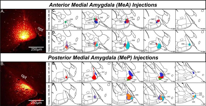
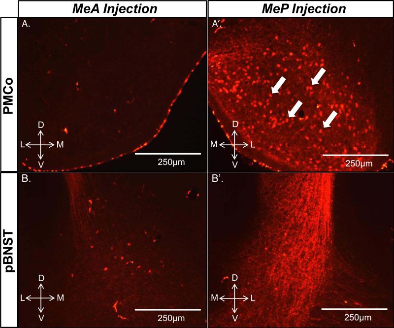
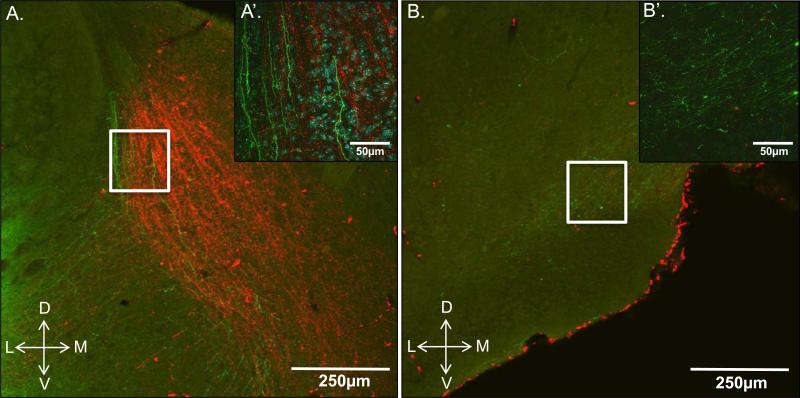
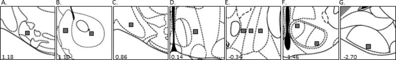
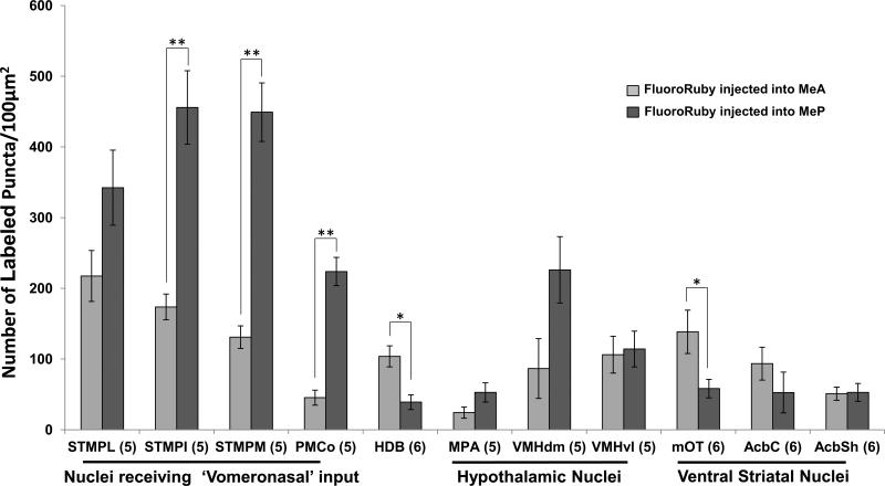
In rodents, many aspects of sociosexual behavior are mediated by chemosignals released by opposite-sex conspecifics. These chemosignals are relayed via the main (MOS) and accessory olfactory systems (AOS) to the medial amygdala (Me). The Me is subdivided into anterior (MeA) and posterior (MeP) subnuclei, and lesions targeting these regions have different effects on proceptive courtship behaviors in female mice. Differential behavioral effects of MeA vs. MeP lesions could reflect a difference in the projections of neurons located in these Me subnuclei. To examine this question, we injected female mice with the anterograde tracer, Fluoro-Ruby into either the MeA or MeP and quantified labeled puncta in 11 forebrain target sites implicated in courtship behaviors using confocal fluorescence microscopy. We found that the MeP more densely innervates the medial and intermediate regions of the posterior bed nucleus of the stria terminalis (pBNST) and the posteromedial cortical amygdala (PMCo), while the MeA more densely innervates the horizontal diagonal band of Broca (HDB) and the medial olfactory tubercle (mOT), a region that may be a component of the circuitry responsible for olfactory-mediated motivated behaviors.
Keywords: Anterograde tracing; Axon terminal; Medial amygdala; Olfactory; Sociosexual behavior.
© 2013 Elsevier B.V. All rights reserved.
Figures

Similar articles
-
Bidirectional connections of the medial amygdaloid nucleus in the Syrian hamster brain: simultaneous anterograde and retrograde tract tracing.J Comp Neurol. 1998 Sep 21;399(2):189-209. doi: 10.1002/(sici)1096-9861(19980921)399:2<189::aid-cne4>3.0.co;2-x. J Comp Neurol. 1998. PMID: 9721903
-
Afferent and efferent connections of the cortical and medial nuclei of the amygdala in sheep.J Chem Neuroanat. 2009 Mar;37(2):87-97. doi: 10.1016/j.jchemneu.2008.09.001. Epub 2008 Sep 12. J Chem Neuroanat. 2009. PMID: 18835351
-
Co-localization of two-color rAAV2-retro confirms the dispersion characteristics of efferent projections of mitral cells in mouse accessory olfactory bulb.Zool Res. 2020 Mar 18;41(2):148-156. doi: 10.24272/j.issn.2095-8137.2020.020. Zool Res. 2020. PMID: 31945810 Free PMC article.
-
Topography of projections from amygdala to bed nuclei of the stria terminalis.Brain Res Brain Res Rev. 2001 Dec;38(1-2):192-246. doi: 10.1016/s0165-0173(01)00079-0. Brain Res Brain Res Rev. 2001. PMID: 11750933 Review.
-
Organization of neural circuits underlying social behavior: A consideration of the medial amygdala.Curr Opin Neurobiol. 2021 Jun;68:124-136. doi: 10.1016/j.conb.2021.02.008. Epub 2021 Apr 30. Curr Opin Neurobiol. 2021. PMID: 33940499 Free PMC article. Review.
Cited by
-
Neural and Hormonal Basis of Opposite-Sex Preference by Chemosensory Signals.Int J Mol Sci. 2021 Aug 2;22(15):8311. doi: 10.3390/ijms22158311. Int J Mol Sci. 2021. PMID: 34361077 Free PMC article. Review.
-
Nanobody-based RFP-dependent Cre recombinase for selective anterograde tracing in RFP-expressing transgenic animals.Commun Biol. 2022 Sep 16;5(1):979. doi: 10.1038/s42003-022-03944-2. Commun Biol. 2022. PMID: 36114373 Free PMC article.
-
Optogenetic Activation of Accessory Olfactory Bulb Input to the Forebrain Differentially Modulates Investigation of Opposite versus Same-Sex Urinary Chemosignals and Stimulates Mating in Male Mice.eNeuro. 2017 Mar 23;4(2):ENEURO.0010-17.2017. doi: 10.1523/ENEURO.0010-17.2017. eCollection 2017 Mar-Apr. eNeuro. 2017. PMID: 28374006 Free PMC article.
-
6-Hydroxydopamine lesions of the anteromedial ventral striatum impair opposite-sex urinary odor preference in female mice.Behav Brain Res. 2014 Nov 1;274:243-7. doi: 10.1016/j.bbr.2014.08.024. Epub 2014 Aug 20. Behav Brain Res. 2014. PMID: 25150042 Free PMC article.
-
Sex and laterality differences in medial amygdala neurons and astrocytes of adult mice.J Comp Neurol. 2016 Aug 15;524(12):2492-502. doi: 10.1002/cne.23964. Epub 2016 Feb 3. J Comp Neurol. 2016. PMID: 26780286 Free PMC article.
References
-
- Blaustein JD, Erskine MS. Feminine sexual behavior: cellular integration of hormonal and afferent information in the rodent brain. In: Pfaff DW, Arnold AP, Al E, editors. Hormones and Behavior. Academic Press; New York: 2002. pp. 139–214.
-
- Bozdagi O, Shan W, Tanaka H, Bensen DL, Huntley GW. Increasing numbers of synaptic puncta during late-phase LTP: N-cadherin is synthesized, recruited to synaptic sites, and required for potentiation. Neuron. 2000;28:245–259. - PubMed
-
- Choi GB, Dong HW, Murphy AJ, Valenzuela DM, Yancopoulos GD, Swanson LW, Anderson DJ. Lhx6 delineates a pathway mediating innate reproductive behaviors from the amygdala to the hypothalamus. Neuron. 2005;46:647–660. - PubMed
Publication types
MeSH terms
Substances
Grants and funding
LinkOut - more resources
Full Text Sources
Other Literature Sources
