. 2014 Mar;34(2):227-34.
doi: 10.1007/s10571-013-0006-9. Epub 2013 Nov 22.

Protective effect of paeoniflorin on Aβ25-35-induced SH-SY5Y cell injury by preventing mitochondrial dysfunction

Affiliations

Protective effect of paeoniflorin on Aβ25-35-induced SH-SY5Y cell injury by preventing mitochondrial dysfunction

Ke Wang et al. Cell Mol Neurobiol. 2014 Mar.

Abstract

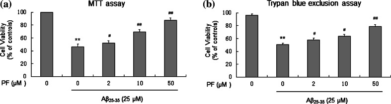
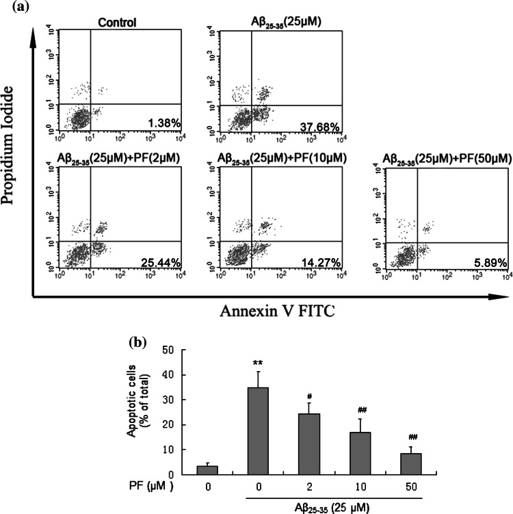
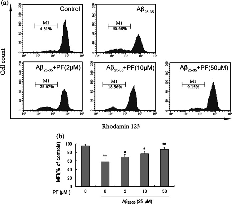
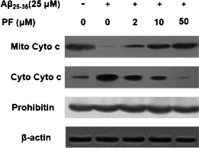
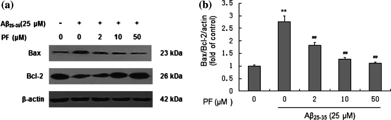
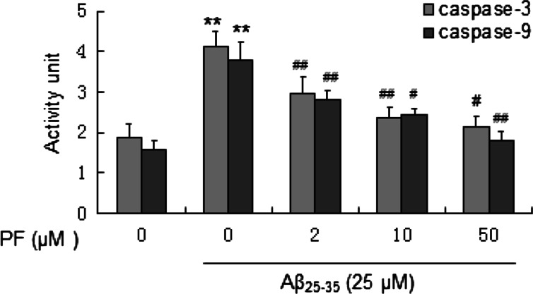
Alzheimer's disease (AD) is a major neurodegenerative brain disorder affecting about 14 million people worldwide. Aβ-induced cell injury is a crucial cause of neuronal loss in AD, thus the suppression of which might be useful for the treatment of this disease. In this study, we aimed to evaluate the effect of paeoniflorin (PF), a monoterpene glycoside isolated from aqueous extract of Radix Paeoniae Alba, on Aβ25-35-induced cytotoxicity in SH-SY5Y cells. The results showed PF could attenuate or restore the viability loss, apoptotic increase, and ROS production induced by Aβ25-35 in SH-SY5Y cells. In addition, PF strikingly inhibited Aβ25-35-induced mitochondrial dysfunction, which includes decreased mitochondrial membrane potential, increased Bax/Bcl-2 ratio, cytochrome c release and activity of caspase-3 and caspase-9. Therefore, our study provided the first experimental evidence that PF could modulate ROS production and apoptotic mitochondrial pathway in model of neuron injury in vitro and which might provide new insights into its application toward Alzheimer's disease therapy.


Conflict of interest statement

Ke Wang and other co-authors have no conflict of interest

Figures

Fig. 1
Fig. 1
Protective effect of PF on Aβ25–35-induced cytotoxicity in SH-SY5Y cells. Cells were pretreated with or without various concentrations of PF (2, 10, 50 μM) for 24 h and then exposed to Aβ25–35 (25 μM) for another 24 h. After incubation, cell viability was evaluated by MTT assay (a) and trypan blue exclusion assay (b). The results are shown as mean ± SEM of three experiments and each included triplicate sets. **p < 0.01 versus control; # p < 0.05; ## p < 0.01 versus Aβ25–35 alone
Fig. 2
Fig. 2
Protective effect of PF on Aβ25–35-induced apoptosis in SH-SY5Y cells. Cells were pretreated with or without various concentrations of PF (2, 10, 50 μM) for 24 h and exposed to Aβ25–35 (25 μM) for another 24 h. After incubation, cell apoptosis were analyzed. a Flow cytometry analysis of cell apoptosis using annexin V-FITC/PI dual-staining. b The bar chart describes the percentage distribution of apoptotic cells. The results are shown as mean ± SEM of three experiments and each included triplicate sets. **p < 0.01 versus control; # p < 0.05; ## p < 0.01 versus Aβ25–35 alone
Fig. 3
Fig. 3
Protective effect of PF on Aβ25–35-induced oxidative stress in SH-SY5Y cells. Cells were pretreated with or without various concentrations of PF (2, 10, 50 μM) for 24 h and exposed to Aβ25–35 (25 μM) for another 24 h. After incubation, oxidative stress was assessed by measuring intracellular ROS level. The results are shown as mean ± SEM of three experiments and each included triplicate sets. **p < 0.01 versus control; # p < 0.05; ## p < 0.01 versus Aβ25–35 alone
Fig. 4
Fig. 4
Protective effect of PF on Aβ25–35-induced reduction of mitochondrial membrane potential in SH-SY5Y cells. Cells were pretreated with or without various concentrations of PF (2, 10, 50 μM) for 24 h and exposed to Aβ25–35 (25 μM) for another 24 h. After incubation, cells were stained with Rh123 for 0.5 h at 37 °C. a Flow cytometry analysis of disturbances of mitochondrial membrane potential using fluorescence probe Rh123. b The bar chart describes the mean relative fluorescent density (MFI) of Rh123. The results are shown as mean ± SEM of three experiments and each included triplicate sets. **p < 0.01 versus control; # p < 0.05; ## p < 0.01 versus Aβ25-35 alone
Fig. 5
Fig. 5
Effect of PF on Aβ25–35-induced cytochrome c release in SH-SY5Y cells. Cells were pretreated with or without various concentrations of PF (2, 10, 50 μM) for 24 h and exposed to Aβ25–35 (25 μM) for another 24 h. The levels of mitochondrial cytochrome c (Mito Cyto c) and cytosolic cytochrome c (Cyto Cyto c) were determined by Western blotting
Fig. 6
Fig. 6
Effect of PF on the expressions of Bax/Bcl-2 proteins in Aβ25–35-induced SH-SY5Y cells. Cells were pretreated with or without various concentrations of PF (2, 10, 50 μM) for 24 h and exposed to Aβ25–35 (25 μM) for another 24 h. a Assessment of Bax and Bcl-2 protein levels in SH-SY5Y cells by Western blotting. b Effect of PF on the ratio of values of Bax/Bcl-2. The results are shown as mean ± SEM of three experiments and each included triplicate sets. **p < 0.01 versus control; ## p < 0.01 versus Aβ25–35 alone
Fig. 7
Fig. 7
Effect of PF on Aβ25–35-induced caspase-3 and caspase-9 activation in SH-SY5Y cells. Cells were pretreated with or without various concentrations of PF (2, 10, 50 μM) for 24 h and exposed to Aβ25–35 (25 μM) for another 24 h. After incubation, the activity of caspase-3 and caspase-9 was assessed. The results are shown as mean ± SEM of three experiments and each included triplicate sets. **p < 0.01 versus control; # p < 0.05; ## p < 0.01 versus Aβ25–35 alone

References

    1. Bertram L, Tanzi RE (2005) The genetic epidemiology of neurodegenerative disease. J Clin Invest 115(6):1449–1457 - PMC - PubMed
    1. Butterfield DA, Reed T, Newman SF, Sultana R (2007) Roles of amyloid β-peptide-associated oxidative stress and brain protein modifications in the pathogenesis of Alzheimer’s disease and mild cognitive impairment. Free Radical Biol Med 43(5):658–677 - PMC - PubMed
    1. Cao BY, Yang YP, Luo WF, Mao CJ, Han R, Sun X, Cheng J, Liu CF (2010) Paeoniflorin, a potent natural compound, protects PC12 cells from MPP+ and acidic damage via autophagic pathway. J Ethnopharmacol 131(1):122–129 - PubMed
    1. Caroppi P, Sinibaldi F, Fiorucci L, Santucci R (2009) Apoptosis and human diseases: mitochondrion damage and lethal role of released cytochrome C as proapoptotic protein. Curr Med Chem 16(31):4058–4065 - PubMed
    1. Chakravarthy B, Gaudet C, Ménard M, Atkinson T, LaFerla FM, Armato U, Whitfield J (2010) Amyloid-β peptides stimulate the expression of the p75^{NTR} neurotrophin receptor in shsy5y human neuroblastoma cells and ad transgenic mice. J Alzheimers Dis 19(3):915–925 - PubMed

Publication types

MeSH terms

LinkOut - more resources