. 2014 Feb;15(1):13-30.
doi: 10.1007/s10162-013-0425-9. Epub 2013 Nov 22.

Permeation of fluorophore-conjugated phalloidin into live hair cells of the inner ear is modulated by P2Y receptors

Affiliations

Permeation of fluorophore-conjugated phalloidin into live hair cells of the inner ear is modulated by P2Y receptors

Benjamin R Thiede et al. J Assoc Res Otolaryngol. 2014 Feb.

Abstract

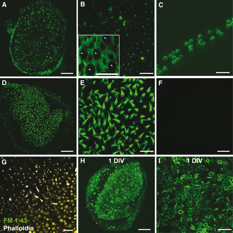
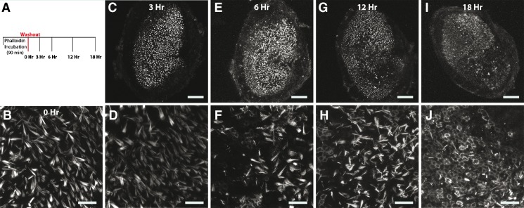
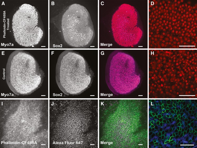
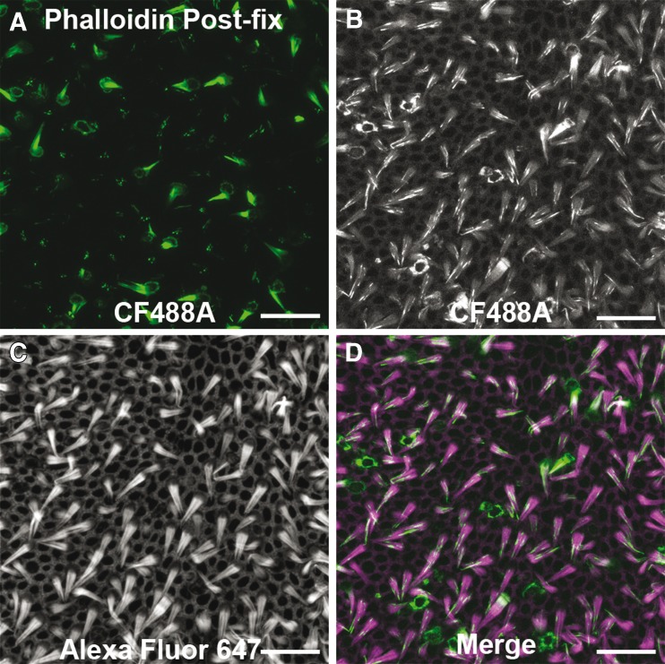
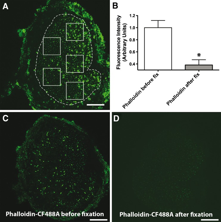
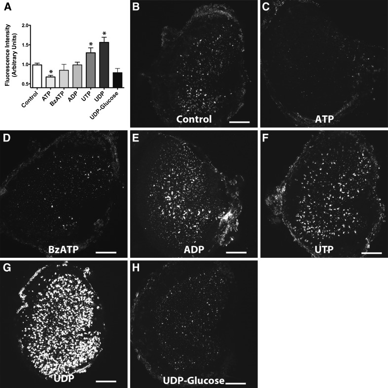
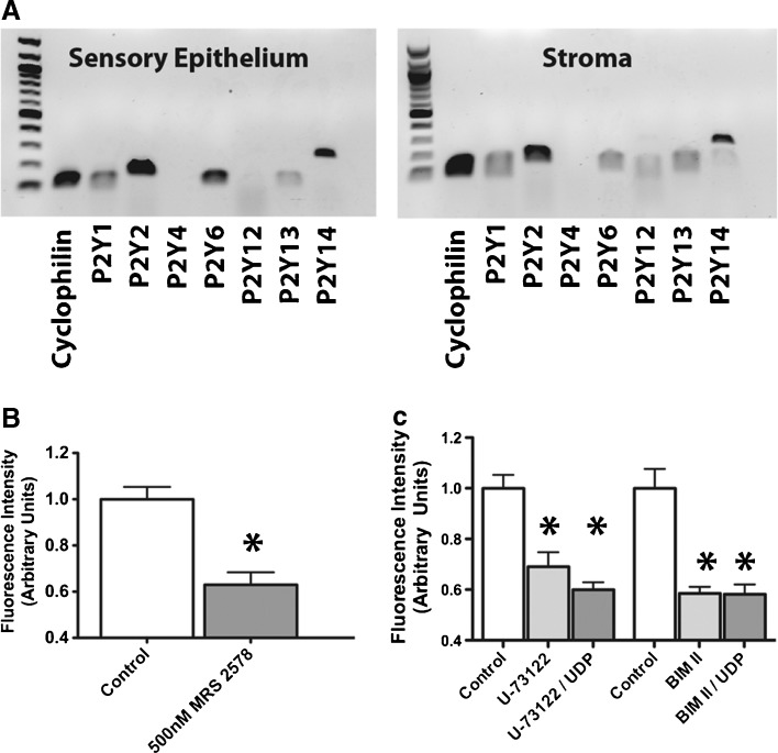
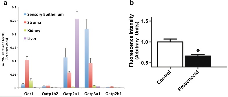
Phalloidin, a toxin isolated from the death cap mushroom, Amanita phalloides, binds to filamentous actin with high affinity, and this has made fluorophore-conjugated phalloidin a useful tool in cellular imaging. Hepatocytes take up phalloidin via the liver-specific organic anion transporting polypeptide 1b2, but phalloidin does not permeate most living cells. Rapid entry of styryl dyes into live hair cells has been used to evaluate function, but the usefulness of those fluorescence dyes is limited by broad and fixed absorption spectra. Since phalloidin can be conjugated to fluorophores with various spectra, we investigated whether it would permeate living hair cells. When we incubated mouse utricles in 66 nM phalloidin-CF488A and followed that by washes in phalloidin-free medium, we observed that it entered a subset of hair cells and labeled entire hair bundles fluorescently after 20 min. Incubations of 90 min labeled nearly all the hair bundles. When phalloidin-treated utricles were cultured for 24 h after washout, the label disappeared from the hair cells and progressively but heterogeneously labeled filamentous actin in the supporting cells. We investigated how phalloidin may enter hair cells and found that P2 receptor antagonists, pyridoxalphosphate-6-azophenyl-2', 4'-disulfonic acid and suramin, blocked phalloidin entry, while the P2Y receptor ligands, uridine-5'-diphosphate and uridine-5'-triphosphaste, stimulated uptake. Consistent with that, the P2Y6 receptor antagonist, MRS 2578, decreased phalloidin uptake. The results show that phalloidin permeates live hair cells through a pathway that requires metabotropic P2Y receptor signaling and suggest that phalloidin can be transferred from hair cells to supporting cells in culture.


Figures

FIG. 1
FIG. 1
Phalloidin permeates into live cells of the inner ear. A Maximum intensity Z-projection image of fluorescence observed after freshly dissected whole mount mouse utricles treated with phalloidin-CF488A for 20 min (37 °C) were imaged live. B Higher magnification from (A). Fluorescence can be observed throughout the entire hair bundle in only a few hair cells and in a discrete, punctate location within the stereocilia in the majority of hair cells. Inset depicts characteristic circumferential F-actin belts found among supporting cells (P86 utricle). Five presumptive HCs, which characteristically have circular apical surfaces, have been labeled with arrowheads while the remaining cells display the thick circumferential F-actin belts characteristic of mouse utricle supporting cells. C Maximum intensity Z-projection image of fluorescence observed after whole mount mouse cochlea treated with phalloidin-CF488A for 20 min and imaged live. D Example image from a live utricle treated with phalloidin-CF488A for 90 min. E Higher magnification from (D) depicts phalloidin uptake throughout the entire hair bundle in nearly all hair cells with this longer treatment of phalloidin. F No fluorescence observed in mouse utricles treated with the CF488A fluorophore alone. G Hair cells are able to take up FM 1–43 after Alexa Fluor 647 phalloidin treatment. Mouse utricles were incubated with Alexa Fluor 647 phalloidin for 20 min. The Alexa Fluor 647 phalloidin was then washed out with DMEM/F-12. Utricles were then treated with FM 1–43 for 10 s. FM 1–43 was washed out, and the utricles were imaged live. H, I Fluorescence observed in the circumferential F-actin belts of supporting cells and a few stereocilia after utricles were maintained 1 DIV (24 h) after a phalloidin treatment. I High magnification region from (H). Scale bars in A, D, F, and H are 100 μm; B, C, E, G, I are 20 μm; inset in B is 10 μm.
FIG. 2
FIG. 2
Time series of fluorescence observed after culturing utricles that had been treated with phalloidin-CF488A. Fluorescence disappears from hair cells and appears within circumferential F-actin belts of supporting cells in culture. A Experimental outline. Utricles were treated with phalloidin-CF488A for 90 min, then the label was washed out three times with DMEM/F-12, and then the media was replaced with DMEM/F-12 containing 1 % FBS. Utricles were then maintained in culture at 37 °C. Utricles were mounted and imaged live immediately (0 h; B), or after 3 h (C, D), 6 h (E, F), 12 h (G, H), or 18 h in culture (I, J). Around 6 h, the label begins to disappear from hair cells and supporting cells become fluorescent (F), and by 18 h, nearly all supporting cells are fluorescent (J). Scale bars in B, C, E, G, and I are 100 μm; scale bars in D, F, H, and J are 20 μm.
FIG. 3
FIG. 3
Hair cell density was not affected in phalloidin-treated utricles maintained in culture 24 h. P2 mouse utricles were treated 90 min with 66 nM phalloidin-CF488A (AD) and then maintained in culture for 24 h. The resulting hair cell density was compared against control P2 utricles that were maintained in culture for 24 h but were not treated with phalloidin-CF488A (EH). After culturing, utricles were immunolabeled with myosin VIIa (A, E) and Sox2 (B, F). The merged images are also displayed in C, G. Higher magnification images of myosin VIIA immunofluorescence from utricles that were treated with phalloidin-CF488A (D) and utricles that were not phalloidin-treated (H). There were no differences in HC density per 2,500 μm2 in these utricles. IL Maximum intensity Z-projection images of utricles that were treated live with phalloidin-CF488A, then maintained in culture for 7 days, fixed, and additionally stained with Alexa Fluor 647 phalloidin. I Utricles that were treated with phalloidin-CF488A maintained the fluorescence after 7 days in culture. J Staining with Alexa Fluor 647 phalloidin reveals that these utricles still have HCs. K Merge. L Higher magnification of region showing fluorescence from phalloidin-CF488A (green) and Alexa Fluor 647 phalloidin. Scale bars in AC, EG, and IK are 100 μm; scale bars in D, H are 50 μm; scale bar in L is 20 μm.
FIG. 4
FIG. 4
Phalloidin permeation in live cells of inner ear is fixable. Example image of a utricle treated live with phalloidin-CF488A for 30 min then fixed with 4 % paraformaldehyde (A). BD CF488A fluorescence maintained after phalloidin-CF488A-treated utricles were fixed as in (A), and then counterstained with Alexa Fluor 647 phalloidin. Permeabilizing the post-fixed tissue to stain with Alexa Fluor 647 phalloidin causes the CF488A fluorescence to become more diffuse. Scale bars in AD are 20 μm.
FIG. 5
FIG. 5
Phalloidin permeation into live cells of inner ear involves an active pathway. A Mean fluorescence intensity was measured in five separate 100 × 100 μm regions as depicted in the example image. B We observed more fluorescence in utricles that were treated with phalloidin-CF488A before fixation compared with utricles treated with phalloidin-CF488A after fixation (Student’s t test, p = 0.014). C Example micrograph of utricle treated with 66 nM phalloidin-CF488A and then fixed. D Example confocal micrograph of utricle fixed with paraformaldehyde for 20 min then treated with 66 nM phalloidin-CF488A. Scale bars are 100 μm.
FIG. 6
FIG. 6
Effects of P2 receptor blockers on fluorophore-conjugated phalloidin permeation. Blockers of P2 receptors inhibit phalloidin uptake. A Example image of a control living utricle treated with phalloidin-CF488a as described. B Example image of a living utricle treated with P2 antagonist PPADS (100 μm, n = 8) and then incubated with PPADS and phalloidin-CF488A. Living PPADS-treated utricle from (B) was fixed and counterstained with phalloidin-Alexa Fluor 568 to visualize the entire F-Actin network and ensure that hair cells were still present (C, D). E Example image of a living utricle treated with P2 antagonist suramin (100 μm, n = 4). F PPADS-treated living utricle was co-incubated for 10 s with PPADS and 5 μM FM 1-43 and then imaged showing that PPADS does not inhibit mechanotransduction. G–I Washing out PPADS restores the ability of hair cells to take up phalloidin-CF488A. G Example image of a utricle treated with 100 μm PPADS for 90 min at 37 °C. H After PPADS treatment and several washes in DMEM/F-12, some utricles were maintained in 66 nM phalloidin-CF488A for 90 min at 37 °C and imaged live. Phalloidin was able to enter the hair cells in these utricles at levels comparable to control utricles treated 180 min in 66 nM phalloidin-CF488A at 37 °C (I). Scale bars in AC, E, and GI are 100 μm; scale bar in D, and F are 50 μm.
FIG. 7
FIG. 7
Effects of P2 receptor agonists on fluorophore-conjugated phalloidin permeation A Mean fluorescent intensity of phalloidin-CF488A detected after pretreating with the various P2 receptor agonists. Shown are example maximum intensity Z-projection images of control (n = 21) (B), ATP (n = 12) (C), BzATP (n = 9) (D), ADP (n = 11) (E), UTP (n = 11) (F), UDP (n = 6) (G), and UDP-glucose (n = 4) (H) treated utricles. Both UTP and UDP significantly enhanced uptake of phalloidin-CF488A (one-way ANOVA with Newman-Keuls multiple comparisons post-test, p < 0.05) and ATP significantly blocked phalloidin-CF488A entry (one-way ANOVA with Newman-Keuls multiple comparisons post-test, p < 0.05). Scale bars are 100 μm.
FIG. 8
FIG. 8
P2Y receptor subunit expression and effects of P2Y6, PLC, and PKC inhibitors on fluorophore-conjugated phalloidin uptake (A) RT-PCR amplification of P2Y receptors in the sensory epithelium and stroma of P5 mouse utricles. The 100 bp ladder shown. B 500 nM MRS 2578, a P2Y6 receptor antagonist, decreased fluorophore-conjugated phalloidin uptake (n = 4). C Summary of effect of U-73122 (PLC antagonist) and BIM II (PKC antagonist) on phalloidin-CF488A permeation. We observed decreased fluorescence from phalloidin-CF488A treatment in utricles treated with 10 μM U-73122 (n = 5) or 50 μM BIM II (n = 6). Utricles treated with UDP and either U-73122 (n = 4) or BIM II (n = 4) also had significantly decreased fluorescence compared with controls.
FIG. 9
FIG. 9
Mouse utricle expresses Oat1 and Oatps. A mRNA expression levels of four Oatps, including Oatp1b2, which mediates phalloidin uptake in rodent hepatocytes, and Oat1 in the neonatal mouse sensory epithelium, stroma, liver, and kidneys (n = 3). B Probenecid treatments decreased fluorescence by 33.9 %.

References

    1. Alharazneh A, Luk L, Huth M, Monfared A, Steyger PS, Cheng AG, Ricci AJ. Functional hair cell mechanotransducer channels are required for aminoglycoside ototoxicity. PLoS One. 2011;6:e22347. doi: 10.1371/journal.pone.0022347. - DOI - PMC - PubMed
    1. Balak KJ, Corwin JT, Jones JE. Regenerated hair cells can originate from supporting cell progeny: evidence from phototoxicity and laser ablation experiments in the lateral line system. J Neurosci. 1990;10:2502–2512. - PMC - PubMed
    1. Baranova A, Ivanov D, Petrash N, Pestova A, Skoblov M, Kelmanson I, Shagin D, Nazarenko S, Geraymovych E, Litvin O, Tiunova A, Born TL, Usman N, Staroverov D, Lukyanov S, Panchin Y. The mammalian pannexin family is homologous to the invertebrate innexin gap junction proteins. Genomics. 2004;83:706–716. doi: 10.1016/j.ygeno.2003.09.025. - DOI - PubMed
    1. Burnette DT, Schaefer AW, Ji L, Danuser G, Forscher P. Filopodial actin bundles are not necessary for microtubule advance into the peripheral domain of Aplysia neuronal growth cones. Nat Cell Biol. 2007;9:1360–1369. doi: 10.1038/ncb1655. - DOI - PubMed
    1. Burnette DT, Ji L, Schaefer AW, Medeiros NA, Danuser G, Forscher P. Myosin II activity facilitates microtubule bundling in the neuronal growth cone neck. Dev Cell. 2008;15:163–169. doi: 10.1016/j.devcel.2008.05.016. - DOI - PMC - PubMed

Publication types

MeSH terms

LinkOut - more resources