. 2014 Jan 9;123(2):228-38.
doi: 10.1182/blood-2013-05-506568. Epub 2013 Nov 21.

Cell intrinsic and extrinsic factors synergize in mice with haploinsufficiency for Tp53, and two human del(5q) genes, Egr1 and Apc

Affiliations

Cell intrinsic and extrinsic factors synergize in mice with haploinsufficiency for Tp53, and two human del(5q) genes, Egr1 and Apc

Angela Stoddart et al. Blood. .

Abstract

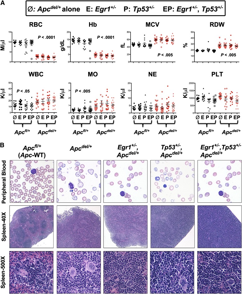
Therapy-related myeloid neoplasms (t-MN) are a late complication of the successful use of cytotoxic therapy for patients with cancer. A heterozygous deletions of the long arm of chromosome 5 [del(5q)], observed in 40% of patients, is associated with prior exposure to alkylating agents, and a high frequency of TP53 loss or mutation. In previous studies, we demonstrated that haploinsufficiency of 2 del(5q) genes, Egr1, and Apc, individually play a role in the pathogenesis of hematologic disease in mice. We now show that loss of one copy of Egr1 or Tp53 in an Apc haploinsufficient background (Apc (del/+)) accelerated the development of a macrocytic anemia with monocytosis, early features of t-MN. The development of anemia was significantly accelerated by treatment of mice with the alkylating agent, N-ethyl-N-nitrosourea (ENU), regardless of the levels of expression of Egr1 and Tp53. Transplantation of either wild type; Egr1(+/-); Tp53(+/-); Apc(del/+); or Egr1(+/-), Apc(del/+) bone marrow cells into lethally irradiated Apc(del/+) recipients resulted in rapid development of anemia that was further accelerated by administration of ENU to recipients, demonstrating that the Apc(del/+)-induced anemia was cell extrinsic and potentiated by ENU mutagenesis. These data emphasize the synergistic role of cell intrinsic and cell extrinsic (microenvironment) factors in the pathogenesis of t-MN, and raise awareness of the deleterious effects of cytotoxic therapy on the stromal microenvironment.


Figures

Figure 1
Figure 1
Loss of 1 Allele of Egr1, Tp53, or both genes equally accelerates Apc del/+-induced fatal anemia. Kaplan-Meier survival curve of Apcfl/+ control mice and Apcdel/+mice, crossed with Egr1+/− (A), Tp53+/− (B), or Egr1+/− and Tp53+/− (C). Percent survival (time to sacrifice of moribund animals) is plotted vs time in days, post-pIpC treatment (to induce Apc deletion). The number of mice in each cohort is shown. Egr1+/−, Apcdel/+; Tp53+/−, Apcdel/+; and Egr1+/−, Tp53+/−, Apcdel/+ mice all had significantly decreased survival compared with Apcdel/+ mice (P < .0001). The deletion of a single allele of Apc was induced in Mx1-Cre+Apcfl/+ mice treated with 3 doses of pI-pC, referred to as Apcdel/+mice. Control Mx1-CreApcfl/+ mice are referred as Apcfl/+mice.
Figure 2
Figure 2
All mice with Apc haploinsufficiency develop a macrocytic anemia with monocytosis. (A) CBC of Apcdel/+and Apcfl/+mice alone or crossed with Egr1+/−(E), or Tp53+/− (P), or Egr1+/−, Tp53+/− (EP) are shown. CBCs of Apcdel/+ mice were taken at the time of sacrifice, when the mice were severely anemic and moribund. Blood counts of Apcfl/+ control mice were taken during a similar timeframe, but mice were not moribund. (B) Representative peripheral blood smears (×1000) and spleen sections at ×40 and ×500 from anemic Apcdel/+mice alone or crossed and of Apcfl/+ control mice. Images were obtained using an Olympus microscope (Model BX51), equipped with an Optronics 3CCD 1080p digital camera, and processed with Adobe Photoshop. MO, monocytes; NE, neutrophils; PLT, platelets; WBC, white blood cells.
Figure 3
Figure 3
Anemia in all Apc haploinsufficient mice is caused by a block at an early erythroblast stage. (A) Splenocytes were isolated from Apcfl/+ control mice, Apcdel/+ mice, or Apcdel/+mice crossed with Egr1+/−(E), Tp53+/− (P), or Egr1+/−, Tp53+/− (EP), and flow cytometric analysis of immature erythroblasts (CD71+Ter119+), mature erythroid cells (CD71Ter119+), granulocytes (Gr1+Mac1+), B cells (CD19+), and T cells (CD4+ or CD8+) was performed when mice displayed severe anemia. (B) Flow cytometric analysis of spleen cells from a representative, anemic Apc haploinsufficient mouse. The 4r erythroid cell populations from least to most differentiated are indicated as R1 to R4. R1, pro-erythroblasts; R2, basophilic erythroblasts; R3, polychromatophilic erythroblasts; R4, orthochromatophilic erythroblasts.
Figure 4
Figure 4
Anemia and block in erythropoiesis is detected earlier in Egr1+/, Apcdel/+ double heterozygous mice compared with Apcdel/+ mice. (A) Peripheral blood counts of Apcfl/+, Apcdel/+, and Apcdel/+, Egr1+/− mice 3 months after induction. (B) Flow cytometric analysis of spleen cells isolated from mice, 3 months postinduction. Ter119high cells were analyzed using the CD71 and FSC parameters. Ery.A (Ter119highCD71highFSChigh) are basophilic, and Ery.B (Ter119highCD71highFSClow) are late basophilic and polychromatic, whereas Ery.C (Ter119highCD71lowFSClow) are orthochromatic erythroblasts and reticulocytes. A representative plot is shown on the right. Data represents the relative erythroid populations and standard error of the mean for 3 mice for each genotype. (C) The frequency of apoptosis in the R2 (CD71+Ter119+) population was measured by Annexin V staining. A representative contour plot is shown on the right. Apcdel/+ and Egr1+/−, Apcdel/+ mice consistently displayed a trend toward less apoptosis in the CD71+Ter119+ population compared with controls. For all experiments, the average and standard error of the mean of 3 mice per genotype is shown. HCT, hemocrit.
Figure 5
Figure 5
ENU-treated mice with Apc haploinsufficiency develop a fatal macrocytic anemia at an accelerated pace, regardless of Egr1 or Tp53 expression. (A) Kaplan-Meier survival curve of Apcdel/+mice, or mice crossed with Egr1+/−; Tp53+/−; or Egr1+/− and Tp53+/−. Mice were treated with 3 doses of pI-pC at 2 months of age, and ENU, where indicated, at 3 months. All mice treated with ENU died at a significantly faster rate (P < .0001). (B) CBC are shown for ENU-treated Apcfl/+and Apcdel/+mice or Apcdel/+mice crossed with Egr1+/−(E), Tp53+/− (P), or Egr1+/−, Tp53+/− (EP). CBCs of Apcdel/+ mice were taken at the time of sacrifice, when the mice were severely anemic and moribund. Blood counts of ENU-treated Apcfl/+ control mice were taken during a similar timeframe, but mice were not moribund. MO, monocytes; PLT, platelets; WBC, white blood cells.
Figure 6
Figure 6
The fatal anemia is induced by an Apc-haploinsufficient BM microenvironment (A) Kaplan-Meier survival curve of Apcfl/+ (WT) or Apcdel/+ recipients transplanted with WT, Egr1+/; Tp53+/−; Apcdel/+; or Egr1+/−, Apcdel/+BM cells. Median survival for Apcdel/+recipients was 130, 118, 139, 130, and 139 days, respectively. All Apcfl/+and Apcdel/+mice were treated with pIpC at 2 months, and recipients were lethally irradiated and transplanted 4 weeks postinduction. (B) Kaplan-Meier survival curve of Apcfl/+ (WT) or Apcdel/+ recipients transplanted with Apc-WT (CD45.1/CD45.2) BM cells. A representative dot plot shows that Apcdel/+ BM are CD45.2+, whereas Apcdel/+ mice are reconstituted with CD45.1+ CD45.2+ BM cells. (C) PCR analysis of splenocytes isolated from Apcdel/+ recipients transplanted with WT or Egr1+/− BM cells. Analysis of Egr1 and Apc clearly indicates that splenocytes are donor-derived. A PCR control on the right indicates expected sizes of deleted and WT bands. (D) Analysis of deletion of Apc, as determined by PCR analysis of DNA from stromal cells: Apcfl/fl (Control), Apcdel/del (Apc-null), Apcdel/+(Apc-het), and WT. In the sample shown, a PCR product from the floxed Apc allele was still visible in Apcdel/del and Apcdel/+ samples, in addition to the anticipated deleted and WT bands, suggesting that deletion in stromal cells may not always occur in every cell. This is in contrast to hematopoietic cells, where the deletion occurs in all cells. (E) Western blot analysis of extracts from spleen cells and stromal cell cultures using β-catenin antibody and actin as a loading control. Apc deletion had occurred in virtually all cells in the stromal cells used for this analysis (not shown).
Figure 7
Figure 7
The fatal anemia is slightly accelerated by pretreatment of Apcdel/+ recipients with ENU. (A) A model showing that Apcfl/+and Apcdel/+ recipients (CD45.2+) were treated with pIpC at 2 months, ENU at 2.5 months (where indicated), and transplanted 4 weeks post-pIpC treatment with CD45.1/CD45.2 WT BM. (B) Kaplan-Meier survival curve of Apcfl/+ (WT) or Apcdel/+ recipients transplanted with CD45.1/CD45.2 WT BM. Recipients treated with ENU are indicated (dashed line). (C) CBC of untreated and ENU-treated Apcfl/+and Apcdel/+recipient mice. RBC, Hb, and MCV were significantly different for both untreated and ENU-treated Apcdel/+recipient mice compared with Apcfl/+-recipient controls. Monocytosis was significant only for the ENU-treated Apcdel/+ vs Apcfl/+-recipient mice. MO, monocytes.

Similar articles

Cited by

References

    1. Stoddart A, McNerney ME, Bartom E, et al. Genetic pathways leading to therapy-related myeloid neoplasms. Mediterr J Hematol Infect Dis. 2011;3(1):e2011019. - PMC - PubMed
    1. Pedersen-Bjergaard J, Andersen MK, Andersen MT, Christiansen DH. Genetics of therapy-related myelodysplasia and acute myeloid leukemia. Leukemia. 2008;22(2):240–248. - PubMed
    1. Smith SM, Le Beau MM, Huo D, et al. Clinical-cytogenetic associations in 306 patients with therapy-related myelodysplasia and myeloid leukemia: the University of Chicago series. Blood. 2003;102(1):43–52. - PubMed
    1. Schanz J, Tüchler H, Solé F, et al. New comprehensive cytogenetic scoring system for primary myelodysplastic syndromes (MDS) and oligoblastic acute myeloid leukemia after MDS derived from an international database merge. J Clin Oncol. 2012;30(8):820–829. - PMC - PubMed
    1. Boultwood J, Pellagatti A, McKenzie AN, Wainscoat JS. Advances in the 5q- syndrome. Blood. 2010;116(26):5803–5811. - PubMed

Publication types

MeSH terms

Substances