. 2013 Dec;73(6):969-82; discussion 982-3.
doi: 10.1227/NEU.0000000000000141.

A novel data-driven approach to preoperative mapping of functional cortex using resting-state functional magnetic resonance imaging

Affiliations

A novel data-driven approach to preoperative mapping of functional cortex using resting-state functional magnetic resonance imaging

Timothy J Mitchell et al. Neurosurgery. 2013 Dec.

Abstract

Background: Recent findings associated with resting-state cortical networks have provided insight into the brain's organizational structure. In addition to their neuroscientific implications, the networks identified by resting-state functional magnetic resonance imaging (rs-fMRI) may prove useful for clinical brain mapping.

Objective: To demonstrate that a data-driven approach to analyze resting-state networks (RSNs) is useful in identifying regions classically understood to be eloquent cortex as well as other functional networks.

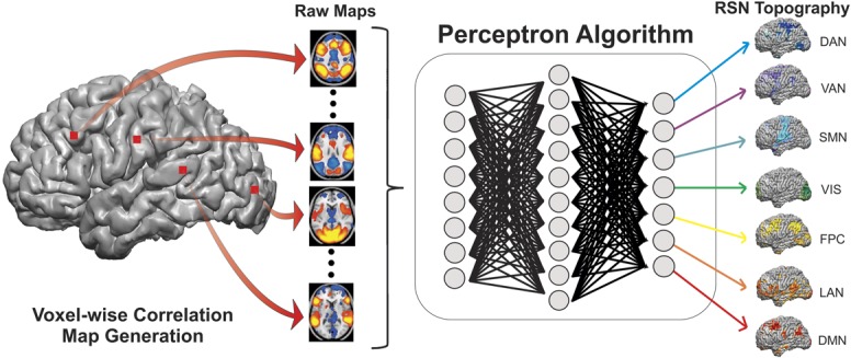
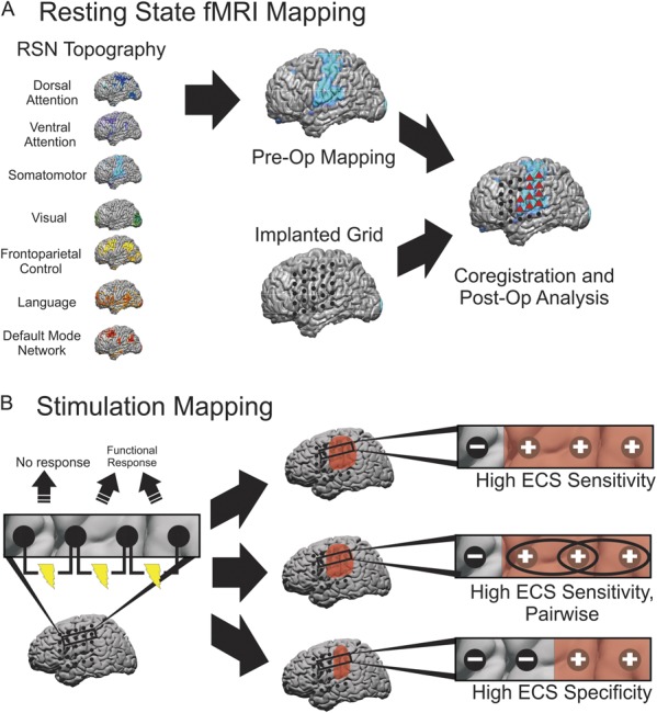
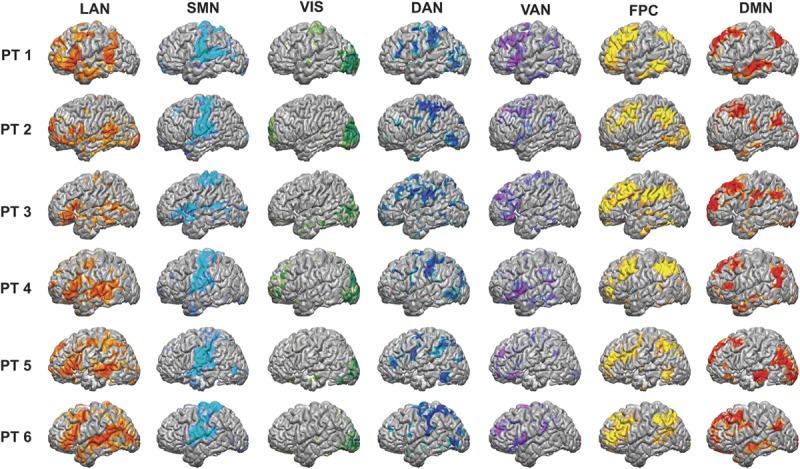
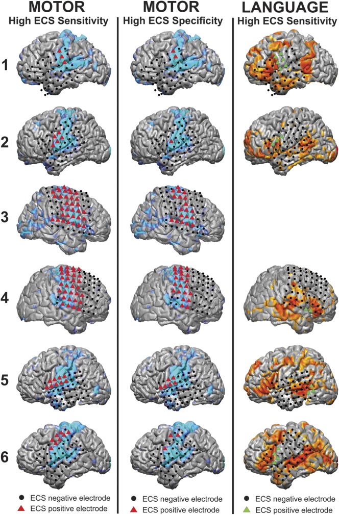
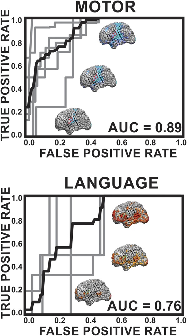
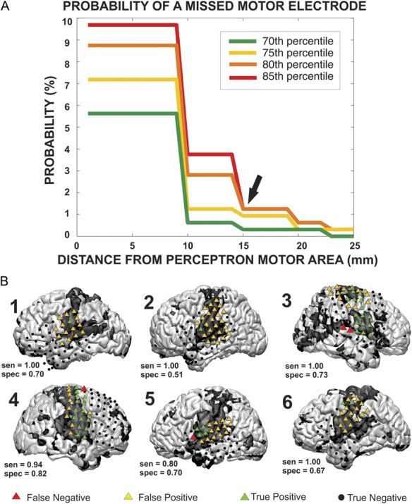
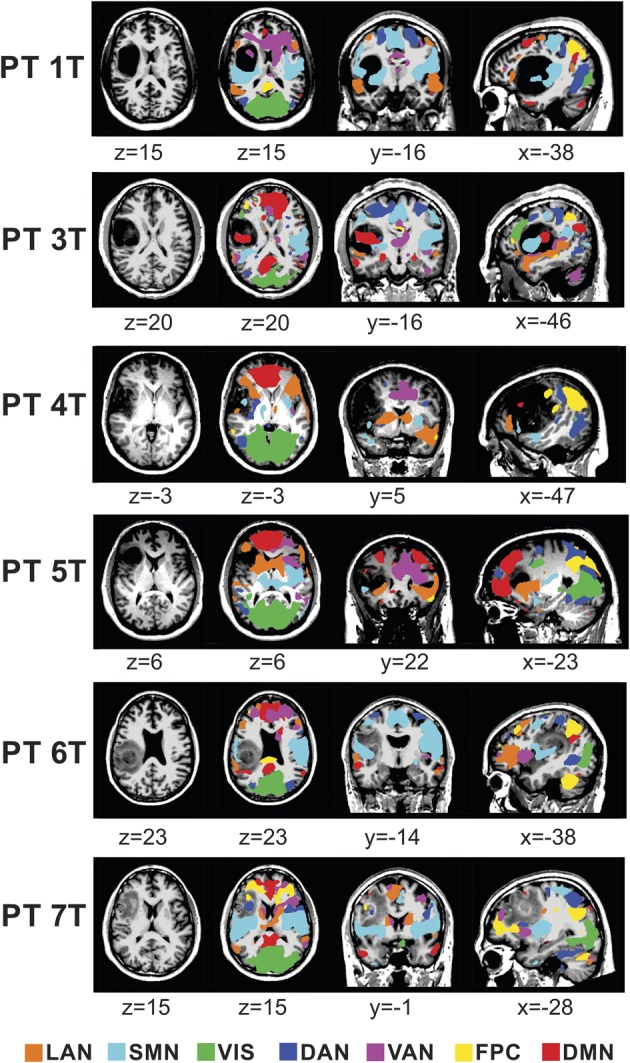
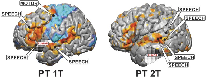
Methods: This study included 6 patients undergoing surgical treatment for intractable epilepsy and 7 patients undergoing tumor resection. rs-fMRI data were obtained before surgery and 7 canonical RSNs were identified by an artificial neural network algorithm. Of these 7, the motor and language networks were then compared with electrocortical stimulation (ECS) as the gold standard in the epilepsy patients. The sensitivity and specificity for identifying these eloquent sites were calculated at varying thresholds, which yielded receiver-operating characteristic (ROC) curves and their associated area under the curve (AUC). RSNs were plotted in the tumor patients to observe RSN distortions in altered anatomy.

Results: The algorithm robustly identified all networks in all patients, including those with distorted anatomy. When all ECS-positive sites were considered for motor and language, rs-fMRI had AUCs of 0.80 and 0.64, respectively. When the ECS-positive sites were analyzed pairwise, rs-fMRI had AUCs of 0.89 and 0.76 for motor and language, respectively.

Conclusion: A data-driven approach to rs-fMRI may be a new and efficient method for preoperative localization of numerous functional brain regions.


Figures

FIGURE 1
FIGURE 1
Voxel-wise classification of resting state networks (RSNs) using the multilayer perceptron (MLP) algorithm. Voxel-wise correlation maps were obtained from resting state functional magnetic resonance imaging data and masked to include only gray matter voxels. The masked images were then passed into the MLP algorithm (see text), which produced RSN maps for the language network (LAN), somatomotor network (SMN), visual network (VIS), dorsal attention network (DAN), ventral attention network (VAN), frontoparietal control (FPC), and default mode network (DMN).
FIGURE 2
FIGURE 2
Schematic for classifying electrodes as motor or language positive using resting state functional magnetic resonance imaging and the gold standard electrocortical stimulation (ECS) in epileptic patients. A, 7 resting-state network (RSN) maps were constructed using the multilayer perceptron (MLP) algorithm for each patient. From these 7 networks, motor and language maps were investigated further; after coregistering the electrode grid to the magnetic resonance data, gray matter voxels located within 30 mm of each electrode were averaged with a weight that was inversely proportional to the square of its distance from the electrode. Electrodes greater than a minimum threshold were then classified as motor or language positive for the MLP (red triangles). This threshold was varied to obtain receiver-operating characteristic curves. B, ECS was used as the gold standard for identifying motor and language cortex. All electrodes that did not evoke a functional response were classified as ECS negative. For the high ECS sensitivity method of classifying ECS electrodes, any site evoking a motor or language response was classified as ECS positive. This method was also used to perform a pairwise analysis. For the high specificity method of classifying ECS electrodes, electrodes that were associated with a functional response were classified as ECS positive, provided that they were not also involved in a stimulated pair that did not evoke a response, in which case they were still labeled ECS negative.
FIGURE 3
FIGURE 3
Resting-state network (RSN) maps produced by the multilayer perceptron (MLP) algorithm for the 6 epilepsy patients in the study. For each patient, the language network (LAN), somatomotor network (SMN), visual network (VIS), dorsal attention network (DAN), ventral attention network (VAN), frontoparietal control (FPC), and default mode network (DMN) were all mapped. The raw outputs of the MLP are estimates of the probability of class membership. For the purposes of visualization, the values displayed here are percentiles after rank ordering the data for each RSN across the cortex. PT, patient.
FIGURE 4
FIGURE 4
Visualization of the results for motor and language cortex using both electrocortical stimulation (ECS) and the multilayer perceptron (MLP) in the 6 epilepsy patients. ECS results are shown with colored triangles for ECS-positive sites and black circles for ECS-negative sites, whereas the MLP color maps are shown on the cortex surface. In the left column, the high ECS sensitivity method was used to classify motor electrodes as ECS positive (red triangles) and compared with the MLP results (light blue). In the middle column, the high ECS specificity method was used to classify motor electrodes. In the right column, the high ECS sensitive method was used to classify language electrodes as ECS positive (green triangles), with the MLP results displayed in orange. Patient 3 had no ECS-positive language sites and was thus excluded from the analysis.
FIGURE 5
FIGURE 5
Receiver-operating characteristic (ROC) curves for epilepsy patients comparing motor and language electrode identification by the multilayer perceptron (MLP) algorithm with the gold standard electrocortical stimulation (ECS). In this pairwise analysis, an MLP pair was considered positive if either 1 or both pairs of cortical regions were positive for motor or language because this implies that eloquent cortex was present in the area of at least 1 of the ECS electrodes. The gray lines represent ROC curves for the individual patients, and the black line is an average over the patients. The area under the curve (AUC) listed is for the average over the patients. The brains shown are the anatomic representations of these networks for a given threshold; namely, a higher threshold (the lower brain) has elevated specificity and lower sensitivity and a lower threshold (the upper brain) has lower specificity and higher sensitivity.
FIGURE 6
FIGURE 6
The method used to define a no-cut area in epilepsy patients, in which the probability of damage to motor cortex is substantial. A, to define the area, several multilayer perceptron (MLP) thresholds (70th, 75th, 80th, 85th percentiles) were used to classify electrodes as covering motor cortex, and the no-cut zone was expanded around each of the motor electrodes. The probability of a missed motor electrode, which could result in motor deficits, was plotted against the radius of expansion. B, a visualization of the method performed at the 85% and at a radius of expansion of 15 mm. Red triangles mark motor cortex as determined by electrocortical stimulation that were missed by the MLP method. sen, sensitivity; spec, specificity.
FIGURE 7
FIGURE 7
Resting-state network (RSN) maps produced by the multilayer perceptron algorithm for 6 tumor patients in the study. The language network (LAN), somatomotor network (SMN), visual network (VIS), dorsal attention network (DAN), ventral attention network (VAN), frontoparietal control (FPC), and default mode network (DMN) were mapped in a winner-take-all format in the area of the tumor. For the purposes of visualization, the values displayed here are percentiles after rank ordering the data for each RSN across the cortex and subsequently smoothed using a gaussian filter with a standard deviation of 6 mm. PT, patient.
FIGURE 8
FIGURE 8
Visualization of the results for identifying eloquent cortex using both electrocortical stimulation (ECS) and the multilayer perceptron (MLP) in 2 tumor patients. ECS results are shown with black circles identifying positive motor and speech sites. The MLP color maps are shown on the cortex surface, with blue representing the somatomotor network and orange representing the language network. Patient 2T had no motor-positive sites identified by ECS so the MLP-identified somatomotor network is not shown. PT, patient.
Figure
Figure
No available caption

References

    1. Keles GE, Chang EF, Lamborn KR, et al. Volumetric extent of resection and residual contrast enhancement on initial surgery as predictors of outcome in adult patients with hemispheric anaplastic astrocytoma. J Neurosurg. 2006;105(1):34-40 - PubMed
    1. Keles GE, Lamborn KR, Berger MS. Low-grade hemispheric gliomas in adults: a critical review of extent of resection as a factor influencing outcome. J Neurosurg. 2001;95(5):735-745 - PubMed
    1. McGirt MJ, Chaichana KL, Gathinji M, et al. Independent association of extent of resection with survival in patients with malignant brain astrocytoma. J Neurosurg. 2009;110(1):156-162 - PubMed
    1. Lacroix M, Abi-Said D, Fourney DR, et al. A multivariate analysis of 416 patients with glioblastoma multiforme: prognosis, extent of resection, and survival. J Neurosurg. 2001;95(2):190-198 - PubMed
    1. Sanai N, Mirzadeh Z, Berger MS. Functional outcome after language mapping for glioma resection. N Engl J Med. 2008;358(1):18-27 - PubMed

Publication types