. 2014 May;82(5):867-78.
doi: 10.1002/prot.24464. Epub 2013 Nov 22.

A molten globule intermediate of the von Willebrand factor A1 domain firmly tethers platelets under shear flow

Affiliations

A molten globule intermediate of the von Willebrand factor A1 domain firmly tethers platelets under shear flow

Alexander Tischer et al. Proteins. 2014 May.

Abstract

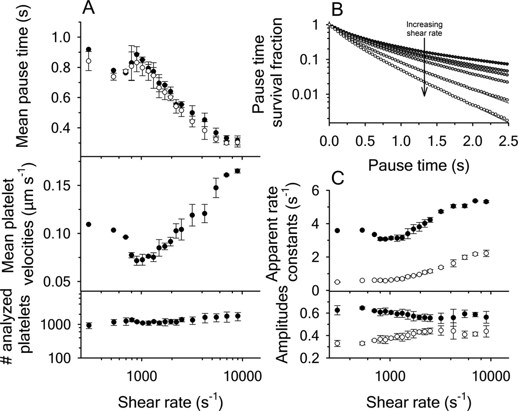
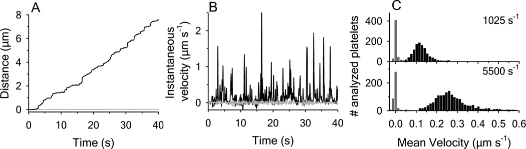
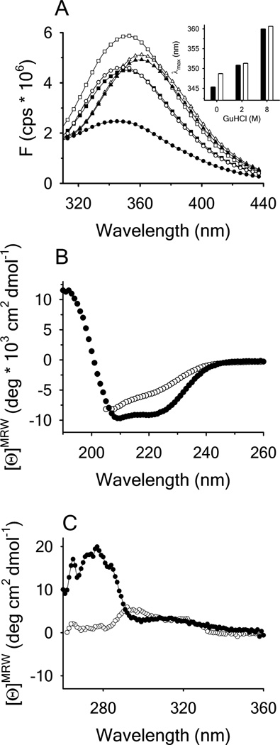
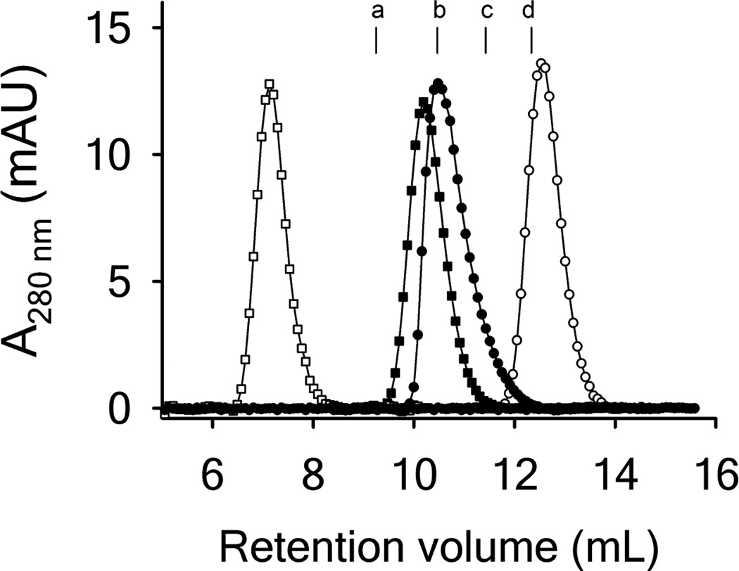
Clinical mutations in patients diagnosed with Type 2A von Willebrand disease (VWD) have been identified that break the single disulfide bond linking N- and C-termini in the vWF A1 domain. We have modeled the effect of these mutations on the disulfide-bonded structure of A1 by reducing and carboxy-amidating these cysteines. Solution biophysical studies show that loss of this disulfide bond induces a molten globule conformational state lacking global tertiary structure but retaining residual secondary structure. The conformational dependence of platelet adhesion to these native and molten globule states of A1 is quantitatively compared using real-time high-speed video microscopy analysis of platelet translocation dynamics under shear flow in a parallel plate microfluidic flow chamber. While normal platelets translocating on surface-captured native A1 domain retain the catch-bond character of pause times that increase as a function of shear rate at low shear and decrease as a function of shear rate at high shear, platelets that interact with A1 lacking the disulfide bond remain stably attached and do not translocate. Based on these findings, we propose that the shear stress-sensitive regulation of the A1-GPIb interaction is due to folding the tertiary structure of this domain. Removal of the tertiary structure by disrupting the disulfide bond destroys this regulatory mechanism resulting in high-strength interactions between platelets and vWF A1 that are dependent only on residual secondary structure elements present in the molten globule conformation.

Keywords: Von Willebrand disease type 2; Von Willebrand factor; disulfide bond; molten globule; platelet adhesiveness; protein folding; rheology; shear stress; thermodynamics.


Figures

Figure 1
Figure 1
Platelet translocation on disulfide-intact A1 domain as a function of shear rate. (A, upper panel) Mean pause times derived from the statistical average of platelet distributions (filled circles) and from biexponential fitting of the pause time survival fraction using equation (1) (open circles), mean platelet velocities (center panel) and the amount of platelets analyzed (lower panel). (B) Survival fraction decay functions of the platelet pause times with increasing shear rates. Rates are: 800, 1320, 1500, 1950, 2500, 4250 and 9000 s−1 from top to bottom. Curves were fit to a biexponential functions. (C) Apparent rate constants k1 (black circles) and k2 (open circles) and the fractional amplitudes associated with k1 (black circles) and k2 (open circles). Error bars represent the mean standard deviation from four independent experiments.
Figure 2
Figure 2
Comparison of platelet translocations on disulfide-intact A1 with RCAM A1. (A) Traveled distance of a single platelet as a function of time at 1025 s−1; disulfide-intact A1 (black line), RCAM A1 (grey line). (B) Comparison of the instantaneous velocities obtained for a single platelet interacting with disufide intact A1 (black line) and RCAM A1 (grey line) at 1025 s−1. (C) Histograms of the mean velocity of platelets translocating on disulfide-intact A1 (black bars) and RCAM A1 (gray bars) at shear rates of 1025 and 5500 s−1. Total number of platelets analyzed were 526 and 422 for RCAM A1 at 1025 and 5500 s−1 respectively; and 1132 and 1687 for disulfide-intact A1 at 1025 and 5500 s−1 respectively. Data are representative of four independent experiments.
Figure 3
Figure 3
(A) Fluorescence emission spectra of disulfide-intact A1 (closed symbols) and RCAM A1 (open symbols) in the presence of 0M (circles), 2M (squares) and 8M GdnHCl (triangles) with excitation λ = 280nm. Inset: Wavelength of maximum fluorescence intensity (λmax) for disulfide-intact A1 (black bars) and RCAM A1 (white bars). (B) Far UV CD spectra of disulfide-intact A1 (closed circles) in buffer and RCAM A1 (open circles) in 0.5M GdnHCl. (C) Near UV CD spectra for disulfide-intact A1 (closed circles) and RCAM A1 in buffer (open circles).
Figure 4
Figure 4
GdnHCl denaturation (A) and urea denaturation (B) of disulfide-intact A1 (closed circles) and RCAM A1 (open circles) monitored by circular dichroism at λ = 222nm.
Figure 5
Figure 5
Thermal denaturation. (A) disulfide-intact A1 in 1M urea (closed circles) and RCAM A1 in 1M urea (closed squares) monitored by circular dichroism at λ = 222nm. (B) disulfide-intact A1 in buffer (closed circles), 9M urea (open circles) and RCAM A1 in buffer (closed squares) monitored by intrinsic protein fluorescence emission at λem = 359nm with excitation at λex = 280nm. (C) disulfide-intact A1 in buffer (closed circles) and RCAM A1 in buffer (closed squares) monitored by ANS fluorescence emission at λ = 495nm with excitation at λex = 350nm. Inset: Spectra of A1 and RCAM A1 in the presence of ANS at 20°C before (closed circles and squares) and after the thermal scan (open circles and squares).
Figure 6
Figure 6
(A) Acrylamide quenching of tryptophan fluorescence in disulfide-intact A1 at 20°C (closed circles) and 1°C (closed squares), RCAM A1 at 20°C (open symbols) and 1µM NATA at 20°C (grey circles) in buffer. (B) Resulting Stern-Volmer quenching constants derived from the slope of F0/F as a function of acrylamide molarity at the indicated concentrations of GdnHCl. Disulfide-intact A1 (black bars), RCAM A1 (white bars) and NATA in buffer (grey bar).
Figure 7
Figure 7
Size exclusion chromatograms for native disulfide-intact A1 (closed symbols) and RCAM A1 (open symbols) in buffer (circles) and 0.25M GdnHCl (squares). The column was equilibrated with PGA buffer and calibrated with several proteins as indicated by their peak maxima (a - Ovoalbumin from chicken (44.0kDa), b - bovine Carboanhydrase (29kDa), c - Myoglobin from horse (17.0kDa), d cytochrome c from horse (12.4kDa). Void volume = 6.85mL with blue dextran (2000kDa). Included volume 17.54mL with vitamin B12 (1.35kDa).

References

    1. Woods AI, Sanchez-Luceros A, Kempfer AC, et al. C1272F: a novel type 2A von Willebrand's disease mutation in A1 domain; its clinical significance. Haemophilia. 2012;18(1):112–116. - PubMed
    1. Penas N, Perez A, Gonzalez-Boullosa R, Batlle J. C1272S: a new candidate mutation in type 2A von Willebrand disease that disrupts the disulfide loop responsible for the interaction of VWF with platelet GP Ib-IX. Am J Hematol. 2004;75(2):73–77. - PubMed
    1. Meyer D, Fressinaud E, Gaucher C, et al. Gene defects in 150 unrelated French cases with type 2 von Willebrand disease: from the patient to the gene. INSERM Network on Molecular Abnormalities in von Willebrand Disease. Thromb. Haemost. 1997;78(1):451–456. - PubMed
    1. Lavergne JM, De Paillette L, Bahnak BR, et al. Defects in type IIA von Willebrand disease: a cysteine 509 to arginine substitution in the mature von Willebrand factor disrupts a disulphide loop involved in the interaction with platelet glycoprotein Ib-IX. Br J Haematol. 1992;82(1):66–72. - PubMed
    1. Azuma H, Hayashi T, Dent JA, Ruggeri ZM, Ware J. Disulfide bond requirements for assembly of the platelet glycoprotein Ib-binding domain of von Willebrand factor. J Biol Chem. 1993;268(4):2821–2827. - PubMed

Publication types

LinkOut - more resources