Review
. 2013 Oct;3(4):1553-67.
doi: 10.1002/cphy.c130003.

Mechanical properties of respiratory muscles

Affiliations
Review

Mechanical properties of respiratory muscles

Gary C Sieck et al. Compr Physiol. 2013 Oct.

Abstract

Striated respiratory muscles are necessary for lung ventilation and to maintain the patency of the upper airway. The basic structural and functional properties of respiratory muscles are similar to those of other striated muscles (both skeletal and cardiac). The sarcomere is the fundamental organizational unit of striated muscles and sarcomeric proteins underlie the passive and active mechanical properties of muscle fibers. In this respect, the functional categorization of different fiber types provides a conceptual framework to understand the physiological properties of respiratory muscles. Within the sarcomere, the interaction between the thick and thin filaments at the level of cross-bridges provides the elementary unit of force generation and contraction. Key to an understanding of the unique functional differences across muscle fiber types are differences in cross-bridge recruitment and cycling that relate to the expression of different myosin heavy chain isoforms in the thick filament. The active mechanical properties of muscle fibers are characterized by the relationship between myoplasmic Ca2+ and cross-bridge recruitment, force generation and sarcomere length (also cross-bridge recruitment), external load and shortening velocity (cross-bridge cycling rate), and cross-bridge cycling rate and ATP consumption. Passive mechanical properties are also important reflecting viscoelastic elements within sarcomeres as well as the extracellular matrix. Conditions that affect respiratory muscle performance may have a range of underlying pathophysiological causes, but their manifestations will depend on their impact on these basic elemental structures.


Figures

Figure 1
Figure 1
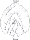
Innervation of the cat diaphragm muscle. Phrenic nerve axons derived from the C4 segment of the cervical spinal cord innervate ventral aspects of the costal and crural regions of the diaphragm muscle, whereas axons derived from C6 innervate more dorsal aspects. Reproduced from ref. (378); used with permission.
Figure 2
Figure 2
Muscle fibers contain myofibrils (a), each comprising sarcomeres arranged in series which give muscle a striated appearance visible also in transmission electron micrographs. Thick and thin filaments in the sarcomere are composed of myosin (red) and actin (yellow), respectively, and their interaction provides the basis for force generation and contraction. In cross section, myosin and actin filaments are organized in a myofilament lattice, clearly visible with electron microscopy. Reproduced from ref. (167); used with permission.
Figure 3
Figure 3
Muscle fibers from the rat diaphragm muscle express a single myosin heavy chain (MyHC) isoform with the exception of MyHC2X and MyHC2B in some fibers. Modified from ref. (130)
Figure 4
Figure 4
Motor units are classified according to their contractile and fatigue properties as slow-twitch (type S) and as fast-twitch units, which display fatigue-resistant (type FR), fatigue-intermediate (type FInt) and fatigable (type FF) characteristics. Expression of MyHC isoforms by muscle fibers corresponds with motor unit properties. Contraction speeds also vary across motor unit types. Reproduced from ref. (156); used with permission.
Figure 5
Figure 5
Cross-bridges cycle between a strongly bound and an unbound state during force generation and contraction. Cross-bridge cycling determines rates of cross-bridge attachment (fapp) and detachment (gapp). Illustration copyrighted by the Mayo Clinic and Foundation and reproduced from ref. (398) with permission.
Figure 6
Figure 6
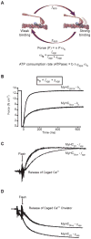
Force measurements in single muscle fibers during maximal activation in rigor solution (without ATP and with free ionized Ca2+ concentration of 100 μM, i.e., pCa 4.0), pCa4.0 solution (with ATP) and pCa9.0 solution (free ionized Ca2+ concentration of 1 nM). Resting and activated stiffness were determined by imposing sinusoidal length oscillations (0.2% Lo) at 2kHz. Reproduced from ref. (174); used with permission.
Figure 7
Figure 7
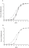
Force development in single diaphragm muscle fibers expressing slow (open symbol) and fast (closed symbols – in A: MyHC2A: ▼, MyHC2X: ■, MyHC2B and/or MyHC2X: ◆) isoforms of MyHC. Force depends on myoplasmic Ca2+ concentrations (pCa; -log[Ca2+]). Reproduced with permission from ref. (130).
Figure 8
Figure 8
Muscle force generation depends on sarcomere length and the overlap between thick and thin filaments, which determines the fraction of cross-bridges that can form (αfs). Reproduced from ref. (167); used with permission.
Figure 9
Figure 9
Titin cDNA sequences for splice variants expressed in rabbit psoas and soleus muscles. Sequences predict differences in the I-band region of titin. Estimated protein molecular weight is show on the right. The longer segments in soleus contribute to lower titin-based passive tension. Titin-based passive tension is similar for diaphragm and soleus (329). Thus, we anticipate similar titin sequences for both muscles. Reproduced from ref. (125); used with permission from Springer®.
Figure 10
Figure 10
Force-length relationship of rat diaphragm muscle during lengthening and shortening cycles. Results are from cycles of sinusoidal oscillations at 2 Hz and loops are displayed in clockwise orientation. Lo is optimal length and cycle strain is the amplitude of oscillations as percentage of Lo. Note that passive force during lengthening is higher than during shortening (hysteresis). The amount of hysteresis depends on resting length of the diaphragm. Viscous work (area within each loop) increases at longer lengths. Reproduced from ref. (423); used with permission.
Figure 11
Figure 11
Movement history-dependence (thixotropy) of length-tension relationship in diaphragm single fibers. Raw tracings of fiber length (A) and tension (B) in a chemically permeabilized fiber from mouse diaphragm (LF Ferreira, KS Campbell, and MB Reid; unpublished observations). Data collected during maximal calcium activation (pCa 4.5) at 15°C. Fiber was stretched by 35 μm from the optimal fiber length (879 μm; sarcomere length 2.586 μm). C) Relationship between tension and fiber length – data re-plotted from panel A. Hysteresis is greater in the first lengthening-shortening cycle than in subsequent cycles. The initial portions of the first and second cycles are traced by blue and red lines, respectively. D, Relationship between changes (Δ) in tension and fiber length shows a decrease in stiffness with a prior lengthening-shortening cycle, i.e., slope of relationship for second cycle (red circles) is approximately 30% lower than slope of first cycle (blue circles). Solid black lines are best fit from linear regression. For details on protocol and methods see Campbell & Moss (68) and Hardin et al. (175).
Figure 12
Figure 12
Schematic illustration of proteins of the extracellular matrix, costamere, and intermediate filaments. The extracellular matrix and basement membrane include laminin, collagen IV, and intermediate filament (IF) proteins which include desmin (yellow) and keratins (K8/K19; red) plus other proteins not shown (synemin, paranemin, syncoilin). Desmin surrounds the Z-disks connecting myofibrils to each other and to the sarcolemma. Most IF proteins are linked to costameric proteins; keratin-containing IF are located around the M-line and also link to costameres. Ank, ankyrin; ANT, adenine nucleotide translocator; CK, creatine kinase; DG, dystroglycan; K, keratin; MLP, striated muscle-specific LIM protein; SP, sarcospan; SG, sarcoglycan; SR, sarcoplasmic reticulum. Reproduced from ref. (69); used with permission from Elsevier.
Figure 13
Figure 13
Force (normalized to percent maximum tetanic force) generated by cat diaphragm motor units at different frequencies of stimulation. Results are for individual motor units classified by their contractile and fatigue properties (see text for details). The steepest portion of the force-frequency curve occurs between 10 and 30 Hz for all types of motor units in the diaphragm muscle (116), consistent with onset and peak discharge frequencies of ~8 Hz and ~25 Hz respectively for type S and FR units (top arrows) and ~15 Hz and ~60 Hz respectively for type FInt and FF units (bottom arrows), reported in (376).
Figure 14
Figure 14
Contribution of neuromuscular transmission failure to diaphragm muscle fatigue. Diaphragm muscle-phrenic nerve preparations are stimulated electrically every 1 s via a suction electrode to the phrenic nerve (40 Hz in 330-ms trains) and direct muscle stimulation via plate electrodes is superimposed intermittently every 15 s. A. Representative measurement of force developed by a mouse diaphragm muscle with repetitive stimulation. Reduced force generation by the diaphragm muscle reflects fatigue. The difference in force elicited by nerve and muscle stimulation reflects neuromuscular transmission failure. B. Neuromuscular transmission failure (mean ± SE) measured in diaphragm muscles from adult rats and mice. *, statistically significant difference. Reproduced from ref. (374); used with permission.

References

    1. Adelman WJ, Palti Y, Senft JP. Potassium ion accumulation in a periaxonal space and its effect on the measurement of membrane potassium ion conductance. J Membr Biol. 1973;13:387–410. - PubMed
    1. Aldrich TK, Shander A, Chaudhry I, Nagashima H. Fatigue of isolated rat diaphragm: role of impaired neuromuscular transmission. J Appl Physiol. 1986;61:1077–1083. - PubMed
    1. Aldrich TK. Transmission fatigue of the rabbit diaphragm. Respir Physiol. 1987;69:307–319. - PubMed
    1. Allan DW, Greer JJ. Embryogenesis of the phrenic nerve and diaphragm in the fetal rat. J Comp Neurol. 1997;382:459–468. - PubMed
    1. Allen DG. Skeletal muscle function: role of ionic changes in fatigue, damage and disease. Clin Exp Pharmacol Physiol. 2004;31:485–493. - PubMed

Publication types

Substances

LinkOut - more resources