. 2013 Nov 12;8(11):e78496.
doi: 10.1371/journal.pone.0078496. eCollection 2013.

Increasing the yield in targeted next-generation sequencing by implicating CNV analysis, non-coding exons and the overall variant load: the example of retinal dystrophies

Affiliations

Increasing the yield in targeted next-generation sequencing by implicating CNV analysis, non-coding exons and the overall variant load: the example of retinal dystrophies

Tobias Eisenberger et al. PLoS One. .

Erratum in

  • PLoS One. 2014;9(11):e108840

Abstract

Retinitis pigmentosa (RP) and Leber congenital amaurosis (LCA) are major causes of blindness. They result from mutations in many genes which has long hampered comprehensive genetic analysis. Recently, targeted next-generation sequencing (NGS) has proven useful to overcome this limitation. To uncover "hidden mutations" such as copy number variations (CNVs) and mutations in non-coding regions, we extended the use of NGS data by quantitative readout for the exons of 55 RP and LCA genes in 126 patients, and by including non-coding 5' exons. We detected several causative CNVs which were key to the diagnosis in hitherto unsolved constellations, e.g. hemizygous point mutations in consanguineous families, and CNVs complemented apparently monoallelic recessive alleles. Mutations of non-coding exon 1 of EYS revealed its contribution to disease. In view of the high carrier frequency for retinal disease gene mutations in the general population, we considered the overall variant load in each patient to assess if a mutation was causative or reflected accidental carriership in patients with mutations in several genes or with single recessive alleles. For example, truncating mutations in RP1, a gene implicated in both recessive and dominant RP, were causative in biallelic constellations, unrelated to disease when heterozygous on a biallelic mutation background of another gene, or even non-pathogenic if close to the C-terminus. Patients with mutations in several loci were common, but without evidence for di- or oligogenic inheritance. Although the number of targeted genes was low compared to previous studies, the mutation detection rate was highest (70%) which likely results from completeness and depth of coverage, and quantitative data analysis. CNV analysis should routinely be applied in targeted NGS, and mutations in non-coding exons give reason to systematically include 5'-UTRs in disease gene or exome panels. Consideration of all variants is indispensable because even truncating mutations may be misleading.


Conflict of interest statement

Competing Interests: TE, CN, CD, AB, CB and HJB are employees of Bioscientia, which is part of a publicly traded diagnostic company. There are no patents, products in development or marketed products to declare. This does not alter the authors' adherence to all the PLOS ONE policies on sharing data and materials, as detailed online in the guide for authors.

Figures

Figure 1
Figure 1. Mutational spectrum in RP and LCA patients.
Percentages refer to patients with mutations in the respective gene that are considered causative. The distribution of causative mutations across many genes, each contributing a relatively small fraction to the mutational spectrum, confirms the extensive genetic heterogeneity of retinal dystrophies. Note that the three patients that were found to carry X-linked mutations are not contained in the schemes A – B. A. arRP. B. adRP. Note that the percentages refer to a relatively small adRP cohort in this study. C. LCA. D. Functional categorization of genes that were found to carry causative mutations in our study. Mutations in genes encoding components of the photoreceptor’s connecting cilium and associated structures were predominant.
Figure 2
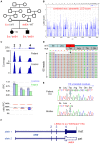
Figure 2. Evidence for mutations in non-coding exon 1 of EYS contributing to arRP.
Compound-heterozygosity for truncating mutations in the coding sequence of EYS and mutations of exon 1 co-segregate with arRP in two families. A. Patient 57 carries a deletion of exon 1 in trans to a nonsense mutation, whereas patient 93 and his sister B. carry a mutation of the donor splice site and a truncating frameshift mutation. C. Scheme of the EYS gene with non-coding (light grey) and coding (black) exons. Mutations of patients 57 (red) and 93 (blue) are indicated. The heterozygous deletion of exon 1 in patient 57 was detected by quantitative analysis of NGS data. The coverage plot illustrates the statistical readout, with the absolute coverage deduced from unique read count (scale bar, upper panel) and as calculated by the CNV analysis mode in SeqNext (JSI Medical Systems, lower panel). Normalized relative coverage (relative product coverage, RPC) of every target region of interest (ROI) of patient sample (green, RPC P.) and average relative target coverage of control samples (blue, RPC C.). Error bars: standard deviation of control samples. Ratio RPC was calculated from patient’s versus controls’ RPC; ratios below 75% indicate a heterozygous deletion. Electropherograms show the confirmation of the three point mutations by Sanger sequencing (arrows: position of the mutations).
Figure 3
Figure 3. Hemizygosity of a CRX mutation in a recessive consanguineous LCA family.
A. Compound-heterozygosity for a potentially protein-extending no-stop mutation (c.899A>G/p.(*300Trpext*118); here designated as Ext) abrogating the natural termination codon in exon 4 and a deletion of the same exon (delE4) in trans in patient 110 and her brother. B. Graphical view of the LOD score calculation from genomewide SNP mapping for this family previous to NGS testing: Genomewide homozygosity mapping prior to NGS did not identify a clear candidate locus. The combined maximum parametric LOD score of 2.4 was not obtained. C. Scheme of the CRX gene and coverage plots for CNV analysis from NGS data (Illumina MiSeq), indicating a heterozygous deletion of exon 4 (upper panel, absolute coverage based on read count; lower panel, SeqNext CNV analysis). See legend to Figure 2C. D. Schematic representation of the mapped sequencing reads for the no-stop mutation (Integrative Genomics Viewer). The mutation (arrow) was present in all 65 reads covering this region of the gene and therefore appeared homozygous. E. Electropherograms from Sanger sequencing of the no-stop mutation with hemizygosity in patient 110 (upper panel) and heterozygosity in her mother (lower panel). F. Summary of the disease-causing genetic constellation in patient 110 and her brother (superimposition on parental alleles).
Figure 4
Figure 4. Different arRP scenarios implicating truncating RP1 mutations with diverse impact on disease.
A. Pedigree of patient 25 whose arRP is caused by two truncating recessive RP1 alleles. In addition, the patient carries a heterozygous CDH23 nonsense mutation that has been reported in USH1 patients but is probably unrelated to disease here. B. LCA in patient 124 is due to homozygosity for the founder mutation p.Gln301* in TULP1. Heterozygosity for the RP1 nonsense mutation p.Glu1750* likely reflects accidental carriership. It likely represents a recessive loss-of-function allele. Dotted horizontal line: likely consanguinity. C. Compound heterozygosity for two truncating PROM1 mutations can be considered pathogenic in arRP patient 55. The RP1 nonsense mutation p.Gln2102* locates near the C-terminus and likely represents an NMD-insensitive non-pathogenic variant. D. Scheme of the RP1 protein and overview of truncating RP1 mutations reported in this study (mutations shown in A – C in red). The four classes of RP1 truncating mutations are displayed. Class I, NMD-sensitive truncations; class II, NMD-insensitive truncating mutations representing the majority of pathogenic truncation mutations in RP1 (dominant negative pathomechanism); class III, NMD-insensitive truncation mutations representing loss-of-function arRP mutations; class IV, NMD-insensitive, non-pathogenic truncations located 3′ of p.1816. CP, “critical position”: 65-residue region between p.1751 and p.1816 containing a yet undefined protein residue before which truncation causes disease.

References

    1. Wright AF, Chakarova CF, Abd El-Aziz MM, Bhattacharya SS (2010) Photoreceptor degeneration: genetic and mechanistic dissection of a complex trait. Nat Rev Genet 11: 273–284. - PubMed
    1. Frick KD, Roebuck MC, Feldstein JI, McCarty CA, Grover LL (2012) Health services utilization and cost of retinitis pigmentosa. Arch Ophthalmol 130: 629–634. - PubMed
    1. den Hollander AI, Black A, Bennett J, Cremers FP (2010) Lighting a candle in the dark: advances in genetics and gene therapy of recessive retinal dystrophies. J Clin Invest 120: 3042–3053. - PMC - PubMed
    1. Hartong DT, Berson EL, Dryja TP (2006) Retinitis pigmentosa. Lancet 368: 1795–1809. - PubMed
    1. Sheffield VC, Stone EM (2011) Genomics and the eye. N Engl J Med 364: 1932–1942. - PubMed