

Expression and activation of caspase-6 in human fetal and adult tissues
- PMID: 24265764
- PMCID: PMC3827169
- DOI: 10.1371/journal.pone.0079313
Expression and activation of caspase-6 in human fetal and adult tissues
Abstract
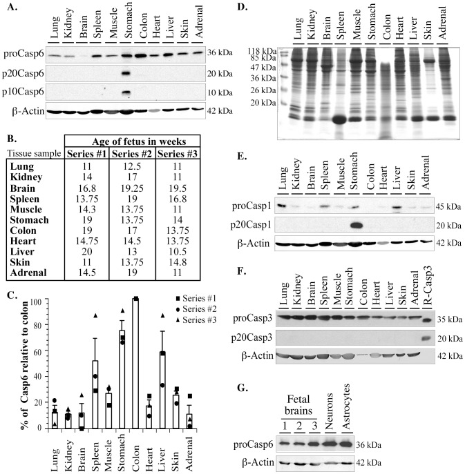
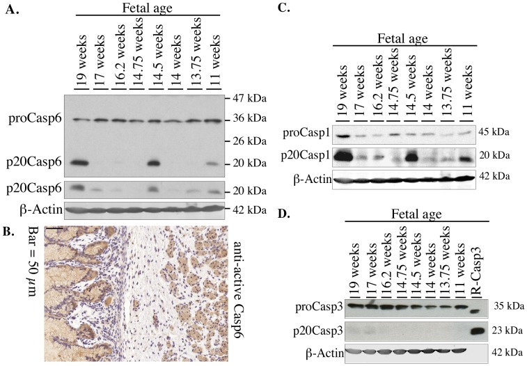
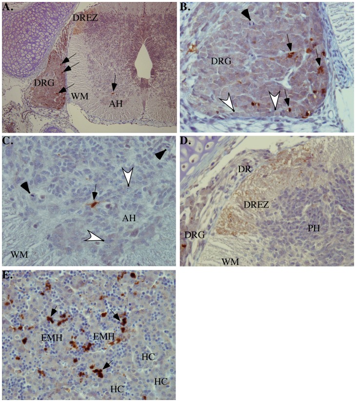
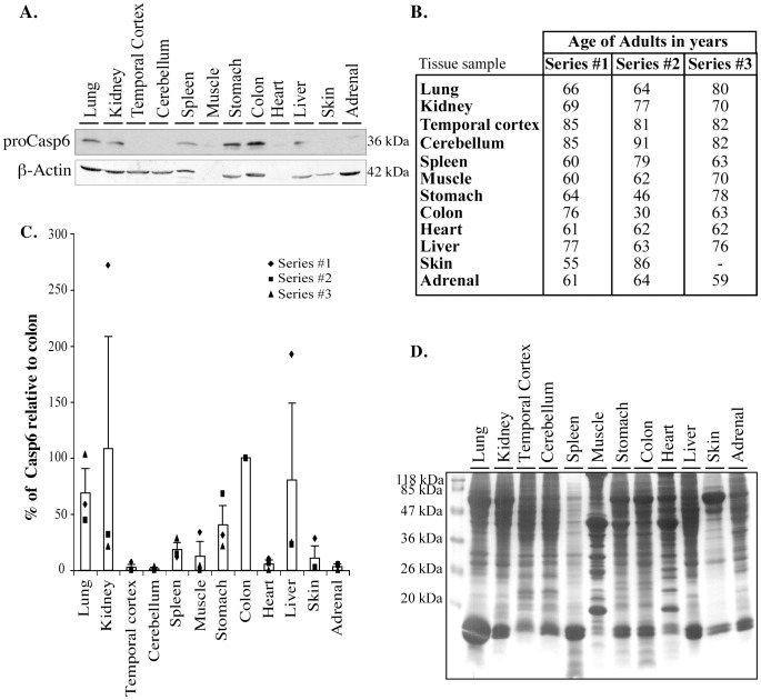
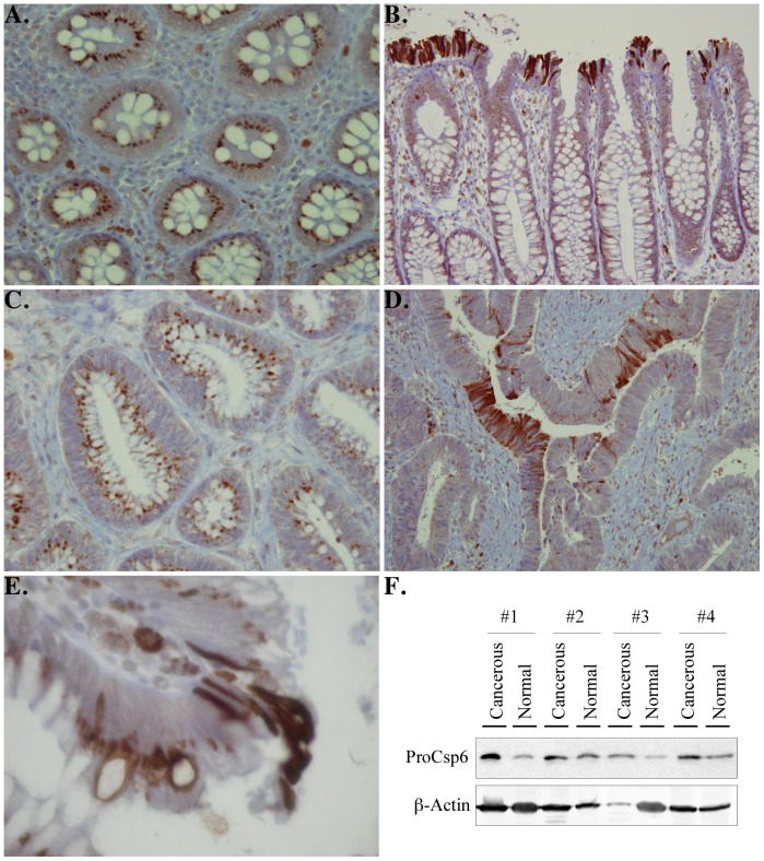
Caspase-6 is an effector caspase that has not been investigated thoroughly despite the fact that Caspase-6 is strongly activated in Alzheimer disease brains. To understand the full physiological impact of Caspase-6 in humans, we investigated Caspase-6 expression. We performed western blot analyses to detect the pro-Caspase-6 and its active p20 subunit in fetal and adult lung, kidney, brain, spleen, muscle, stomach, colon, heart, liver, skin, and adrenals tissues. The levels were semi-quantitated by densitometry. The results show a ubiquitous expression of Caspase-6 in most fetal tissues with the lowest levels in the brain and the highest levels in the gastrointestinal system. Caspase-6 active p20 subunits were only detected in fetal stomach. Immunohistochemical analysis of a human fetal embryo showed active Caspase-6 positive apoptotic cells in the dorsal root ganglion, liver, lung, kidney, ovary, skeletal muscle and the intestine. In the adult tissues, the levels of Caspase-6 were lower than in fetal tissues but remained high in the colon, stomach, lung, kidney and liver. Immunohistological analyses revealed that active Caspase-6 was abundant in goblet cells and epithelial cells sloughing off the intestinal lining of the adult colon. These results suggest that Caspase-6 is likely important in most tissues during early development but is less involved in adult tissues. The low levels of Caspase-6 in fetal and adult brain indicate that increased expression as observed in Alzheimer Disease is a pathological condition. Lastly, the high levels of Caspase-6 in the gastrointestinal system indicate a potential specific function of Caspase-6 in these tissues.
Conflict of interest statement
Figures
Similar articles
-
Development and regulation of glucose-6-phosphatase gene expression in rat liver, intestine, and kidney: in vivo and in vitro studies in cultured fetal hepatocytes.Diabetes. 1998 Jun;47(6):882-9. doi: 10.2337/diabetes.47.6.882. Diabetes. 1998. PMID: 9604863
-
Profiling transcript levels for steroidogenic enzymes in fetal tissues.J Steroid Biochem Mol Biol. 2003 Nov;87(2-3):181-9. doi: 10.1016/j.jsbmb.2003.07.006. J Steroid Biochem Mol Biol. 2003. PMID: 14672738
-
Bcl-xL is a negative regulator of caspase-3 activation in immature neurons during development.Brain Res Dev Brain Res. 1999 Aug 5;116(1):69-78. doi: 10.1016/s0165-3806(99)00076-0. Brain Res Dev Brain Res. 1999. PMID: 10446348
-
Caspase-6 and neurodegeneration.Trends Neurosci. 2011 Dec;34(12):646-56. doi: 10.1016/j.tins.2011.09.001. Epub 2011 Oct 22. Trends Neurosci. 2011. PMID: 22018804 Review.
-
Activation and regulation of caspase-6 and its role in neurodegenerative diseases.Annu Rev Pharmacol Toxicol. 2015;55:553-72. doi: 10.1146/annurev-pharmtox-010814-124414. Epub 2014 Oct 17. Annu Rev Pharmacol Toxicol. 2015. PMID: 25340928 Review.
Cited by
-
The macrophage cytoskeleton acts as a contact sensor upon interaction with Entamoeba histolytica to trigger IL-1β secretion.PLoS Pathog. 2017 Aug 24;13(8):e1006592. doi: 10.1371/journal.ppat.1006592. eCollection 2017 Aug. PLoS Pathog. 2017. PMID: 28837696 Free PMC article.
-
Exploring the significance of caspase-cleaved tau in tauopathies and as a complementary pathology to phospho-tau in Alzheimer's disease: implications for biomarker development and therapeutic targeting.Acta Neuropathol Commun. 2024 Feb 28;12(1):36. doi: 10.1186/s40478-024-01744-9. Acta Neuropathol Commun. 2024. PMID: 38419122 Free PMC article. Review.
-
Increased Caspase-6 activity in the human anterior olfactory nuclei of the olfactory bulb is associated with cognitive impairment.Acta Neuropathol Commun. 2016 Dec 8;4(1):127. doi: 10.1186/s40478-016-0400-x. Acta Neuropathol Commun. 2016. PMID: 27931265 Free PMC article.
-
Does Caspase-6 Have a Role in Perinatal Brain Injury?Dev Neurosci. 2015;37(4-5):321-37. doi: 10.1159/000375368. Epub 2015 Mar 24. Dev Neurosci. 2015. PMID: 25823427 Free PMC article.
-
Rare CASP6N73T variant associated with hippocampal volume exhibits decreased proteolytic activity, synaptic transmission defect, and neurodegeneration.Sci Rep. 2021 Jun 16;11(1):12695. doi: 10.1038/s41598-021-91367-0. Sci Rep. 2021. PMID: 34135352 Free PMC article.
References
-
- Thornberry NA, Rano TA, Peterson EP, Rasper DM, Timkey T, et al. (1997) A combinatorial approach defines specificities of members of the caspase family and granzyme B. Functional relationships established for key mediators of apoptosis. J Biol Chem 272: 17907–17911. - PubMed
-
- Rickers A, Peters N, Badock V, Beyaert R, Vandenabeele P, et al. (1999) Cleavage of transcription factor SP1 by caspases during anti-IgM-induced B-cell apoptosis. Eur J Biochem 261: 269–274. - PubMed
-
- Samejima K, Svingen PA, Basi GS, Kottke T, Mesner PW Jr, et al. (1999) Caspase-mediated cleavage of DNA topoisomerase I at unconventional sites during apoptosis. J Biol Chem 274: 4335–4340. - PubMed
Publication types
MeSH terms
Substances
Grants and funding
LinkOut - more resources
Full Text Sources
Other Literature Sources
