

Virtual tissue alignment and cutting plane definition--a new method to obtain optimal longitudinal histological sections
- PMID: 24266502
- PMCID: PMC3969053
- DOI: 10.1111/joa.12140
Virtual tissue alignment and cutting plane definition--a new method to obtain optimal longitudinal histological sections
Abstract
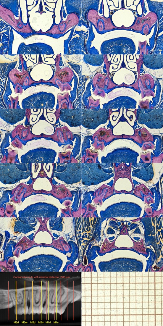
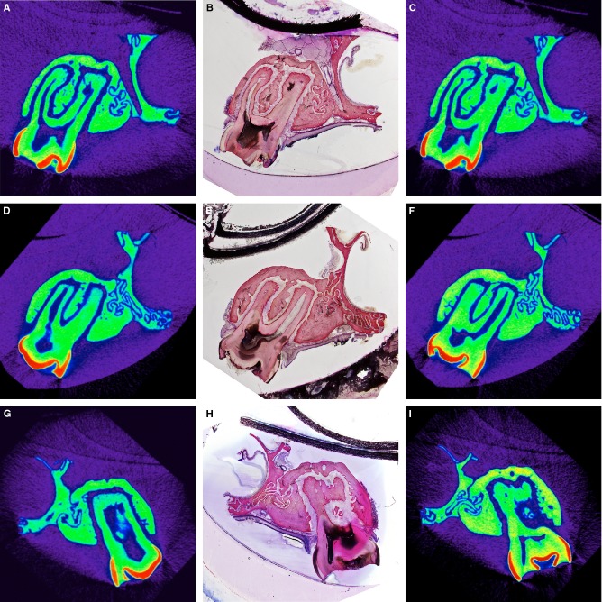
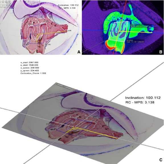
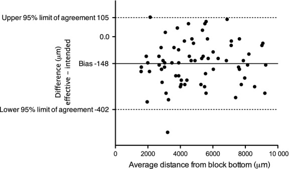
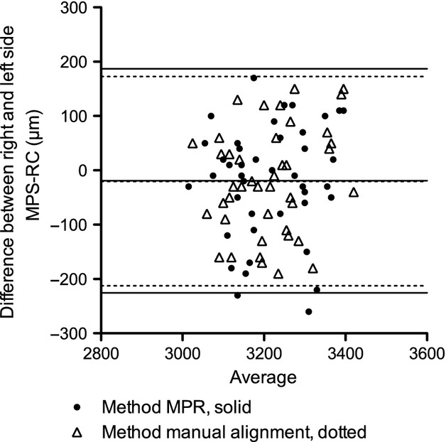
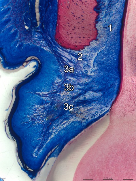
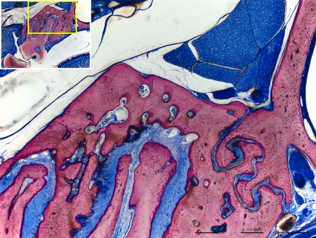
Histomorphometric evaluation of the buccal aspects of periodontal tissues in rodents requires reproducible alignment of maxillae and highly precise sections containing central sections of buccal roots; this is a cumbersome and technically sensitive process due to the small specimen size. The aim of the present report is to describe and analyze a method to transfer virtual sections of micro-computer tomographic (CT)-generated image stacks to the microtome for undecalcified histological processing and to describe the anatomy of the periodontium in rat molars. A total of 84 undecalcified sections of all buccal roots of seven untreated rats was analyzed. The accuracy of section coordinate transfer from virtual micro-CT slice to the histological slice, right-left side differences and the measurement error for linear and angular measurements on micro-CT and on histological micrographs were calculated using the Bland-Altman method, interclass correlation coefficient and the method of moments estimator. Also, manual alignment of the micro-CT-scanned rat maxilla was compared with multiplanar computer-reconstructed alignment. The supra alveolar rat anatomy is rather similar to human anatomy, whereas the alveolar bone is of compact type and the keratinized gingival epithelium bends apical to join the junctional epithelium. The high methodological standardization presented herein ensures retrieval of histological slices with excellent display of anatomical microstructures, in a reproducible manner, minimizes random errors, and thereby may contribute to the reduction of number of animals needed.
Keywords: bone; histology; morphometric measurements; orthodontics; periodontal ligament; rat.
© 2013 Anatomical Society.
Figures
Similar articles
-
Root and periodontal tissue development after allogenic tooth transplantation between rat littermates.Oral Dis. 2011 May;17(4):379-86. doi: 10.1111/j.1601-0825.2010.01761.x. Epub 2010 Oct 28. Oral Dis. 2011. PMID: 21029265
-
A histological and micro-CT investigation in to the effect of NGF and EGF on the periodontal, alveolar bone, root and pulpal healing of replanted molars in a rat model - a pilot study.Prog Orthod. 2014 Jan 6;15(1):2. doi: 10.1186/2196-1042-15-2. Prog Orthod. 2014. PMID: 24393534 Free PMC article.
-
Regional structural characteristics of bovine periodontal ligament samples and their suitability for biomechanical tests.J Anat. 2008 Mar;212(3):319-29. doi: 10.1111/j.1469-7580.2008.00856.x. J Anat. 2008. PMID: 18304207 Free PMC article.
-
[Cementum formation in rat molar roots].Kaibogaku Zasshi. 2000 Aug;75(4):365-9. Kaibogaku Zasshi. 2000. PMID: 11025944 Review. Japanese.
-
Role of the epithelial cell rests of Malassez in the development, maintenance and regeneration of periodontal ligament tissues.Periodontol 2000. 2013 Oct;63(1):217-33. doi: 10.1111/prd.12023. Periodontol 2000. 2013. PMID: 23931062 Review.
Cited by
-
Definition of a sectioning plane and place for a section containing hoped-for regions using a spare counterpart specimen.Sci Rep. 2022 Aug 3;12(1):13342. doi: 10.1038/s41598-022-17380-z. Sci Rep. 2022. PMID: 35922656 Free PMC article.
-
Photothermal Antibacterial Effect of Gold Nanostars Coating on Titanium Implant and Its Osteogenic Performance.Int J Nanomedicine. 2025 May 9;20:5983-5999. doi: 10.2147/IJN.S519183. eCollection 2025. Int J Nanomedicine. 2025. PMID: 40370805 Free PMC article.
-
Structure and collagen crimp patterns of functionally distinct equine tendons, revealed by quantitative polarised light microscopy (qPLM).Acta Biomater. 2018 Apr 1;70:281-292. doi: 10.1016/j.actbio.2018.01.034. Epub 2018 Feb 2. Acta Biomater. 2018. PMID: 29409868 Free PMC article.
-
The Effect of Coronal Implant Design and Drilling Protocol on Bone-to-Implant Contact: A 3-Month Study in the Minipig Calvarium.Materials (Basel). 2021 May 18;14(10):2645. doi: 10.3390/ma14102645. Materials (Basel). 2021. PMID: 34070127 Free PMC article.
-
Impact of Orthodontic Forces on Plasma Levels of Markers of Bone Turnover and Inflammation in a Rat Model of Buccal Expansion.Front Physiol. 2021 May 25;12:637606. doi: 10.3389/fphys.2021.637606. eCollection 2021. Front Physiol. 2021. PMID: 34113259 Free PMC article.
References
-
- Bagi CM, Wilkie D, Georgelos K, et al. Morphological and structural characteristics of the proximal femur in human and rat. Bone. 1997;21:261–267. - PubMed
-
- Bland JM, Altman DG. Measuring agreement in method comparison studies. Stat Meths Med Res. 1999;8:135–160. - PubMed
-
- Bosshardt DD, Sculean A, Windisch P, et al. Effects of enamel matrix proteins on tissue formation along the roots of human teeth. J Periodontal Res. 2005;40:158–167. - PubMed
-
- Danz JC, Dalstra M, Bosshardt DD, et al. A rat model for orthodontic translational expansive tooth movement to investigate its effect on the periodontium. Orthod Craniofac Res. 2013;16:223–233. - PubMed
MeSH terms
LinkOut - more resources
Full Text Sources
Other Literature Sources
