Review
. 2013 Nov 22:8:35.
doi: 10.1186/1750-1326-8-35.

Drosophila melanogaster as a model organism for Alzheimer's disease

Affiliations
Review

Drosophila melanogaster as a model organism for Alzheimer's disease

Katja Prüßing et al. Mol Neurodegener. .

Abstract

Drosophila melanogaster provides an important resource for in vivo modifier screens of neurodegenerative diseases. To study the underlying pathogenesis of Alzheimer's disease, fly models that address Tau or amyloid toxicity have been developed. Overexpression of human wild-type or mutant Tau causes age-dependent neurodegeneration, axonal transport defects and early death. Large-scale screens utilizing a neurodegenerative phenotype induced by eye-specific overexpression of human Tau have identified several kinases and phosphatases, apoptotic regulators and cytoskeleton proteins as determinants of Tau toxicity in vivo. The APP ortholog of Drosophila (dAPPl) shares the characteristic domains with vertebrate APP family members, but does not contain the human Aβ42 domain. To circumvent this drawback, researches have developed strategies by either direct secretion of human Aβ42 or triple transgenic flies expressing human APP, β-secretase and Drosophila γ-secretase presenilin (dPsn). Here, we provide a brief overview of how fly models of AD have contributed to our knowledge of the pathomechanisms of disease.


Figures

Figure 1
Figure 1
Genetic tools in Drosophila. In Drosophila the UAS/Gal4 expression system has been used extensively to express endogenous and exogenous sequences in the tissue of interest [39]. This is implemented using two different lines. The so-called driver line contains a Gal4 coding sequence inserted downstream of a promoter of an endogenous Drosophila gene. Gal4 is a transcription factor originating from Saccharomyces cerevisiae[40]. It specifically binds to promoter elements termed upstream activating sequence (UAS), thus activating expression of the downstream target sequence [40,41]. A collection of Gal4 driver lines which display a great variety of Gal4 expression in numerous tissues and organs is available to the public [42]. Frequently used are the glass multimer reporter (GMR) driver inducing retinal expression [43] and the elav driver inducing pan-neuronal expression [44]. After crossbreeding both, the Gal4 driver and the UAS line, the UAS target sequences will be expressed in a spatiotemporal manner (depending on the Gal4 driver used). EP-elements are randomly inserted in the fly genome and contain UAS sites. Depending on the orientation EP-elements might facilitate activation (same orientation) or inactivation (reverse orientation) of neighboring genes in a Gal4-dependent manner. There are various collections of EP strains available allowing misexpression of a large number of fly genes [45,46]. So-called RNAi lines express short inverted repeat sequences under UAS control. The sequence of the inverted repeat corresponds to an endogenous gene. Gal4-dependent expression of the inverted repeat results in the formation short hairpin RNAs (shRNAs). The presence of shRNAs initiates a series of cellular mechanisms eventually resulting in silencing of the corresponding endogenous gene by RNA interference (RNAi) [47].
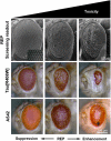
Figure 2
Figure 2
Exemplified rough eye phenotypes (REP) used as readout for modifier screens. Scanning electron micrographs (top) of fly eyes are shown. The Drosophila compound eye consists of a stereotypic array of about 800 omatidia (left). These hexagonal structures are highly ordered and display regular spacing of hairs called interomatidial bristles (inset). Expression of disease-linked proteins/peptides in the eye can cause a REP (middle). The rough appearance of the eye can be caused by loss of interomatidial bristles, fusion of omatidia, necrotic tissue, dints in the retina and is often accompanied by loss of pigmentation and reduced eye size. An enhancement in severity (left) is easily observable by more pronounced REP characteristics. Usually, such REPs are sensitive towards genetic interactions, causing either a suppression (left) or an enhancement (right), changing the overall eye appearance towards a more wild-type like appearance (suppression) or by increasing the rough appearance of the eye (enhancement), respectively. Exemplary light micrographs show REPs induced by expression of either Tau[R406W] (middle) or Aβ42 (bottom). These REPs are sensitive towards genetic modification like suppression (left) and enhancement (right) and can be/have been used for screening approaches.

References

    1. Ferri CP, Prince M, Brayne C, Brodaty H, Fratiglioni L, Ganguli M, Hall K, Hasegawa K, Hendrie H, Huang Y. et al.Global prevalence of dementia: a Delphi consensus study. Lancet. 2005;366:2112–2117. - PMC - PubMed
    1. Chow VW, Mattson MP, Wong PC, Gleichmann M. An overview of APP processing enzymes and products. Neuromolecular Med. 2010;12:1–12. doi: 10.1007/s12017-009-8104-z. - DOI - PMC - PubMed
    1. Burdick D, Soreghan B, Kwon M, Kosmoski J, Knauer M, Henschen A, Yates J, Cotman C, Glabe C. Assembly and aggregation properties of synthetic Alzheimer’s A4/beta amyloid peptide analogs. J Biol Chem. 1992;267:546–554. - PubMed
    1. Iqbal K, Liu F, Gong CX, Grundke-Iqbal I. Tau in Alzheimer disease and related tauopathies. Curr Alzheimer Res. 2010;7:656–664. doi: 10.2174/156720510793611592. - DOI - PMC - PubMed
    1. Hardy JA, Higgins GA. Alzheimer’s disease: the amyloid cascade hypothesis. Science. 1992;256:184–185. doi: 10.1126/science.1566067. - DOI - PubMed

Substances