. 2013 Nov 20;80(4):1077-89.
doi: 10.1016/j.neuron.2013.08.019.

Attentional modulation of cell-class-specific gamma-band synchronization in awake monkey area v4

Affiliations

Attentional modulation of cell-class-specific gamma-band synchronization in awake monkey area v4

Martin Vinck et al. Neuron. .

Abstract

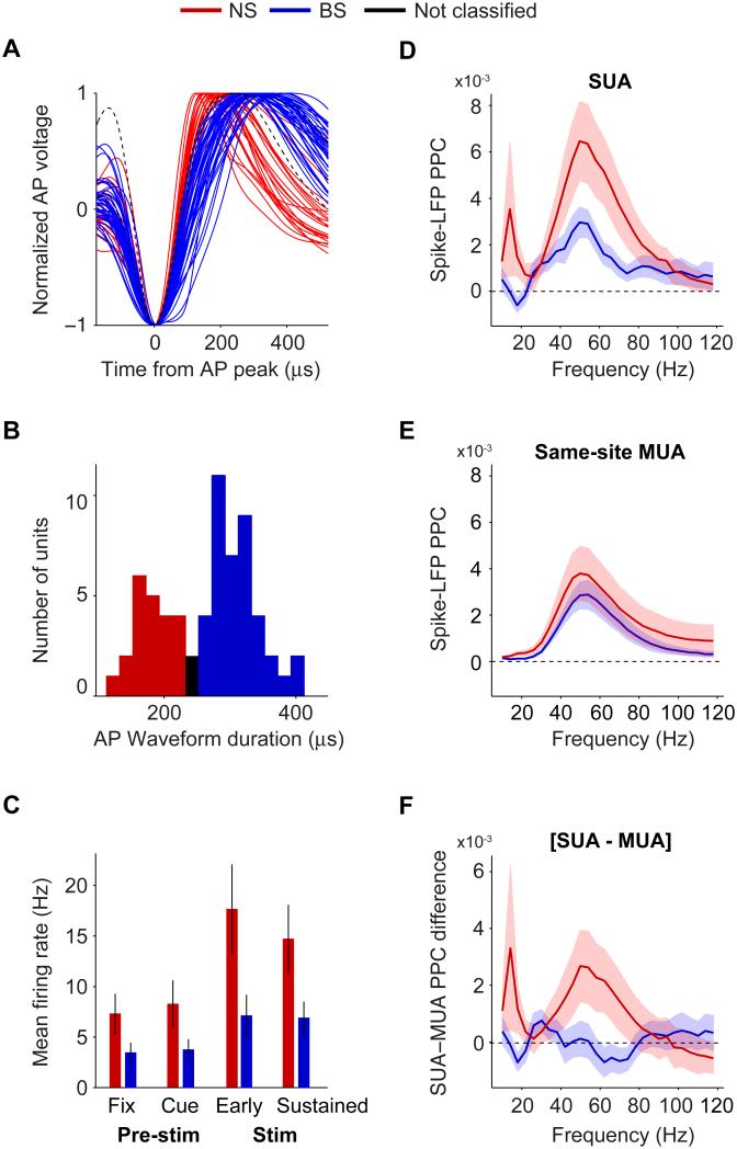
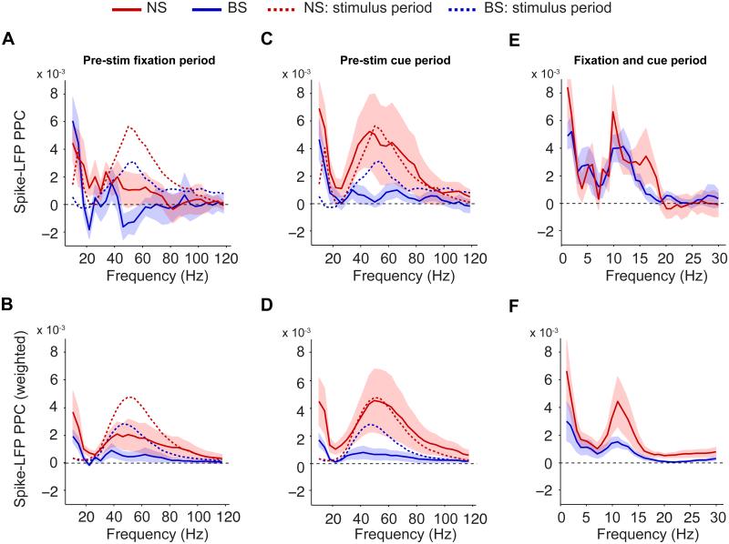
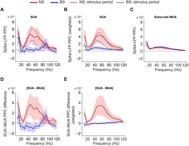
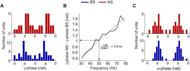
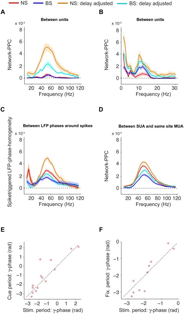
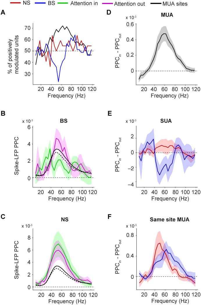
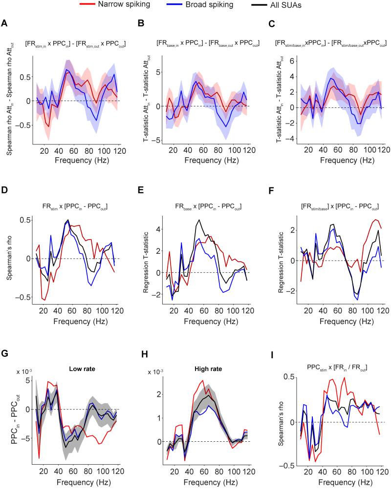
Selective visual attention is subserved by selective neuronal synchronization, entailing precise orchestration among excitatory and inhibitory cells. We tentatively identified these as broad (BS) and narrow spiking (NS) cells and analyzed their synchronization to the local field potential in two macaque monkeys performing a selective visual attention task. Across cells, gamma phases scattered widely but were unaffected by stimulation or attention. During stimulation, NS cells lagged BS cells on average by ∼60° and gamma synchronized twice as strongly. Attention enhanced and reduced the gamma locking of strongly and weakly activated cells, respectively. During a prestimulus attentional cue period, BS cells showed weak gamma synchronization, while NS cells gamma-synchronized as strongly as with visual stimulation. These analyses reveal the cell-type-specific dynamics of the gamma cycle in macaque visual cortex and suggest that attention affects neurons differentially depending on cell type and activation level.


Figures

Fig 1
Fig 1. Classification of NS and BS cells based on AP waveforms, and their differences in gamma locking during visual stimulation
(A) Normalized voltage vs. time from AP peak for all average waveforms of the isolated single units. (B) Histogram of AP peak-to-trough durations. (C) Average firing rate for different task periods (vertical lines indicate SEMs). Left-to-right: pre-stimulus fixation, cue, early onset (0-0.3 s after grating stimulus onset), and sustained stimulus period. (D) Average spike-LFP PPC spectra. (E) Average PPC for the same-site MUAs corresponding to either the NS or BS cells. (F) Average SUA-MUA PPC difference [PPCSUA – PPCMUA] vs. frequency. (D-F) Shadings indicate SEMs. See Fig S1 and S2.
Fig 2
Fig 2. Precision of pre-stimulus phase locking
(A) Average spike-LFP PPC spectra for the pre-stimulus fixation period. Dashed lines indicate average PPC for sustained stimulus period. (B) Same as (A), but now shown the weighted PPC group average, with the relative weight of a unit proportional to its spike count. (C) Same as (A), but now for the cue period. (D) Same as (B), but now for cue period. (E) Same as (A and C), but now for the complete pre-stimulus period (fixation onset to stimulus onset) and low frequencies. (F) Same as (B and D), but now for complete pre-stimulus period and low frequencies. (A-F) Shadings indicate SEMs. See Fig S1, S3 and S4.
Fig 3
Fig 3. Comparison of MUA and SUA phase locking in the pre-stimulus cue period
(A) Average spike-LFP PPC spectra for the pre-stimulus fixation period, including eight cells that were recorded with a block design, i.e. without an ‘uncued’ fixation period. Dashed lines indicate average PPC for sustained stimulus period. (B) Same as (A), but now shown the weighted PPC group average. (C) Average spike-LFP PPC spectra for the same-site MUAs corresponding to either the NS or BS cells, in the cue period. (D) Average SUA-MUA PPC difference for the cue period. (E) Same as (D), but now shown the weighted average of the SUA-MUA PPC difference, similar to (Fig 2B, D and F). See Fig S3 and S4.
Fig 4
Fig 4. Differences in locking phase between NS and BS cells
(A) Histogram of mean spike-LFP gamma phases across units. Only units for which the gamma PPC exceeded zero are shown. (B) Mean gamma phase delay vs. frequency. Dashed black line indicates linear regression fit (Pearson R=0.93, p<0.001). (C) Histogram of preferred alpha phases in the complete pre-stimulus period (for units with an alpha PPC exceeding zero). See Fig S1 and S2.
Fig 5
Fig 5. Diversity of gamma phases across population
(A) Network-PPC vs. frequency. The network-PPC indicates to what extent spikes from different neurons have similar phases. Shown in orange (NS) and cyan (BS) the delay-adjusted network-PPC that is obtained by setting the mean spike-LFP phase equal for all neurons. (B) Same as (A), but now for the low frequencies in the pre-stimulus period. (C) Same as (B), but now shown the spike-triggered LFP phase diversity, and its delay-adjusted version. The spike-triggered LFP phase diversity quantifies to what extent the distribution of spike-LFP phases measured relative to one (LFP) electrode is similar to the distribution of spike-LFP phases measured relative to another (LFP) electrode. (D) Same as (A-B), but now shown the network-PPC for same-site MUA and SUA. In this case, the network-PPC indicates to what extent the same-site MUA and the corresponding SUA have similar phases or not. (A-D) Shadings indicate SEMs. (E) Mean gamma phase in stimulus period vs. cue period, for NS cells with a spike-LFP PPC exceeding zero in both periods. (F) Same as (E), but now for stimulus vs. fixation period. See Fig S3.
Fig 6
Fig 6. Effect of attention on MUA-LFP and SUA-LFP PPC
(A) Percentage of SUAs (red, blue) and MUAs (black) for which the PPC was higher with attention inside than outside the RF. (B) Average BS cell PPC vs. frequency, separate for attention inside and outside the RF. Solid black and dashed black line correspond to MUA PPC with attention inside and outside the RF, respectively. (C) Same as (B), now for NS cells. (D) Frequency vs. the average difference in MUA-LFP PPC between attention inside and outside the RF. (E) Same as (D), but now for NS and BS cells. (F) Same as (E), but now for the same-site MUAs corresponding to either the NS (red) or BS (blue) cells. (B-F) Shadings indicate SEMs. See Fig S1 and S5.
Fig 7
Fig 7. Relationships between PPC, firing rate and selective attention
(A) Difference between attention conditions in Spearman correlations of PPC and stimulus period firing rate. Shadings indicate SEMs. (B) Same as (A), but now shown the difference between attention conditions in the T-statistic of the baseline firing rate predictor. This T-statistic was derived from a multiple regression of PPC onto baseline firing rate and relative stimulus firing rate to baseline. (C) Same as (B), but now for the relative stimulus firing rate to baseline. (D) Spearman correlation between stimulus driven firing rate and the attentional modulation of SUA PPC [PPCin - PPCout] vs. frequency. (E) Same as (D), but now shown the T-statistic of the baseline firing rate predictor. (F) Same as (E), but now for relative stimulus firing rate to baseline. (G-H) Average difference in PPC between attention conditions for units with low (G) and high (H) average firing rate (median split). (I) Spearman correlation between PPC and attentional modulation of SUA firing rate [FRin / FRout] vs. frequency. See Fig S1, S6 and S7.

References

    1. Barthó P, Hirase H, Monconduit L, Zugaro M, Harris KD, Buzsáki G. Characterization of neocortical principal cells and interneurons by network interactions and extracellular features. J. Neurophysiol. 2004;92:600–608. - PubMed
    1. Bartos M, Vida I, Jonas P. Synaptic mechanisms of synchronized gamma oscillations in inhibitory interneuron networks. Nat. Rev. Neurosci. 2007;8:45–56. - PubMed
    1. Bollimunta A, Chen Y, Schroeder CE, Ding M. Neuronal mechanisms of cortical alpha oscillations in awake-behaving macaques. J. Neurosci. 2008;28:9976–9988. - PMC - PubMed
    1. Börgers C, Kopell N. Effects of noisy drive on rhythms in networks of excitatory and inhibitory neurons. Neural Computation. 2005;17:557–608. - PubMed
    1. Bosman C, Schoffelen J, Brunet N, Oostenveld R, Bastos A, Womelsdorf T, Rubehn B, Stieglitz T, De Weerd P, Fries P. Attentional stimulus selection through selective synchronization between monkey visual areas. Neuron. 2012;75:875–888. - PMC - PubMed

Publication types