. 2013 Nov 19;105(10):2289-300.
doi: 10.1016/j.bpj.2013.09.044.

β-Catenin serves as a clutch between low and high intercellular E-cadherin bond strengths

Affiliations

β-Catenin serves as a clutch between low and high intercellular E-cadherin bond strengths

Saumendra Bajpai et al. Biophys J. .

Abstract

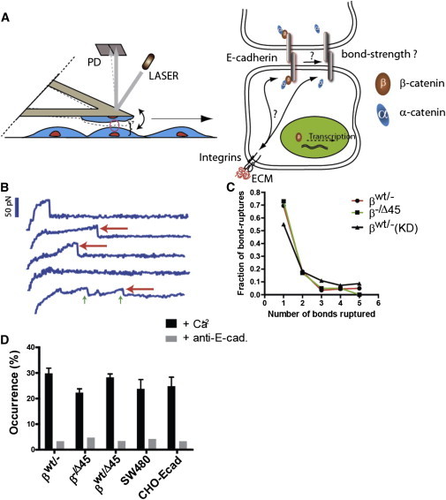
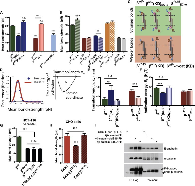
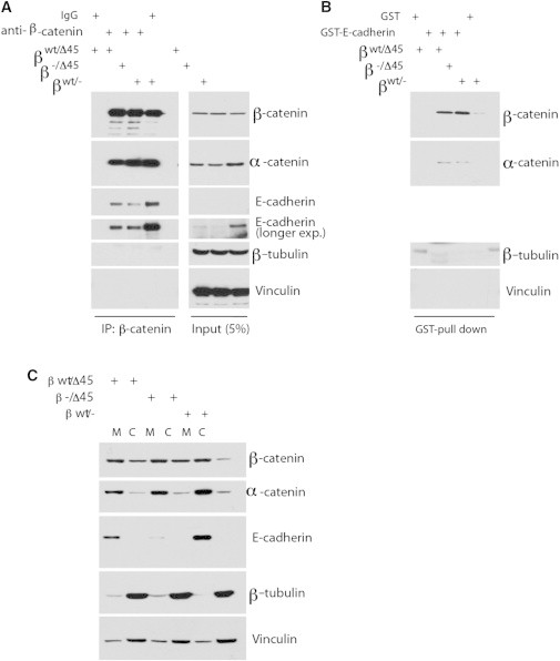
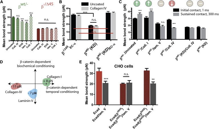
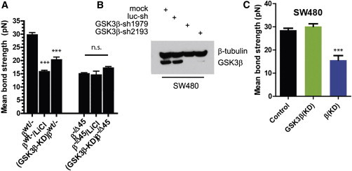
A wide range of invasive pathological outcomes originate from the loss of epithelial phenotype and involve either loss of function or downregulation of transmembrane adhesive receptor complexes, including Ecadherin (Ecad) and binding partners β-catenin and α-catenin at adherens junctions. Cellular pathways regulating wild-type β-catenin level, or direct mutations in β-catenin that affect the turnover of the protein have been shown to contribute to cancer development, through induction of uncontrolled proliferation of transformed tumor cells, particularly in colon cancer. Using single-molecule force spectroscopy, we show that depletion of β-catenin or the prominent cancer-related S45 deletion mutation in β-catenin present in human colon cancers both weaken tumor intercellular Ecad/Ecad bond strength and diminishes the capacity of specific extracellular matrix proteins-including collagen I, collagen IV, and laminin V-to modulate intercellular Ecad/Ecad bond strength through α-catenin and the kinase activity of glycogen synthase kinase 3 (GSK-3β). Thus, in addition to regulating tumor cell proliferation, cancer-related mutations in β-catenin can influence tumor progression by weakening the adhesion of tumor cells to one another through reduced individual Ecad/Ecad bond strength and cellular adhesion to specific components of the extracellular matrix and the basement membrane.


Figures

Figure 1
Figure 1
The regulation of Ecad-mediated intercellular adhesion at single-molecule resolution. (A) Schematic of the methodology used to measure Ecad/Ecad bond strength between adjoining live cells at single-molecule resolution. An AFM loaded with living cells is brought into contact with cells plated on uncoated or ECM-coated dishes. The cartoon on the right depicts the central question investigated in this work: how do changes in intracellular β-catenin and changes in extracellular ECM affect intercellular adhesion via modulation of single Ecad/Ecad bond strength. (B) Representative force-displacement curves obtained during the controlled retraction of the AFM cantilever at a speed of 20 μm/s. Red arrows mark bond rupture events; the height of the fall corresponds to the bond strength expressed in pico-Newton. Green arrows mark multiple rupture events. Only the last bond rupture was counted toward the determination of mean bond strength. (C) Distribution of occurrence of single and multiple rupture events followed Poisson statistics, shown for HCT116 cells expressing only WT (βWT/−), only ΔS45 β-catenin (β−/Δ45), and with β-catenin depleted (βWT/−(KD)). (D) The frequency of bond rupture with and without function-blocking antibody, together showing that the recorded rupture events predominantly reflect the rupture of Ecad/Ecad bonds. All error bars designate SEM.
Figure 2
Figure 2
β-catenin modulates the strength of single intercellular Ecad/Ecad bonds. (A) Strength of Ecad/Ecad bonds is significantly weakened in HCT116 cells expressing only a mutant β-catenin with S45 deleted (β−/Δ45, plain red) and in HCT116 cells wherein β-catenin is knocked down (βWT/−(KD), plain blue) as compared to cells expressing only WT β-catenin (βWT/−, plain green). Chimeric fusion of α-catenin to Ecad is sufficient to rescue the strength of single Ecad/Ecad bonds, (βWT/−(KD)EC-α, threaded blue), and (β−/Δ45EC-α). Loss of α-catenin leads to significantly weaker bonds (βWT/−-α-cat(KD, light blue). Results depict one-way ANOVA variance analysis using Bonferroni’s multiple comparison test, with ∗∗∗ implying α = 0.05 (95% confidence intervals). n > 140 for each case; number of independent experiments = 3 or more. (B) Strengthening of single Ecad/Ecad bonds over time depends on the state of β-catenin. For cells expressing WT β-catenin (βWT/−, green bars), Ecad/Ecad bonds strengthened over 300 ms, whereas for cells depleted of β-catenin (blue bars), Ecad/Ecad bonds weakened overt 300 ms (p = 0.0064; n = minimum 140 cells, one-way ANOVA analysis using Bonferroni’s multiple-comparison test with α = 0.05). Cells expressing Ecad-α-catenin chimera (β−/Δ45EC-α, orange bars), cells expressing mutant β-catenin (β−/Δ45, red bars), and parental HCT116 cells did not exhibit any time-dependent behavior. (C) Illustration showing the state of β-catenin when stronger or weaker E-cad/Ecad bonds are formed. (D) Thermodynamic analysis of rupture-force distribution based on the fit of bond strength distribution by the Hummer-Dudko model (left panel (20),) give rise to computation of transition length (x) and activation energy (Ea). Fit shows that (E) transition length increases and (F) free energy of activation decrease significantly for mutated or depleted β-catenin. ∗∗∗ Designates p < 0.001, unpaired Student’s t-test. A minimum of 140 ruptures were analyzed for each case, with N > 3. (G) In parental HCT116 colon-carcinoma cells, heterozygous expression of both WT β-catenin and mutated β-catenin leads to weakened Ecad/Ecad bonds between adjoining cells (p = 0.0072; n >140 cells each, one-way ANOVA analysis using Bonferroni’s multiple-comparison test with α = 0.05). Genetic depletion (shRNAi) of both β-catenin and known β-catenin kinase GSK3β has the same effect. n.s: not significant. (H) In CHO cells exogenously expressing Ecad, expression of S45 deletion-mutated β-catenin leads to the formation of weaker Ecad/Ecad bonds (p = 0.0093; n >140 cells each, one-way ANOVA analysis using Bonferroni’s multiple-comparison test with α = 0.05), whereas expression of phosphomimetic S > D mutated β-catenin induces no significant (n.s.) weakening of Ecad/Ecad bonds. Immunoprecipitation results (I) exhibit no significant change in Ecad/β-catenin binding affinity in CHO cells expressing ΔS45 mutant β-catenin. All error bars designate SEM.
Figure 3
Figure 3
Affinity of WT and mutant β-catenin to E-cad remains unaltered. (A) β-catenin mutant binds to Ecad similarly to WT. Immunoprecipitation was performed using β-catenin antibody or mouse IgG charged protein G beads and cell lysates from HCT-derived cells as described in Methods, Western blotted as indicated. (B). Bacterially ecto-expressed E-cad cytoplasmic tail binds to β-catenin mutant. Bacterially expressed GST or GST-Ecad proteins were charged to Glutathione-agrose beads, pulldowns were performed as described and Western blotted. (C) Less cytosolic (lane C) β-catenin in WT/− than in mutant cells, as compared to membrane-bound β-catenin (lane M). Cells were fractionated and Western blotted.
Figure 4
Figure 4
Ecad/Ecad bond strength is differentially modulated by cell adhesion to specific ECM molecules through β-catenin. (A) Ecad/Ecad bonds between HCT116 cells expressing only WT β-catenin (βWT/−) or only ΔS45-mutated β-catenin (β−/Δ45) and plated on substrates coated with different ECM ligands (Coll. IV – human collagen IV; EHS –Engelbreth-Holm-Swarm MatriGel basement membrane matrix; lam V. – human laminin V; Coll. I – rat-tail collagen I). Whereas exposure to varied ECM ligands induced change in Ecad/Ecad bond strength in (βWT/−) cells, no such response was observed for β−/Δ45cells. n = minimum 140 cells (N > 3 independent experiments) analyzed with one-way ANOVA using Bonferroni’s multiple-comparison test and α = 0.05. Unless otherwise marked, all significance is relative to (βWT/−) behavior. (B) Collagen IV does not affect single Ecad/Ecad bonds, even when α-catenin is directly fused to Ecad (βWT/−(KD)EC-α and β−/Δ45EC-α), mimicking the Ecad/Ecad bonds formed between cells with β-cateninß-catenin depleted (βWT/−(KD). n = minimum 140 cells (N > 3 independent experiments) analyzed with one-way ANOVA using Bonferroni’s multiple-comparison test and α = 0.05. (C) Response to increasing duration of cell-cell contact, to 300 ms. Cells expressing WT β-catenin (βWT/−) exhibit time-dependent weakening of Ecad/Ecad bonds when placed on collagen IV, strengthening when plated on collagen I, and no change when coated in laminin V. Cells with depleted (βWT/−(KD) do not show any time-dependent response. ∗∗∗Designates p < 0.001 under the Michelin grade scale. n = minimum 140 cells (N > 3 independent experiments) analyzed with one-way ANOVA using Bonferroni’s multiple-comparison test and α = 0.05. (D) Illustration showing the different roles played by different ECM ligands. Although Collagen IV and Collagen I induce a temporal but no sensory response, laminin V induces a sensory response, but no temporal response. Numbers depict the weakening or strengthening of Ecad/Ecad bonds. (E) Similar to βWT/− cells, CHO cells expressing either WT β-catenin or phosphomimetic S > D mutated β-catenin exhibit significant weakening of Ecad/Ecad bonds upon exposure to laminin V-coated substrate, whereas simultaneous expression of WT (endogenous) and ΔS45-mutated (exogenous) β-catenin induces no laminin V-specific response. All bars designate mean ± SEM, and n > 140 for each condition.
Figure 5
Figure 5
The kinase activity of GSk3β regulates the strength of Ecad/Ecad bonds but only in the presence of β-catenin. (A) Upon shRNAi depletion of GSk3β, HCT116 cells expressing WT β-catenin (GSK3β-KD)βWT/−) exhibit significantly weaker Ecad/Ecad bonds, whereas HCT116 cells expressing mutant β-catenin (GSk3β-KD)β−/Δ45) do not show this weakening. Inhibition of GSk3β (using LiCl, βWT/−/LiCl and β−/ΔS45/LiCl) also weakened individual intercellular Ecad/Ecad bonds. ∗∗∗ Designates p < 0.001, unpaired Student’s t-test. (B and C) In SW480 cells with endogenous APC mutation, genetic depletion of GSK3β (B) induces no change in the Ecad/Ecad bond strength (C), whereas depletion of endogenous WT β-catenin causes significant weakening of Ecad/Ecad bonds. All bars designate mean ± SEM, and n > 140 for each case.

References

    1. Giampieri S., Manning C., Sahai E. Localized and reversible TGFbeta signalling switches breast cancer cells from cohesive to single cell motility. Nat. Cell Biol. 2009;11:1287–1296. - PMC - PubMed
    1. Hidalgo-Carcedo C., Hooper S., Sahai E. Collective cell migration requires suppression of actomyosin at cell-cell contacts mediated by DDR1 and the cell polarity regulators Par3 and Par6. Nat. Cell Biol. 2011;13:49–58. - PMC - PubMed
    1. Zhu B., Chappuis-Flament S., Leckband D. Functional analysis of the structural basis of homophilic cadherin adhesion. Biophys. J. 2003;84:4033–4042. - PMC - PubMed
    1. Chou J.J., Gaemers S., Bax A. A simple apparatus for generating stretched polyacrylamide gels, yielding uniform alignment of proteins and detergent micelles. J. Biomol. NMR. 2001;21:377–382. - PubMed
    1. Zimrin A.B., Villeponteau B., Maciag T. Models of in vitro angiogenesis: endothelial cell differentiation on fibrin but not matrigel is transcriptionally dependent. Biochem. Biophys. Res. Commun. 1995;213:630–638. - PubMed

Publication types

LinkOut - more resources