. 2013 Dec 2;23(23):2434-9.
doi: 10.1016/j.cub.2013.10.057. Epub 2013 Nov 21.

Heart-specific stiffening in early embryos parallels matrix and myosin expression to optimize beating

Affiliations

Heart-specific stiffening in early embryos parallels matrix and myosin expression to optimize beating

Stephanie Majkut et al. Curr Biol. .

Abstract

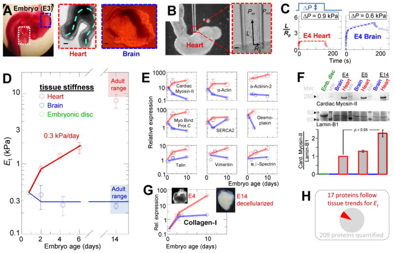
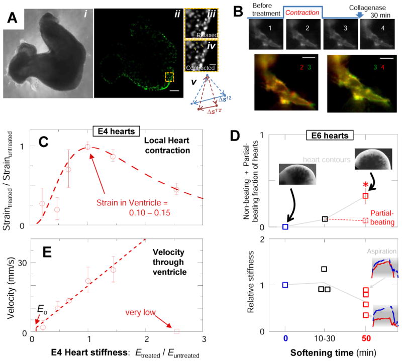
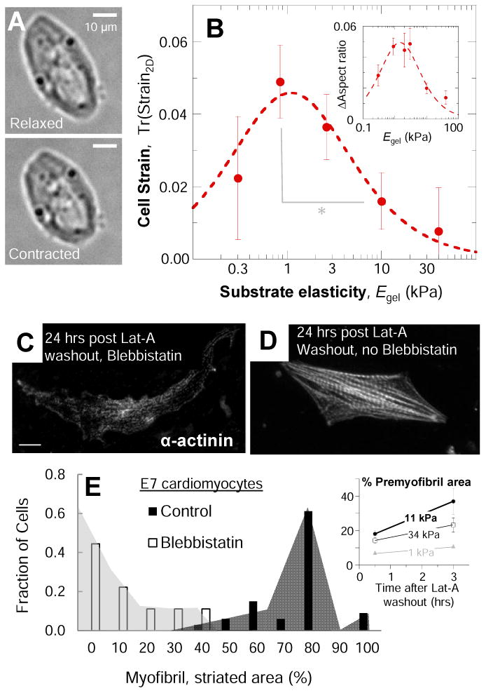
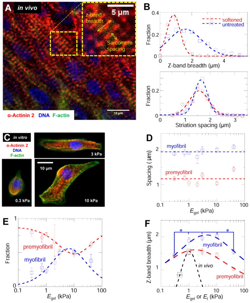
In development and differentiation, morphological changes often accompany mechanical changes [1], but it is unclear whether or when cells in embryos sense tissue elasticity. The earliest embryo is uniformly pliable, while adult tissues vary widely in mechanics from soft brain and stiff heart to rigid bone [2]. However, cell sensitivity to microenvironment elasticity is debated based in part on results from complex three-dimensional culture models [3]. Regenerative cardiology provides strong motivation to clarify any cell-level sensitivities to tissue elasticity because rigid postinfarct regions limit pumping by the adult heart [4]. Here, we focus on the spontaneously beating embryonic heart and sparsely cultured cardiomyocytes, including cells derived from pluripotent stem cells. Tissue elasticity, Et, increases daily for heart to 1-2 kPa by embryonic day 4 (E4), and although this is ~10-fold softer than adult heart, the beating contractions of E4 cardiomyocytes prove optimal at ~Et,E4 both in vivo and in vitro. Proteomics reveals daily increases in a small subset of proteins, namely collagen plus cardiac-specific excitation-contraction proteins. Rapid softening of the heart's matrix with collagenase or stiffening it with enzymatic crosslinking suppresses beating. Sparsely cultured E4 cardiomyocytes on collagen-coated gels likewise show maximal contraction on matrices with native E4 stiffness, highlighting cell-intrinsic mechanosensitivity. While an optimal elasticity for striation proves consistent with the mathematics of force-driven sarcomere registration, contraction wave speed is linear in Et as theorized for excitation-contraction coupled to matrix elasticity. Pluripotent stem cell-derived cardiomyocytes also prove to be mechanosensitive to matrix and thus generalize the main observation that myosin II organization and contractile function are optimally matched to the load contributed by matrix elasticity.


Figures

Fig. 1
Fig. 1. Mechanical development of heart and brain tissue parallels expression of abundant cell and matrix proteins
(A) E3 chick embryo with heart tube (white box) and midbrain (blue box) in situ and after isolation. The heart continues to beat ex vivo, with contraction and flow propagating along the dashed line. Scale = 100 μm. (B) Micropipette aspiration of an E3 heart tube in phase contrast microscopy. Scale = 10 μm. (C) Representative aspiration and relaxation curves for E4 heart and brain demonstrate the respective elastic and inelastic responses. (D) Et for heart and brain tissue throughout embryonic development, starting with day-1 embryonic disk, then E2, E4, E6, and E14 heart and brain (n ≥3 measurements each). By the time beating starts, the heart is already 3-fold stiffer than early embryonic tissue and then stiffens at a rate of 0.3 kPa/day. Due to the thick epicardium of E6 hearts and older relative to the inner diameter of our micropipettes, measurements likely underestimate stiffness of the myocardium at those stages due to significant contribution of epicardium. Brain tissue does not stiffen during development and remains viscoelastic with a mean Et = 0.3 kPa. (E) Quantitative mass spectrometry (MS) of cellular proteins extracted from intact embryonic disc (Hamburger-Hamilton stage 3-4), E2, E3, E4, and E10 heart and brain tissue reveals a small set of detected proteins with expression patterns similar to heart or brain mechanics: namely, a general increase in heart and relatively small increase in brain. Expression is relative to average in brain E2-E3 (n≥3 MS measurements). (F) Immunoblot confirms that MS measurements of Cardiac Myosin-II expression increase in heart development. Samples are pooled from 3-4 embryos at each reported stage and were normalized to Lamin-B1 (n ≥3). (G) MS indicates that collagen-I expression increases during heart development, but not greatly during brain development. Inset images: 1% SDS-decellularized E4 and E14 hearts. The insoluble matrices retain the shape of the embryonic hearts, but while E14 matrix (with 80% of MS ion current being collagen-1) appears solid, the E4 matrix appears more reticulated and porous, consistent with relatively less mass. (H) Of the proteins identified by Mass-Spec (Table S1), a small subset had expression levels across tissues and development that paralleled mechanics. Error bars in all figures represent SEM.
Fig. 2
Fig. 2. Effect of extracellular matrix softening and stiffening on heart tube beating
(A) E4 heart imaged by (i) phase contrast or (ii) fluorescence after sparse transfection with GFP. Scale = 100 um. Three GFP-expressing cells used to calculate strain during beating are tracked from their relaxed (iii) to contracted positions (iv, scale = 20 μm). Strain is schematized in (v) per [28]. (B) SIRPA-GFP-expressing cell in transfected E4 heart tissue before and after softening. Overlays of SIRPA-GFP expressing cells over time help visualize cell conformational changes during contraction and softening treatments. Overlay of the same cell pre-treatment while relaxed (green) and contracted (red) (bottom-left) shows less overlap (yellow) than the relaxed cell before and after tissue softening; cells thus maintain morphology and adhesions during softening. (C) Tissue strain during beating of GFP-transfected softened and stiffened E4 heart normalized to that of untreated and the resulting relative strain averaged for atria, ventricles, and outflow tract: softened and stiffened tissues suppress contractions. Typical peak strains throughout the untreated heart tube were 10 ± 4%. The dashed curve is a fit to Eq. 1 with exponent n = 4 ± 1 and Em=1.6 ± 0.2 kPa. (D) E6 hearts treated progressively with collagenase stop beating or only beat partially. Beating is suppressed after 50 min softening treatment, relative to untreated or briefly (10-30 min) treated hearts (p = 0.016). Insets: representative aspiration-relaxation curves for mildly softened or considerably softened hearts. Red indicates untreated tissue and blue indicates treated tissue. (E) Velocity of the contraction wave through the ventricle vs normalized Et. Wave-speeds in untreated ventricle, atria, and outflow tract of 22±4 mm/s, 4±2 mm/s and 2.8±0.7 mm/s, respectively, are consistent with past work [29]. For the most extreme stiffening treatment, contraction does not propagate past the presumptive pacemaker. The velocity in the ventricle increases linearly with tissue stiffness. The dashed line is the theoretical prediction with a single adjustable parameter, namely the ratio of the stress threshold to the magnitude of the force dipole corresponding to a contracting cell. Eo indicates the theoretically predicted stiffness below which a contraction wave should not propagate. Error bars for all figures represent SEM (n ≥3 hearts).
Fig. 3
Fig. 3. Isolated cardiomyocytes are sensitive to matrix stiffness, with striation dependent on actomyosin work
(A) Cardiomyocytes were imaged beating in culture after 18-24 hr in culture. Morphologies in the relaxed and contractile states and contractile strains were measured for beating cells. (B) Edge-strain of cardiomyocytes cultured on PA gels of various stiffnesses, measured as the trace of the 2D strain-matrix of cell edge points during beating, as described in methods. In beating, cell and matrix strain is strongly modulated by substrate elasticity with an optimal Egel similar to that of E4 heart and much lower than that measured for more mature cells ([6, 8]). Softer and stiffer substrates impede beating of cultured cells. The Lorentzian fit gives Em = 1.3 ± 0.3 kPa, consistent with the tissue elasticity of E4 hearts (Et = 1.3 ± 0.4 kPa in Fig. 1). (C-D) Representative image of E7 cardiomyocytes recovering from latrunculin-A (Lat-A) treatment in the presence (C) or absence (D) of blebbistatin, which does not affect cell viability [12]. (E) Myofibril assembly was measured as the percentage of cell area covered by mature myofibrils (sarcomere spacing > 1.5 um). Inhibition of beating and actomyosin contractility by blebbistatin reduces the amount of new myofibrils formed following Lat-A washout and causes mature myofibrils to disassemble per (C). Inset shows that for these late embryo cardiomyocytes, the optimal elasticity for rapid recovery of striated pre-myofibrils is close to that of adult heart (Et ∼ 10-15 kPa). Error bars are SEM (n ≥3 cells). (*) p < 0.05.
Fig. 4
Fig. 4. Sarcomere breadth changes in softened heart and in isolated cardiomyocytes on compliant substrates
(A) Untreated and softened whole E4 hearts were immunostained for sarcomeric α-actinin-2, F-actin, and DNA and imaged by confocal microscopy. Sarcomere spacing and Z-disc breadth (inset) were measured to assess any structural changes. (B) Z-disc breadth is significantly decreased in the 47%-softened heart relative to untreated controls. The decreased registry of myofibrils suggests a decreased coupling between adjacent myofibrils during contraction. Sarcomere spacing is consistent with mature myofibril sarcomere spacing and is not significantly different in the softened and untreated hearts. (C) E4 cardiomyocytes cultured on gels were stained in the same way as the whole hearts of figure A. Figure shows typical E4 cardiomyocytes on gels with stiffnesses of 0.3, 3.0 and 10 kPa. (D) Striation spacing was bimodal in distribution, indicating mature myofibrils (sarcomere spacing > 1.8 um) as well as pre-myofibrils (sarcomere spacing < 1.4 um). (E) Fraction of each type of striation per cell with myofibrils maximal on gels where pre-myofibrils are minimal. We fit the fraction of myofibrils with fm = f (Eq. 1), (blue dashed line) and pre-myofibrils using fp=1&−fm with Em = 9 ± 2 kPa. (F) Z-disc breadth for myofibrils and pre-myofibrils were maximized on substrates of intermediate stiffness. Fits to Eq. 1 yield Em = 1.7 ± 0.3 kPa for pre-myofibrils and Em = 4.2 ± 0.6 kPa for myofibrils. Error bars are SEM (n ≥3 hearts or cells).

References

    1. Gonzalez-Rodriguez D, Guevorkian K, Douezan S, Brochard-Wyart F. Soft matter models of developing tissues and tumors. Science. 2012;338(6109):910–917. - PubMed
    1. Swift J, Ivanovska I, Buxboim A, Harada T, Dingal P, Pinter J, Pajerowski J, Spinler K, Shin J, Tewari M, Rehfeldt F, Speicher D, Discher D. Nuclear Lamin-A scales with tissue stiffness and enhances matrix-directed differentiation. Science. 2013;341(6149) - PMC - PubMed
    1. Khetan S, Guvendiren M, Legant W, Cohen D, Chen C, Burdick J. Degradation-mediated cellular traction directs stem cell fate in covalently crosslinked three-dimensional hydrogels. Nature Materials. 2013 - PMC - PubMed
    1. Chien K, Domian I, Parker K. Cardiogenesis and the complex biology of regenerative cardiovascular medicine. Science. 2008;322(5907):1494–1497. - PubMed
    1. Butcher JT, McQuinn TC, Sedmera D, Turner D, Markwald RR. Transitions in early embryonic atrioventricular valvular function correspond with changes in cushion biomechanics that are predictable by tissue composition. Integrative Physiology. 2007;100:1503–1511. - PubMed

Publication types

MeSH terms

LinkOut - more resources