. 2013 Dec 26;52(6):805-18.
doi: 10.1016/j.molcel.2013.10.023. Epub 2013 Nov 21.

Tsix RNA and the germline factor, PRDM14, link X reactivation and stem cell reprogramming

Affiliations

Tsix RNA and the germline factor, PRDM14, link X reactivation and stem cell reprogramming

Bernhard Payer et al. Mol Cell. .

Abstract

Transitions between pluripotent and differentiated states are marked by dramatic epigenetic changes. Cellular differentiation is tightly linked to X chromosome inactivation (XCI), whereas reprogramming to induced pluripotent stem cells (iPSCs) is associated with X chromosome reactivation (XCR). XCR reverses the silent state of the inactive X, occurring in mouse blastocysts and germ cells. In spite of its importance, little is known about underlying mechanisms. Here, we examine the role of the long noncoding Tsix RNA and the germline factor, PRDM14. In blastocysts, XCR is perturbed by mutation of either Tsix or Prdm14. In iPSCs, XCR is disrupted only by PRDM14 deficiency, which also affects iPSC derivation and maintenance. We show that Tsix and PRDM14 directly link XCR to pluripotency: first, PRDM14 represses Rnf12 by recruiting polycomb repressive complex 2; second, Tsix enables PRDM14 to bind Xist. Thus, our study provides functional and mechanistic links between cellular and X chromosome reprogramming.


Figures

Figure 1
Figure 1. XCR assayed by H3K27me3 staining in blastocysts
Z-series projections of E3.5 and E4.5 female blastocysts immunostained for H3K27me3 (green) and the lineage markers NANOG (red) and GATA4 (blue). (A,B) Wildtype, (C) Prdm14−/−, (D) Tsix−/−, (E) double mutant embryos. Cell lineage territories are outlined in (E) [Epiblast (Epi) = NANOG+ within ICM, primitive endoderm (PE) = GATA4+, trophectoderm (TE) = rest]. Middle and right panels show close-ups of the ICM (white boxes). Arrows, Xi in NANOG+ cells. NANOG+ cells outside the ICM never undergo XCR (stars). Scale bars = 20 μm. See also Figure S1.
Figure 2
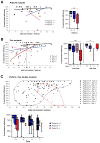
Figure 2. Quantification of XCR in Prdm14- and Tsix-mutant blastocysts
XCR in Prdm14−/− (A), Tsix−/− (B) and double mutant (C) E4.5 blastocysts was scored as %NANOG+ epiblast cells without H3K27me3 foci per embryo. Every data point on the scatter plots represents reactivation efficiency plotted against total number of cells per embryo indicating developmental progression. Trend lines (A,B) follow logarithmic regression. The box plots summarize the data, with boxes demarcating the 25th–75th percentile (only shown if n>3), the median indicated by dotted and the mean by solid lines. Whiskers extend to minimum and maximum values. Statistical significances have been calculated using 1-way ANOVA (** P<0.01). Note: For genotypes, maternal allele is represented first by convention. In (B), black dashed lines separate delayed embryos with low cell number (≤80 cells) from advanced embryos (>80 cells). (C) All embryos including Prdm14/Tsix double-mutants. The highlighted areas encompass Prdm14+/+ (black) or Prdm14−/− (red) embryos of all Tsix genotypes.
Figure 3
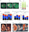
Figure 3. Prdm14−/− embryos show normal cell fates and Mendelian ratios during preimplantation stages, but abnormal postimplantation development
(A, B) Analysis of cell lineage distribution in E4.5 blastocysts from Prdm14+/− intercrosses (2 litters, 11 embryos) by immunostaining (A) for the cell lineage markers NANOG (cyan, Epi), GATA4 (red, PE) and CDX2 (green, TE). Scale bar = 20 μm. Counts in (B) are given in % of cells of each lineage per total blastocyst cell number (error bars = SEM). (C) Genotype distribution of embryos and pups from Prdm14+/− heterozygous intercrosses. The right column represents the expected Mendelian ratio. Resorptions were not genotyped due to lack of embryonic material. n = number of embryos. (D, E) Sex- and genotype distribution from Prdm14+/− het-crosses (D; 16 litters, 75 pups) and Prdm14+/− Tsix−/− female with Prdm14+/− Tsix male intercrosses (E; 14 litters, 49 pups). (F, G) Examples for abnormal Prdm14−/− E10.5 (F) and E12.5 (G) embryos compared to wildtype littermates. The resorption in (G) consists of decidual tissue with no apparent embryonic material. Scale bars = 1 mm.
Figure 4
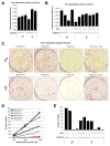
Figure 4. Prdm14 is important for self-renewal of iPSCs
(A, B) Primary iPSC colonies of different Prdm14 and Tsix genotypes. Reprogramming efficiency was scored as % primary colonies formed per input TTFs after 10 days of Dox-induction. Each graph depicts data from one experiment performed in triplicate (error bars = SEM). (C) AP-stainings of replated iPSC colonies. 1000 primary iPSCs for each genotype have been reseeded and grown for 14 days without Dox (independent of viral 4-factor expression). (D) Quantification of AP-positive replated colonies of different Prdm14 genotypes. 100, 500 or 1000 primary iPSCs were seeded and grown without Dox for 7 days. (E) Quantification of AP-positive replated colonies of different Prdm14 and Tsix genotypes grown from 1000 primary iPSCs without Dox for 11 days.
Figure 5
Figure 5. The self-renewal defect of Prdm14−/− iPSCs is partially rescued by maintained exogenous pluripotency factor expression or by derivation and culture in 2i+LIF
(A, B) Replating efficiency of iPSCs depending on exogenous pluripotency factor expression. Primary iPSCs were derived in 11 days with Dox before reseeding and culture for 14 days with (+Dox) or without (−Dox) induction of the lentiviral reprogramming cassette. Replating efficiency was measured as AP-positive colonies per seeded input iPSCs (A). * P<0.05, ** P<0.01 2-way ANOVA with Bonferroni post-tests to compare individual means. In (B), the relative colony number increase of continuous Dox-treatment over Dox-withdrawal is shown. Error bars = SEM. (C, D) Quanititative (q)PCR measurements of gene expression (C) in Prdm14+/+ (black) and Prdm14−/− (red) iPSCs cultured with (+Dox) or without (−Dox) exogenous OKSM factors. (C) Endogenous pluripotency gene expression. * P=0.034 two-tailed Student’s t-test. (D) Gata6 (endodermal differentiation marker) and de novo DNA-methyltransferase expression. ** P=0.00097. Error bars = SEM. (E–H) Comparison of derivation efficiency of Prdm14+/+ vs. Prdm14−/− iPSCs under FBS+LIF (E, F) or 2i+LIF conditions (G, H) using retroviral vectors. (E, G) Relative number of NANOG+ colonies from reprogrammed Prdm14−/− cells (red) compared to Prdm14+/+ (black, set to 100%). ** P=0.008 two-tailed Student’s t-test. Error bars = SEM. (F, H) NANOG-activation and silencing of retroviral vectors (DsRed-negative) in iPSC-colonies as indicators of successful reprogramming (Hoechst = DNA). Scale bar = 4 mm.
Figure 6
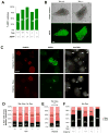
Figure 6. XCR during iPSC reprogramming is perturbed in Prdm14−/− but not in Tsix−/− cells
(A) % iPSC colonies of different Tsix genotypes with XGFP-reactivation during reprogramming (10 days +Dox). (B) Tsix+/+ and Tsix−/− iPSC colonies showing complete XGFP-reactivation during Dox-independent culture after replating (7 days −Dox). Scale bar = 100 μm. (C) RNA immunoFISH of fibroblasts/iPSC during reprogramming (8 days +Dox). Some NANOG/SSEA1 double-positive cells have downregulated Xist (stars), while others (yellow arrows) still show Xist expression similar to NANOG-negative cells (white arrowheads), which were consistently Xist-positive. Scale bar = 10 μm. (D–F) Xist RNA-FISH of NANOG-positive iPSCs of different Tsix- (D, 10d +Dox, 1d −Dox), Prdm14- (E, 10d +Dox), or Prdm14/Tsix- (F, 8d +Dox) genotypes. * P=0.0463 two-tailed Fisher’s exact test.
Figure 7
Figure 7. PRDM14 promotes XCR through repressing the Xist-activator Rnf12 and through Tsix-dependent binding to Xist intron 1
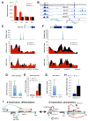
(A) Expression analysis of X-inactivation regulator genes at the Xic by qPCR in undifferentiated Prdm14+/+ and Prdm14−/− ESCs. Expression levels are normalized by Gapdh relative to Prdm14+/+ cells (=100%) ** P=0.0026 two-tailed Student’s t-test. Error bars = SEM. (B) ChIP-Seq data for binding of NANOG, OCT4, SOX2 (Marson et al., 2008) and PRDM14 (Ma et al., 2010) along the Xic, retrieved via the NCBI epigenomics database (http://www.ncbi.nlm.nih.gov/epigenomics). Arrows indicate co-bound pluripotency factor binding sites 5kb (a), 4kb (b) or immediately (c) upstream of Rnf12 and in Xist intron 1 (d). See also Figure S2. (C) ChIP-Seq analysis of the Rnf12 locus. PRDM14 binding (blue) and comparison of SUZ12 and H3K27me3 occupancy between Prdm14+/+ (black) and Prdm14−/− (red) ESCs (Yamaji et al., 2013). (D) ChIP-qPCR with HA-tagged PRDM14 expressed in undifferentiated ESCs at the Rnf12 promoter (*** P=0.0008). Control = non-transfected ESCs. Error bars (D, E, G, H) = SD. (E) ChIP-qPCR for H3K27me3 upstream of Rnf12 in Prdm14+/+ and −/− ESCs. (** P=0.002). (F) ChIP-Seq analysis of the Xist locus (as in B). (G) ChIP-qPCR with PRDM14-HA at Xist intron 1 (as in C). *** P=0.0009 (H) Mus/Cas allelic binding ratio of PRDM14-HA in undifferentiated hybrid Tsix−/+ (purple; Mus = Tsix-, Cas = Tsix+) and Tsix+/+ (black) ESCs (** P=0.0013). (I) Model for the roles of PRDM14 and Tsix during XCR. In differentiated cells and before XCR (left), PRDM14 and Tsix are absent while RNF12 and Jpx RNA activate Xist leading to XCI. During XCR in pluripotent stem cells and during embryogenesis (right), PRDM14 is expressed and binds upstream of Rnf12. In turn PRC2 is recruited and methylates H3K27, which leads to Rnf12 repression. Furthermore, Tsix is expressed facilitating PRDM14-binding to Xist intron 1. The lack of Xist-activators (Rnf12 and Jpx) and repressive effects of Tsix and PRDM14 on Xist lead to Xist repression, an important step for XCR. A more comprehensive model (Figure S3) includes other key pluripotency factors binding at the Xic implicated in Xist-repression (e.g. NANOG, REX1, OCT4, SOX2).

Similar articles

Cited by

References

    1. Anguera MC, Sadreyev R, Zhang Z, Szanto A, Payer B, Sheridan SD, Kwok S, Haggarty SJ, Sur M, Alvarez J, et al. Molecular signatures of human induced pluripotent stem cells highlight sex differences and cancer genes. Cell Stem Cell. 2012;11:75–90. - PMC - PubMed
    1. Barakat TS, Gribnau J. X chromosome inactivation in the cycle of life. Development. 2012;139:2085–2089. - PubMed
    1. Barakat TS, Gunhanlar N, Pardo CG, Achame EM, Ghazvini M, Boers R, Kenter A, Rentmeester E, Grootegoed JA, Gribnau J. RNF12 activates Xist and is essential for X chromosome inactivation. PLoS Genet. 2011;7:e1002001. - PMC - PubMed
    1. Brown CJ, Hendrich BD, Rupert JL, Lafreniere RG, Xing Y, Lawrence J, Willard HF. The human XIST gene: analysis of a 17 kb inactive X-specific RNA that contains conserved repeats and is highly localized within the nucleus. Cell. 1992;71:527–542. - PubMed
    1. Chan YS, Goke J, Lu X, Venkatesan N, Feng B, Su IH, Ng HH. A PRC2-dependent repressive role of PRDM14 in human embryonic stem cells and induced pluripotent stem cell reprogramming. Stem Cells. 2012;31:682–692. - PubMed

Publication types

MeSH terms

LinkOut - more resources