. 2013 Dec 26;52(6):855-65.
doi: 10.1016/j.molcel.2013.10.020. Epub 2013 Nov 21.

Dynamics of leading-strand lesion skipping by the replisome

Affiliations

Dynamics of leading-strand lesion skipping by the replisome

Joseph T P Yeeles et al. Mol Cell. .

Abstract

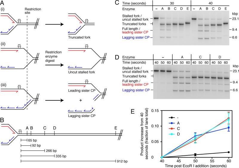
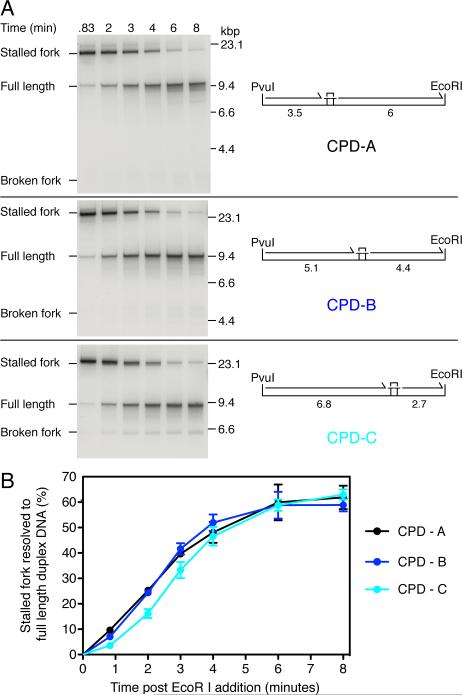
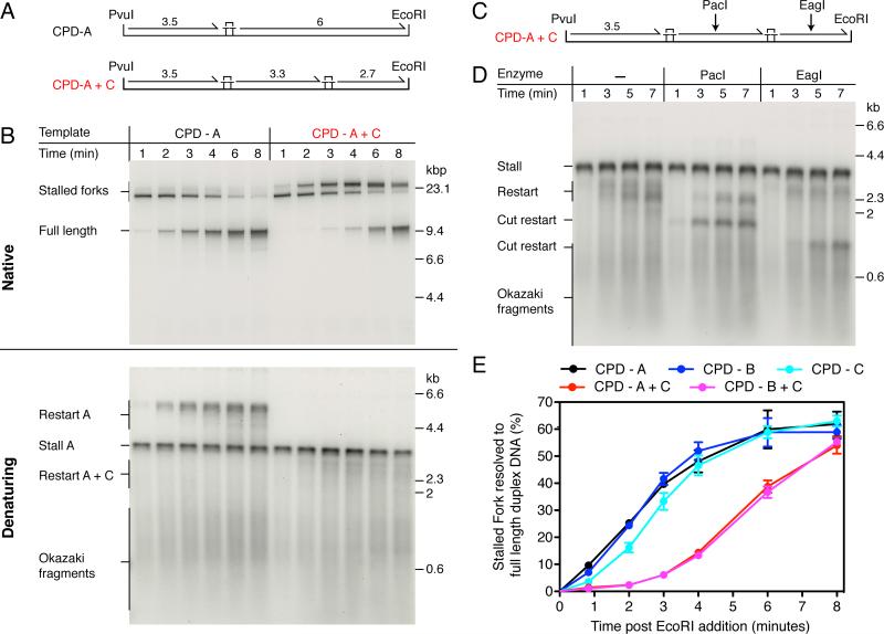
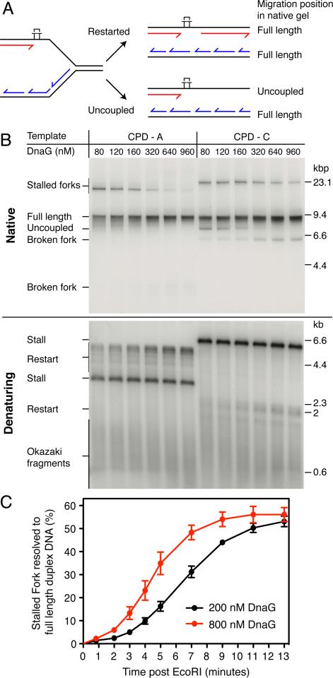
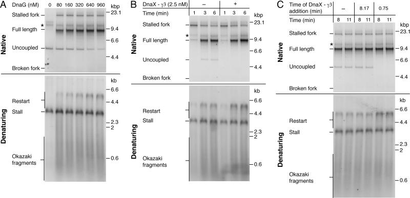
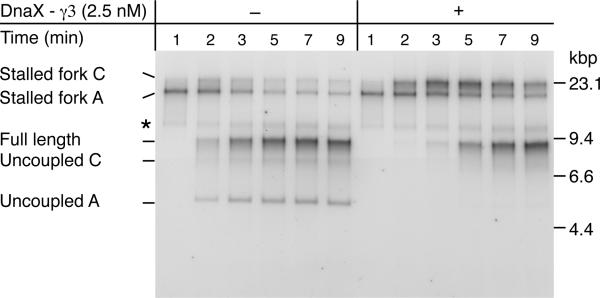
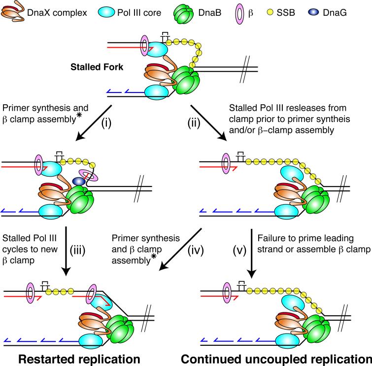
The E. coli replisome stalls transiently when it encounters a lesion in the leading-strand template, skipping over the damage by reinitiating replication at a new primer synthesized downstream by the primase. We report here that template unwinding and lagging-strand synthesis continue downstream of the lesion at a reduced rate after replisome stalling, that one replisome is capable of skipping multiple lesions, and that the rate-limiting steps of replication restart involve the synthesis and activation of the new primer downstream. We also find little support for the concept that polymerase uncoupling, where extensive lagging-strand synthesis proceeds downstream in the absence of leading-strand synthesis, involves physical separation of the leading-strand polymerase from the replisome. Instead, our data indicate that extensive uncoupled replication likely results from a failure of the leading-strand polymerase still associated with the DNA helicase and the lagging-strand polymerase that are proceeding downstream to reinitiate synthesis.


Figures

Figure 1
Figure 1. Lagging-strand Synthesis and Template Unwinding Proceed Slowly After the Replisome Stalls at a Leading-strand Lesion
(A) Illustration of replication fork structures that could be generated following fork stalling. (i) The replisome arrests at the site of damage and little template unwinding occurs downstream. (ii) Template unwinding continues downstream of the damage in the absence of leading- and lagging-strand synthesis. (iii) Template unwinding and lagging-strand synthesis continue downstream of the damage in the absence of leading-strand synthesis. Different fork architectures can be identified by restriction mapping with enzymes that are located downstream of the lesion. A single cleavage event on the lagging-strand sister will generate leading- and lagging-strand sister cleavage products, denoted leading sister CP and lagging sister CP. (B) Schematic illustrating the positions relative to the CPD of the restriction sites used for fork mapping. Enzyme A, KpnI; B, PstI; C, BsaI; D, AhdI and E, ApaLI. (C) Restriction mapping of stalled replication forks. Standard replication reactions were conducted using the CPD-C template (Figures 2A and S2) for the indicated times. Following quenching, stalled forks were digested with the indicated restriction enzymes and analyzed by native gel electrophoresis. Digested products were identified by two-dimensional electrophoresis (Figure S1B). Note that the leading sister CP migrates to a position indistinguishable from that of full-length products (fully replicated lagging strands and restarted leading strands). (D) Pulse-chase reaction followed by restriction mapping. The experiment was conducted as in (C), except that a 25-fold molar excess of unlabelled dGTP (chase) was added 30-sec post EcoRI addition. (E) Quantification of the data shown in panel (D). Data has been normalized by subtracting the initial value (40 sec) from the remaining time points. Error bars represent the standard error of the mean (SEM) from three independent experiments.
Figure 2
Figure 2. The Kinetics of Full-length DNA Production Are Independent of the CPD Location
(A) Pulse-chase analysis of replication on three templates where the location of the CPD was varied as indicated. Reactions were conducted under standard replication conditions with [α-32P]dGTP. Forty seconds post-EcoR I addition, a 25-fold excess (1 mM) of unlabelled dGTP was added to prevent further incorporation of labeled nucleotide and aliquots were withdrawn at the indicated times post-EcoR I cleavage. The maximum lengths (kbp) of the putative leading-strand stall and restart products are illustrated. (B) Quantification of pulse-chase experiments in (A). Error bars represent the SEM from a minimum of three independent experiments.
Figure 3
Figure 3. The Replisome Can Reinitiate Leading-strand Synthesis Downstream from a Second Polymerization-blocking Lesion
(A) Single- and double-damage replication templates. The maximum lengths (kbp) of the putative leading-strand stall and restart products are illustrated. (B) Comparison of replication products generated from the single-damage template, CPD-A, and the double-damage template, CPD-A+C, under standard replication conditions. (C) Illustration showing the position of the PacI and EagI restriction enzyme sites. (D) Replication was conducted with the CPD-A+C template under standard conditions for the indicated times. To aid visualization of the shorter restart product downstream of CPD-C, [α-32P]dATP was substituted with [α-32P]dGTP, as the leading-strand template in this region is cytosine rich relative to the region between CPD-A and CPD-C. Following quenching, the reaction products were digested with the indicated enzymes prior to alkaline gel electrophoresis. (E) Quantification of pulse chase experiments (Figures 2B and S5) comparing the rate of full-length product formation for the single- and double-damage templates. Error bars represent the SEM from a minimum of three independent experiments.
Figure 4
Figure 4. Primase Concentration Influences the Rate of Leading-strand Reinitiation
(A) Illustration of the replication products generated following restarted replication, or complete uncoupled replication downstream from the CPD. (B) Titration of DnaG using standard reaction conditions for 6 min. (C) Quantification of full-length replication products (restarted leading strands and lagging strands) from pulse-chase experiments conducted using 200 nM and 800 nM DnaG on the CPD-A+C template. Note that complete uncoupled replication is not observed at either concentration of DnaG (Figure S6). Error bars represent the SEM from three independent experiments.
Figure 5
Figure 5. The DnaX Complex Enhances the Efficiency By Which Leading-strand Synthesis is Reinitiated Downstream of a Lesion
(A) DnaG titration following column isolation of ERIs. Reactions were conducted for 6 min on the CPD-A template. (B) Column-isolated replication reactions using the CPD-A template and 1 μM DnaG in the presence or absence of DnaX-γ3 complex that was added 45 sec post EcoRI. (C) Column-isolated replication reactions conducted at 250 nM DnaG with the CPD-A template. DnaX-γ3 complexes (2.5 nM) were added at different time points following EcoRI addition. * ERIs that are labeled but not extended.
Figure 6
Figure 6. Leading-strand Synthesis Remains Coupled to DnaB Template Unwinding Following Replication Restart
Column-isolated replication reactions conducted with the CPD-A+C template at 200 nM DnaG in the presence or absence of DnaX-γ3 complex that was added 45 sec post EcoRI. * ERIs that are labeled but not extended.
Figure 7
Figure 7. Model for Stalled Fork Resolution by Leading-strand Reinitiation and Uncoupling
The model is described in the Discussion. * Assembly of the new β-clamp around the leading strand primer can be catalyzed either by the DnaB-associated DnaX complex or an exogenous DnaX complex not associated with the replisome (i.e DnaX-γ3).

References

    1. Dallmann HG, Kim S, Pritchard AE, Marians KJ, McHenry CS. Characterization of the unique C terminus of the Escherichia coli tau DnaX protein. Monomeric C-tau binds alpha AND DnaB and can partially replace tau in reconstituted replication forks. J. Biol. Chem. 2000;275:15512–15519. - PubMed
    1. Downey CD, McHenry CS. Chaperoning of a replicative polymerase onto a newly assembled DNA-bound sliding clamp by the clamp loader. Mol. Cell. 2010;37:481–491. - PMC - PubMed
    1. Flower AM, McHenry CS. The gamma subunit of DNA polymerase III holoenzyme of Escherichia coli is produced by ribosomal frameshifting. Proc. Nat'l. Acad. Sci. U. S. A. 1990;87:3713–3717. - PMC - PubMed
    1. Gao D, McHenry CS. tau binds and organizes Escherichia coli replication proteins through distinct domains. Domain IV, located within the unique C terminus of tau, binds the replication fork, helicase, DnaB. J. Biol. Chem. 2001;276:4441–4446. - PubMed
    1. Gupta MK, Guy CP, Yeeles JT, Atkinson J, Bell H, Lloyd RG, Marians KJ, McGlynn P. Protein-DNA complexes are the primary sources of replication fork pausing in Escherichia coli. Proc. Nat'l. Acad. Sci. U. S. A. 2013;110:7252–7257. - PMC - PubMed

Publication types

MeSH terms

LinkOut - more resources