

Amorphous no more: subdiffraction view of the pericentriolar material architecture
- PMID: 24268653
- PMCID: PMC3991556
- DOI: 10.1016/j.tcb.2013.10.001
Amorphous no more: subdiffraction view of the pericentriolar material architecture
Abstract
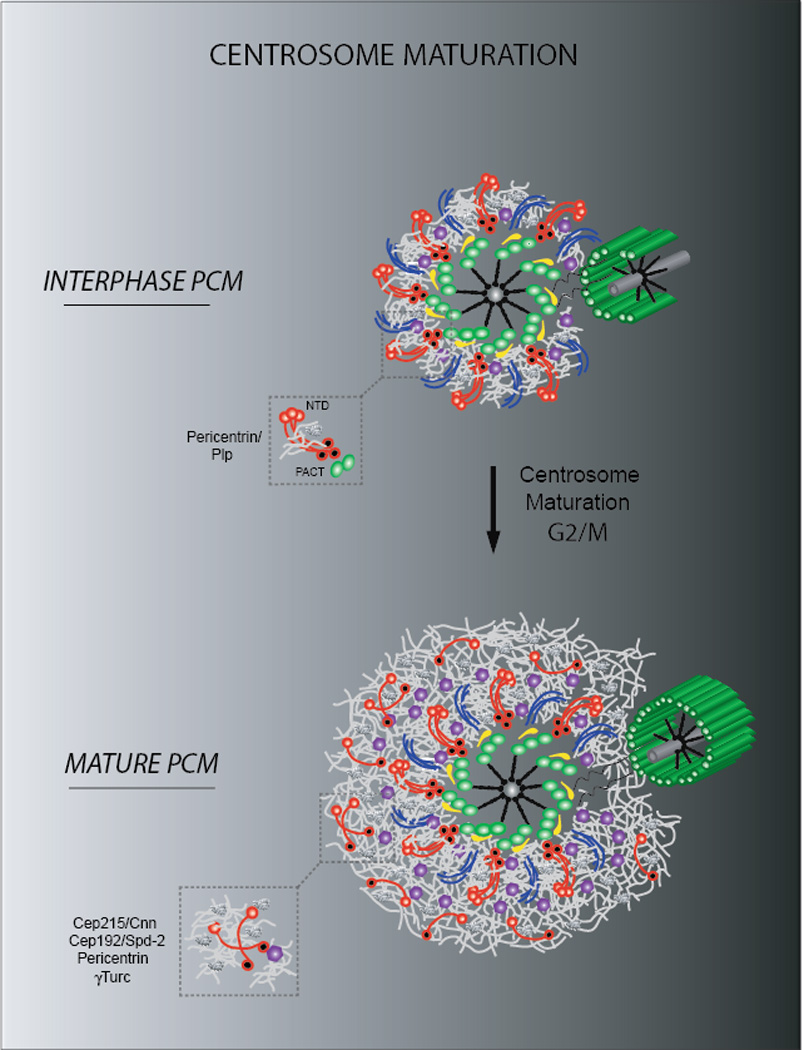
The centrosome influences the shape, orientation and activity of the microtubule cytoskeleton. The pericentriolar material (PCM), determines this functionality by providing a dynamic platform for nucleating microtubules and acts as a nexus for molecular signaling. Although great strides have been made in understanding PCM activity, its diffraction-limited size and amorphous appearance on electron microscopy (EM) have limited analysis of its high-order organization. Here, we outline current knowledge of PCM architecture and assembly, emphasizing recent super-resolution imaging studies that revealed the PCM has a layered structure made of fibers and matrices conserved from flies to humans. Notably, these studies debunk the long-standing view of an amorphous PCM and provide a paradigm to dissect the supramolecular organization of organelles in cells.
Keywords: cell cycle; centrosomes; cilia; mitosis; pericentriolar material; super-resolution microscopy.
Copyright © 2013 Elsevier Ltd. All rights reserved.
Figures


References
-
- Bornens M. The centrosome in cells and organisms. Science. 2012;335(6067):422–426. - PubMed
-
- Cunha-Ferreira I, et al. From zero to many: control of centriole number in development and disease. Traffic. 2009;10(5):482–498. - PubMed
-
- Bornens M. Centrosome composition and microtubule anchoring mechanisms. Curr. Opin. Cell Biol. 2002 Feb;14(1):25–34. - PubMed
Publication types
MeSH terms
Grants and funding
LinkOut - more resources
Full Text Sources
Other Literature Sources
