Case Reports
. 2013 Dec 5;93(6):1108-17.
doi: 10.1016/j.ajhg.2013.10.020. Epub 2013 Nov 21.

Identification of KLHL41 Mutations Implicates BTB-Kelch-Mediated Ubiquitination as an Alternate Pathway to Myofibrillar Disruption in Nemaline Myopathy

Affiliations
Case Reports

Identification of KLHL41 Mutations Implicates BTB-Kelch-Mediated Ubiquitination as an Alternate Pathway to Myofibrillar Disruption in Nemaline Myopathy

Vandana A Gupta et al. Am J Hum Genet. .

Abstract

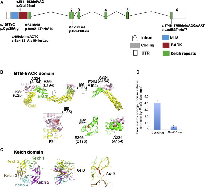
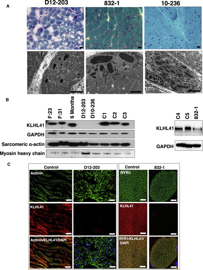
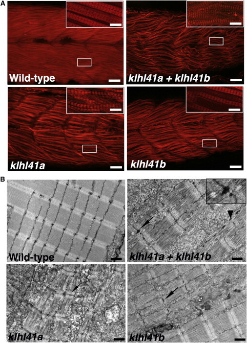
Nemaline myopathy (NM) is a rare congenital muscle disorder primarily affecting skeletal muscles that results in neonatal death in severe cases as a result of associated respiratory insufficiency. NM is thought to be a disease of sarcomeric thin filaments as six of eight known genes whose mutation can cause NM encode components of that structure, however, recent discoveries of mutations in non-thin filament genes has called this model in question. We performed whole-exome sequencing and have identified recessive small deletions and missense changes in the Kelch-like family member 41 gene (KLHL41) in four individuals from unrelated NM families. Sanger sequencing of 116 unrelated individuals with NM identified compound heterozygous changes in KLHL41 in a fifth family. Mutations in KLHL41 showed a clear phenotype-genotype correlation: Frameshift mutations resulted in severe phenotypes with neonatal death, whereas missense changes resulted in impaired motor function with survival into late childhood and/or early adulthood. Functional studies in zebrafish showed that loss of Klhl41 results in highly diminished motor function and myofibrillar disorganization, with nemaline body formation, the pathological hallmark of NM. These studies expand the genetic heterogeneity of NM and implicate a critical role of BTB-Kelch family members in maintenance of sarcomeric integrity in NM.


Figures

Figure 1
Figure 1
Overview of Mutations in KLHL41 and Their Effect on Protein Structure (A) Schematic representation of mutations in KLHL41. Boxes represent exons 1–6. Conserved domains of KLHL41 are indicated as follows: BTB (blue), BACK (red), and Kelch repeats (green). The BTB and BACK domains are encoded by exon 1 and the five Kelch repeats are encoded by exons 1–6. (B and C) Crystal structures of the BTB-BACK domain of human Kelch-like protein (KLHL11) in complex with CUL3 (Protein Data Bank code 4AP2) (B) and the Kelch domain of rat KLHL41 (PDB code 2WOZ) (C). α helices, β strands, and loops are drawn as ribbons, arrows, and threads, respectively. The squared areas correspond to the close-up views in the insets. In (B), the BTB and BACK domains are colored pink and green, respectively, whereas CUL3 is colored yellow, except that Ile96, Ala224, and Glu264 (Cys35, Ala154, and Glu194 in human KLHL41, respectively) are colored red. The side chains of these residues and Glu263 (Glu193 in human KLHL41) are shown as sticks with the indications of amino acid numbers for human KLHL11 and those for human KLHL41 in parentheses. Side chains involved in hydrophobic cores around Ile96 and Ala224 are drawn in van der Waal’s representation. In (C), the Kelch domain is color-coded to indicate each Kelch repeat, except that Ser413 is colored red. The side chain of Ser413 is shown as sticks. Molecular structures are drawn with PyMOL. (D) Predicted free energy changes upon the substitutions of KLHL41 with FoldX software.
Figure 2
Figure 2
Muscle Pathology and Expression of KLHL41 Levels and Localization in Muscle of Affected Individuals (A) Light microscopy of Gomori trichrome stained skeletal muscle from affected individuals with KLHL41 mutations show cytoplasmic nemaline bodies (top panel). Electron microscopy of affected muscles reveals rods of variable frequency and size and severe myofibrillar disarray (bottom panels). (Scale bars represent 2 μm). Affected individuals’ IDs are indicated at top. (B) Immunoblotting analysis of KLHL41 levels in affected and unaffected muscles. A decrease in protein levels was observed in individuals with KLHL41 mutations in comparison to normal control muscles. Immunoblotting with sarcomeric actin or Coomassie staining of myosin heavy chain showed no abnormal accumulation of sarcomeric proteins in affected muscles. Immunostaining for GAPDH was used for loading controls. Lanes: F:23, 23 week control fetus; F:30, 31 week control fetus; 6-month-old control baby, C1–C5 are normal age-matched control muscles. (C) Immunofluorescence for KLHL41 in control and affected individual muscle biopsies showed highly reduced levels of KLHL41 in longitudinally oriented (left) or transverse sections (right) of skeletal muscles from affected individuals. Scale bars represent 50 μm.
Figure 3
Figure 3
Characterization and Knockdown of Zebrafish Orthologs of KLHL41 (A) In situ hybridization of the zebrafish Klhl41 genes shows early expression during myogenesis in developing somites (11 hr after fertilization). Klhl41a is expressed in brain, eyes, and muscle at 1 dpf. Later in development expression is largely restricted to brain and heart (2 dpf), although low levels of expression in axial slow skeletal myofibers cannot be excluded due to limited sensitivity of the assay. Klhl41b expression is localized to skeletal muscle and heart at all developmental stages (1–2 dpf). (B) Knockdown of Klhl41 genes in zebrafish using antisense morpholinos results in myopathic changes. Live microscopy of zebrafish embryos at 3 dpf reveals leaner and smaller bodies in comparison to wild-type (WT) fish. Under polarized microscopy, zebrafish embryos exhibit a reduction in birefringence in morphant fish, quantified in ImageJ as described (WT controls: 100% ± 5.9% klhl41a: 23% ± 3.0%; klhl41b: 31% ± 8.2%; klhl41ab: 16% ± 4.2%). Double knockdown fish show a more severe skeletal muscle phenotype than single morphants. (C) RT-PCR analysis showed knockdown of normal transcripts in the morphant fish. (D) Immunoblot analysis showed reduction in Klhl41 levels in klhl41a, klhl41b, and klhl41ab fish. Klhl41 antibody recognizes both klhl41a and klhl41b and therefore show immunoreactibility to the other gene in the single morphants that is highly reduced in double morphants. (E) Overexpression of human KLHL41 mRNA restores the skeletal muscle phenotypes of klhl41a/b single and double morphants suggesting morpholino specificity. The mRNA concentration used to rescue were as follows: klhl41a (50 pg), klhl41b (75 pg), klhl41a+b (60 pg of each).
Figure 4
Figure 4
Loss of klhl41 Function in Zebrafish Recapitulates the Disease Pathology of Human Nemaline Myopathies (A) Whole-mount staining of 3 dpf zebrafish embryos with phalloidin showed extensive myofibrillar disarray of myofibers in klhl41 morphant fish (scale bar represents 2 μm). Three dpf embryos fixed in 4% paraformaldehyde were incubated with phalloidin (Invitrogen, A12380, 1:40) overnight at 4°C. Skeletal muscles of klhl41-deficient embryos were smaller and exhibited an overall reduction of myofibrillar organization (inset, high magnification). (B) Electron microscopy of klhl41-deficient skeletal muscle revealed thickened Z-lines in klhl41a or klhl41b morphants. In addition, skeletal muscle of double knockdown fish contained electron dense bodies, reminiscent of nascent nemaline rods (arrowhead, nemaline bodies like structures; arrow, thickened Z-lines) (scale bar represents 1 μm).

References

    1. Wallgren-Pettersson C., Sewry C.A., Nowak K.J., Laing N.G. Nemaline myopathies. Semin. Pediatr. Neurol. 2011;18:230–238. - PubMed
    1. Ryan M.M., Schnell C., Strickland C.D., Shield L.K., Morgan G., Iannaccone S.T., Laing N.G., Beggs A.H., North K.N. Nemaline myopathy: a clinical study of 143 cases. Ann. Neurol. 2001;50:312–320. - PubMed
    1. Wallgren-Pettersson C. Nemaline and myotubular myopathies. Semin. Pediatr. Neurol. 2002;9:132–144. - PubMed
    1. Sewry C.A. Pathological defects in congenital myopathies. J. Muscle Res. Cell Motil. 2008;29:231–238. - PubMed
    1. Hutchinson D.O., Charlton A., Laing N.G., Ilkovski B., North K.N. Autosomal dominant nemaline myopathy with intranuclear rods due to mutation of the skeletal muscle ACTA1 gene: clinical and pathological variability within a kindred. Neuromuscul. Disord. 2006;16:113–121. - PubMed

Publication types

MeSH terms