Review
. 2014 Aug;1843(8):1687-97.
doi: 10.1016/j.bbamcr.2013.11.009. Epub 2013 Nov 22.

Sec-secretion and sortase-mediated anchoring of proteins in Gram-positive bacteria

Affiliations
Review

Sec-secretion and sortase-mediated anchoring of proteins in Gram-positive bacteria

Olaf Schneewind et al. Biochim Biophys Acta. 2014 Aug.

Abstract

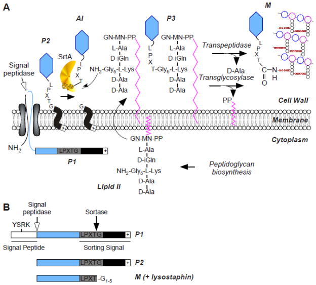
Signal peptide-driven secretion of precursor proteins directs polypeptides across the plasma membrane of bacteria. Two pathways, Sec- and SRP-dependent, converge at the SecYEG translocon to thread unfolded precursor proteins across the membrane, whereas folded preproteins are routed via the Tat secretion pathway. Gram-positive bacteria lack an outer membrane and are surrounded by a rigid layer of peptidoglycan. Interactions with their environment are mediated by proteins that are retained in the cell wall, often through covalent attachment to the peptidoglycan. In this review, we describe the mechanisms for both Sec-dependent secretion and sortase-dependent assembly of proteins in the envelope of Gram-positive bacteria. This article is part of a Special Issue entitled: Protein trafficking and secretion in bacteria. Guest Editors: Anastassios Economou and Ross Dalbey.

Keywords: Cell wall; LPXTG; Leader peptide; Peptidoglycan; Sec; Sortase.


Figures

FIG. 1
FIG. 1
Sortase A-dependent surface display of proteins. (A) Model for preprotein secretion and sorting in S. aureus. Surface proteins are first synthesized in the bacterial cytoplasm as full-length precursors (P1) containing an N-terminal signal sequence and a C-terminal sorting signal. The signal sequence directs the cellular export of the polypeptide through the Sec system and, upon translocation, is cleaved by signal peptidase. The product of this reaction, P2 precursor harboring only the C-terminal sorting signal, is retained within the secretory pathway via its C-terminal hydrophobic domain (black box) and positively charged tail (+). Sortase, a membrane anchored transpeptidase with active site cysteine, cleaves the peptide bond between the threonine (T) and the glycine (G) of the LPXTG motif, generating an acyl intermediate (AI). Lipid II, the peptidoglycan biosynthesis precursor, and its pentaglycine crossbridge (Gly5) amino group attacks the acyl intermediate, linking the C-terminal threonine of surface protein to lipid II (P3 precursor) and regenerating the active site of sortase. P3 precursor functions as a substrate for penicillin binding proteins and is incorporated into the cell wall envelope to generate mature anchored surface protein (M) that is also displayed on the bacterial surface. Note that the pentaglycine cell wall crossbridge is not commonly found in other bacteria nonetheless this pathway is conserved in many Gram-positive bacteria where the functional elements of cell wall crossbridges, LPXTG motif, sortase and penicillin binding proteins are conserved (adapted from [188]). (B) Schematic representation of protein precursors identified by using metabolic labeling of staphylococcal cultures with radiolabeled methionine. Immunoprecipitation of labeled polypeptides identifies the precursor P2 and P1. The short-lived AI and P3 intermediates are not identified. Treatment of the staphylococcal peptidoglycan with lysostaphin (glycyl-glycine endopeptidase) releases the final product, i.e. M, the surface protein anchored to the cell wall.
FIG. 2
FIG. 2
Isd-mediated heme-iron uptake across the cell wall of S. aureus. IsdA, IsdB, and IsdH are anchored to the cell wall by sortase A and function as receptors for hemoprotein ligands, such as hemoglobin (Hb). Upon binding to Isd receptors, heme is released from the hemoproteins and passaged through the cell wall in an IsdC-dependent manner. IsdC carries the NPQTN motif and is the substrate of Sortase B. When S. aureus are incubated with proteinase K, IsdB is completely degraded, IsdA is partially degraded but IsdC remains intact, suggesting that the three proteins are displayed at varying distances from the surface of the envelope. The heme molecule is taken up by the membrane transport system composed of IsdDEF and upon entry into the cytoplasm, is degraded by the heme oxygenases IsdG and IsdI to the release free iron (adapted from [189]).
FIG. 3
FIG. 3
Model for sortase-mediated polymerization of heterodimeric pili. Pilin subunits are typical sortase substrates, containing an N-terminal signal peptide that promotes secretion through the Sec system, and a C-terminal cell wall sorting signal. The pilin-specific sortase (SrtC) cleaves the sorting signals of the Tip and Shaft proteins such as the BcpA and BcpB proteins of Bacillus cereus at the Thr of the LPXTG motif, generating an acyl–enzyme intermediate that is resolved by the free amino group of a conserved lysine residue (K) present in the YPKN pilin motif of an incoming pilin subunit. Nucleophilic attack by the side chain of Lys in the YPKN motif results in intermolecular isopeptide bond between tip and shaft. The remainder of the filament assembles by a sequence of similar transpeptidation reactions. Nucleophilic attack of lipid II on the acyl intermediate formed by the housekeeping sortase (SrtA) and the polymerized pilus results in transfer to the cell wall envelope and terminates pilus assembly. Note: a similar mechanism occurs with heterotrimeric pili for example SpaCAB pili of Corynebacterium diphtheriae. SpaC is the pilus tip and SpA, the shaft protein. A third subunit SpaC serves to anchor the polymerized pilus to the cell wall in SrtA-dependent manner (adapted from [169]).

References

    1. Gram HC. Über die isolierte Färbung der Schizomyceten in Schnitt- und Trockenpräparaten. Fortschritte der Medizin. 1884;2:185–189.
    1. Bishop PJ, Neumann G. The history of the Ziehl-Neelsen stain. Tubercle. 1970;51:196–206. - PubMed
    1. Munoz E, Ghuysen JM, Heymann H. Cell walls of Streptococcus pyogenes, type 14. C polysaccharide-peptidoglycan and G polysaccharide-peptidoglycan complexes. Biochemistry. 1967;6:3659–3670. - PubMed
    1. Coley J, Archibald AR, Baddiley J. A linkage unit joining peptidoglycan to teichoic acid in Staphylococcus aureus H. FEBS Lett. 1976;61:240–242. - PubMed
    1. Schneewind O, Fowler A, Faull KF. Structure of the cell wall anchor of surface proteins in Staphylococcus aureus. Science. 1995;268:103–106. - PubMed

Publication types

MeSH terms