. 2014 Apr;63(2):341-8.
doi: 10.1016/j.parint.2013.11.002. Epub 2013 Nov 22.

A comparative analysis of trypanosomatid SNARE proteins

Affiliations

A comparative analysis of trypanosomatid SNARE proteins

Edwin Murungi et al. Parasitol Int. 2014 Apr.

Abstract

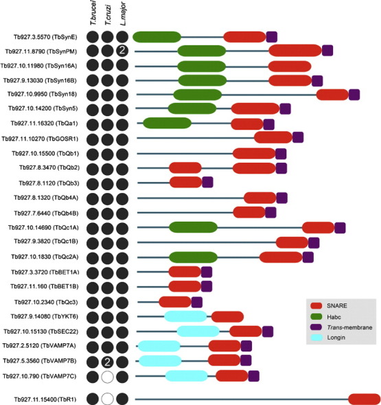
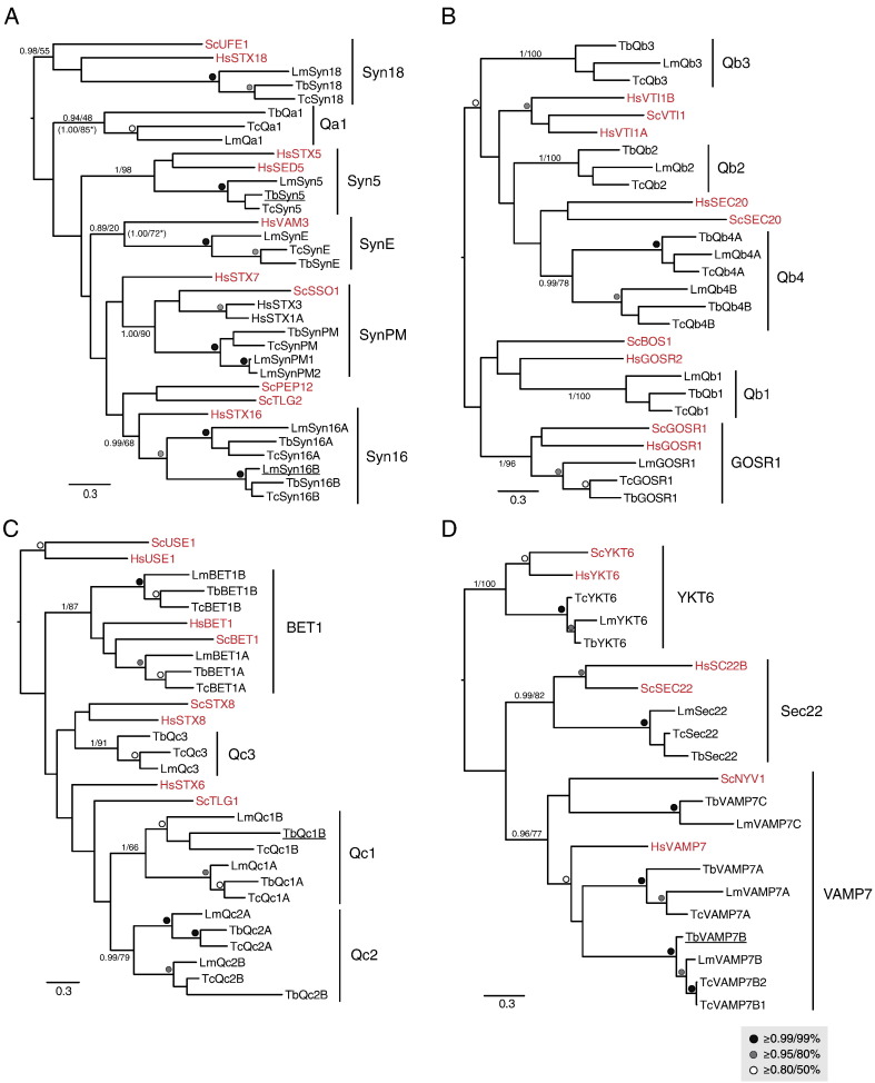
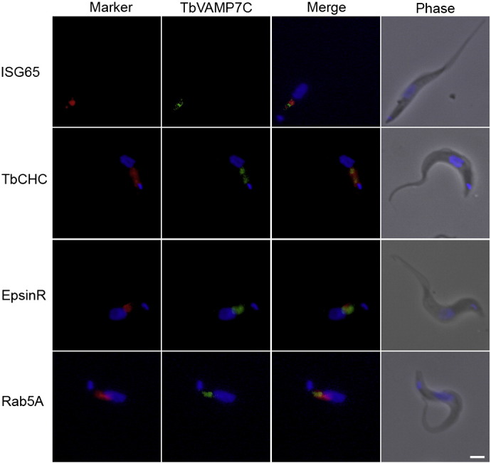
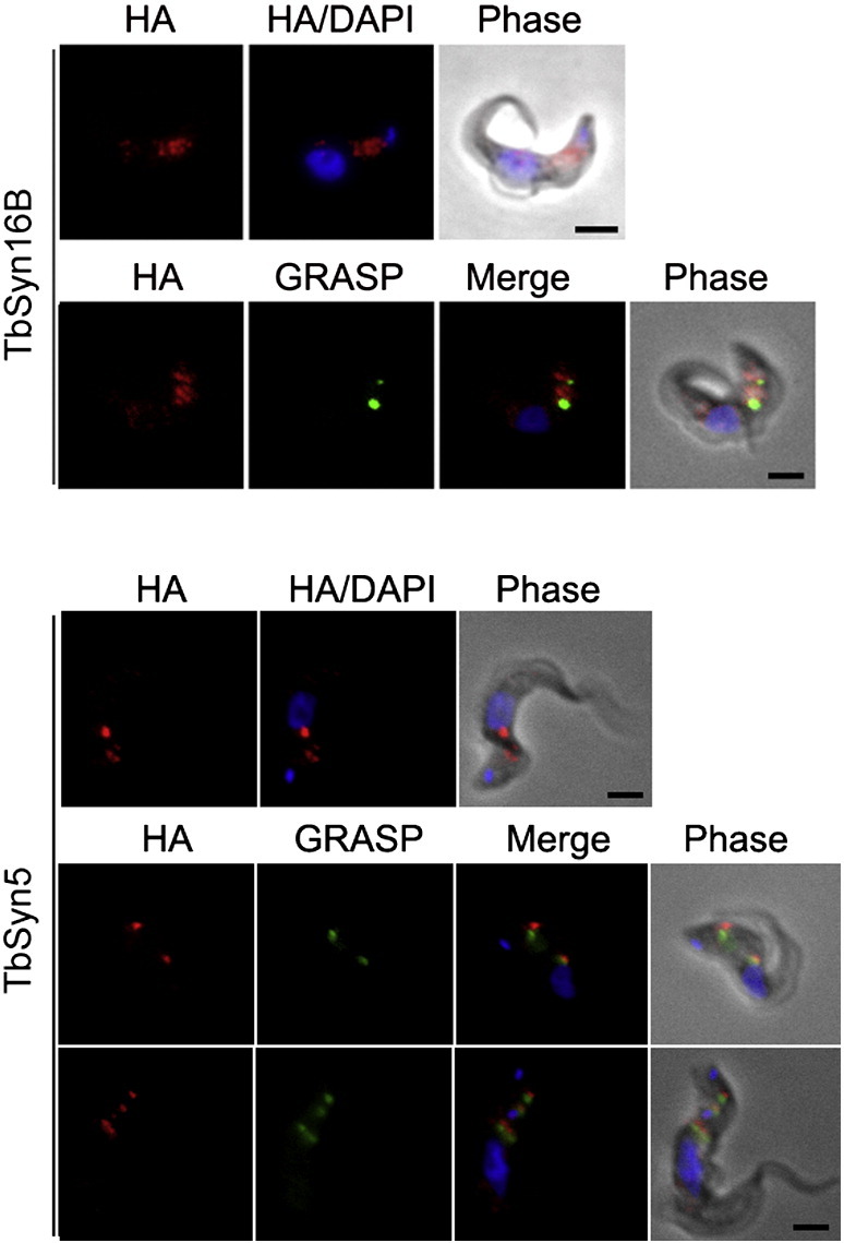
The Kinetoplastida are flagellated protozoa evolutionary distant and divergent from yeast and humans. Kinetoplastida include trypanosomatids, and a number of important pathogens. Trypanosoma brucei, Trypanosoma cruzi and Leishmania spp. inflict significant morbidity and mortality on humans and livestock as the etiological agents of human African trypanosomiasis, Chagas' disease and leishmaniasis respectively. For all of these organisms, intracellular trafficking is vital for maintenance of the host-pathogen interface, modulation/evasion of host immune system responses and nutrient uptake. Soluble N-ethylmaleimide-sensitive factor attachment protein receptors (SNAREs) are critical components of the intracellular trafficking machinery in eukaryotes, mediating membrane fusion and contributing to organelle specificity. We asked how the SNARE complement evolved across the trypanosomatids. An in silico search of the predicted proteomes of T. b. brucei and T. cruzi was used to identify candidate SNARE sequences. Phylogenetic analysis, including comparisons with yeast and human SNAREs, allowed assignment of trypanosomatid SNAREs to the Q or R subclass, as well as identification of several SNAREs orthologous with those of opisthokonts. Only limited variation in number and identity of SNAREs was found, with Leishmania major having 27 and T. brucei 26, suggesting a stable SNARE complement post-speciation. Expression analysis of T. brucei SNAREs revealed significant differential expression between mammalian and insect infective forms, especially within R and Qb-SNARE subclasses, suggesting possible roles in adaptation to different environments. For trypanosome SNAREs with clear orthologs in opisthokonts, the subcellular localization of TbVAMP7C is endosomal while both TbSyn5 and TbSyn16B are at the Golgi complex, which suggests conservation of localization and possibly also function. Despite highly distinct life styles, the complement of trypanosomatid SNAREs is quite stable between the three pathogenic lineages, suggesting establishment in the last common ancestor of trypanosomes and Leishmania. Developmental changes to SNARE mRNA levels between blood steam and procyclic life stages suggest that trypanosomes modulate SNARE functions via expression. Finally, the locations of some conserved SNAREs have been retained across the eukaryotic lineage.

Keywords: Molecular evolution; SNARE; Trypanosoma; Vesicle trafficking.


Figures

None
Graphical abstract
Fig. 1
Fig. 1
Phylogenetic relationships of Trypanosomatid SNAREs. In all panels, the best Bayesian topology is shown, with support values for nodes defining clades of interest given in the order of posterior probabilities (MrBayes) and bootstrap values (PhyML). All values for all other nodes above the threshold of 0.8/50% are iconized as inset. (A) Qa SNARE sub-family analysis. Note the orthology with opisthokont orthologs for Syn18, 5, E, PM, and 16. The Syntaxin16 clade includes two paralogues for each trypanosomatid species. Support values from additional phylogenetic analyses, with long-branching taxa removed, are indicated by asterisks. (B) Qb SNARE sub-family analysis showing orthology with Gos1 and four trypanosomatid Qb clades. (C) Qc SNARE sub-family analysis. Bet1 orthologs plus three additional trypanosomatid clades were reconstructed. (D) R-SNARE analysis. Orthologs for opisthokont sub-families were identified with an expansion in the Vamp7 clades in trypanosomatids. An additional clade of R-SNARE-related trypanosomatid proteins had an unstable position in the phylogeny when the sequences were included (data not shown), therefore, these sequences were removed. Accession numbers for all trypanosomatid sequences are shown in Table S1. Underlined sequences were localized in this study (see Figs. 4–6).
Fig. 2
Fig. 2
Schematic illustration of the structural organization of T. brucei SNAREs and representation among the TriRyps. Red ellipses represent the C-terminal SNARE motif, the trans-membrane domain is represented by dark purple rectangles. The Habc domain is represented by green ellipses while the N-terminus longin domain of R-SNAREs is represented by cyan ellipses. Designations are taken from GeneDB accessions. The N-terminus of the protein is drawn towards the left. Dots represent presence (black) or absence (white) from a detectable ortholog in T. brucei, L. major and T. cruzi. A numeral within a circle represents the presence of more than one ortholog. TbR1 is shown spaced from the main body as this SNARE could not be assigned using phylogenetics, but only on BLAST and domain searches.
Fig. 3
Fig. 3
Steady state mRNA levels of T. brucei SNAREs. Triplicate RNA samples from wild type BSF and PCF cells were subjected to qRT-PCR. BSF and PCF expression levels are represented by red and blue bars respectively. Data normalization for RNA was relative to β-tubulin and telomerase reverse transcriptase (TERT) proteins. Note error bars are absent from the PCF data set as this is set at 1.0 and variance was less than 5% throughout.
Fig. 4
Fig. 4
Subcellular localization of HA-tagged Tb927.10.790 (TbVAMP7C) protein in the bloodstream form of T. brucei. Shown is the localization of Tb927.10.790 (TbVAMP7C) relative to organelle markers ISG65, clathrin, epsinR and Rab5A. The tagged protein was visualized with a mouse monoclonal anti-HA antibody (green). Organelles were stained with rabbit polyclonal antibodies against specific trypanosome marker proteins (red). The nucleus and kinetoplast were stained blue with DAPI. Scale bar: 2 μm.
Fig. 5
Fig. 5
Localization of HA-tagged Tb927.9.3820 (TbQc1B) protein in the bloodstream form T. brucei. Shown is the localization of Tb927.9.3820 (TbQc1B) relative to known organelle markers ISG75, clathrin, epsinR and p67. The tagged protein was visualized with a mouse monoclonal anti-HA antibody (green). Organelles were stained with rabbit polyclonal antibodies against specific trypanosome marker proteins (red). The nucleus and kinetoplast (blue) were stained with DAPI. Scale bar: 2 μm.
Fig. 6
Fig. 6
Localizations of Tb927.10.1420 (TbSyn5) and Tb927.9.13030 (Syn16B) proteins in the bloodstream form of T. brucei. Shown are the localizations of Tb927.10.1420 (TbSyn5) and Tb927.9.13030 (TbSyn16B) relative to DAPI or DAPI and GRASP. The tagged protein was visualized with a mouse monoclonal anti-HA antibody (red). Organelles were stained with rabbit polyclonal antibodies against specific trypanosome marker proteins (green). The nucleus and kinetoplast (blue) were stained with DAPI. Scale bar: 2 μm.

References

    1. Adl S.M., Simpson A.G., Lane C.E., Lukeš J., Bass D., Bowser S.S. The revised classification of eukaryotes. J Eukaryot Microbiol. 2012;59:429–493. - PMC - PubMed
    1. Barrett M.P., Croft S.L. Management of trypanosomiasis and leishmaniasis. Br Med Bull. 2012;104:175–196. - PMC - PubMed
    1. http://www.who.int/trypanosomiasis_african/en/
    1. Ghedin E., Bringaud F., Peterson J., Myler P., Berriman M., Ivens A. Gene synteny and evolution of genome architecture in trypanosomatids. Mol Biochem Parasitol. 2004;134:183–191. - PubMed
    1. El-Sayed N.M., Myler P.J., Bartholomeu D.C., Nilsson D., Aggarwal G., Tran A.N. The genome sequence of Trypanosoma cruzi, etiologic agent of Chagas disease. Science. 2005;309:409–415. - PubMed

Publication types

MeSH terms