. 2014 Feb;12(2):283-94.
doi: 10.1158/1541-7786.MCR-13-0349. Epub 2013 Nov 22.

BMP signaling induces astrocytic differentiation of clinically derived oligodendroglioma propagating cells

Affiliations

BMP signaling induces astrocytic differentiation of clinically derived oligodendroglioma propagating cells

Maya Srikanth et al. Mol Cancer Res. 2014 Feb.

Abstract

Oligodendrogliomas are a type of glioma that lack detailed investigation because of an inability to cultivate oligodendroglioma cells that faithfully recapitulate their salient qualities. We have successfully isolated and propagated glioma stem-like cells from multiple clinical oligodendroglioma specimens. These oligodendroglioma-propagating cells (OligPC) are multipotent and form xenografts with oligodendroglioma features. Bone morphogenetic proteins (BMP) are considered potent inhibitors of oligodendrogliogenesis during development; therefore, the effects of BMP signaling in OligPCs were characterized. BMP pathway components are expressed by OligPCs and canonical signaling via Smad proteins is intact. This signaling potently depletes CD133-positive OligPCs, decreasing proliferation, and inducing astrocytic differentiation. Furthermore, analyses revealed that cytoplasmic sequestration of the oligodendrocyte differentiation factors OLIG1/2 by the BMP signaling effectors ID2 and ID4 is a plausible underlying mechanism. These findings elucidate the molecular pathways that underlie the effects of BMP signaling on oligodendroglioma stem-like cells.

Implications: Stem-like cells are capable of propagating oligodendrogliomas, and BMP signaling potently diminishes their stemness by inducing astrocytic differentiation, suggesting that BMP activation may be effective as a cancer stem cell-targeted therapy.


Conflict of interest statement

Conflicts of Interest:

The authors disclose no potential conflicts of interest.

Figures

Figure 1
Figure 1. Human oligodendroglioma propagating cells (OligPCs) form spheres and express NSC markers
(A) Brightfield image of primary spheres formed by dissociated oligodendroglioma cells in GSC conditions. Adherent cells with oligodendrocytic morphology are evident (inset, arrow). Scale bar: 200μm. (B) Brightfield image of OligPC spheres formed after serial passaging, demonstrating self-renewal. Scale bar: 500μm. (C) OligPCs immunostained for NSC markers sox2 and nestin, with DAPI nuclear counterstain; merged image at far right. Scale bar: 50μm. (D) Flow cytometry for CD133 in two different OligPC lines.
Figure 2
Figure 2. OligPCs are multipotent and readily generate oligodendrocytes
(A) Undifferentiated OligPCs were allowed to attach to PDL coverslips and stained for the oligodendrocyte marker O4, the neuronal marker MAP2, and the astrocytic marker GFAP, with DAPI nuclear counterstain. Scale bar: 50μm. (B) OligPCs were differentiated on PDL/laminin coverslips in media without growth factors and stained for O4, MAP2, and GFAP, merged with DAPI nuclear counterstain at bottom right. A representative confocal z-stack is shown. Scale bar: 200μm. (C) OligPCs differentiated as above and stained for MBP and MAP2. Scale bar: 50μm. (D) OligPCs differentiated as above and stained for multiple oligodendrocyte lineage markers: O4 (left), myelin basic protein (MBP, center), and CNPase (right). Scale bars: 50μm.
Figure 3
Figure 3. OligPCs form tumors that resemble oligodendrogliomas in vivo
(A) Kaplan-Meier survival curves showing tumorigenicity of 2 OligPC lines following orthotopic transplantation in 6 mice each. (B) H&E staining of an OligPC xenograft shows cytoplasmic clearing, spontaneous hemorrhage (arrow), and fine branching vasculature (arrowhead). Dashed line: tumor margin. Magnification: 10×. (C) Immunostaining with a human-specific nestin antibody confirms xenograft origin and demonstrates invading cells (arrowheads) beyond the tumor margin (dashed line). Scale bar: 100μm. (D) Immunostaining with lineage markers reveals robust OLIG2 expression with a relative paucity of GFAP expression in xenografts; merged with DAPI nuclear counterstain at far right. Scale bar: 100μm.
Figure 4
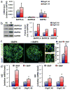
Figure 4. BMP signaling remains intact in OligPCs
(A) Quantitiative RT-PCR for BMP receptor subunits in 2 OligPC lines, relative to GAPDH control. Shown as mean+SEM. (B) Western blot for BMP receptor subunits in 2 OligPC lines, with GAPDH loading control, quantified at right. (C) OligPCs were incubated with or without 100ng/ml BMP4 for 2 hours and immunostained for phospho-Smad-1/5/8, with DAPI nuclear counterstain. Scale bars: 50μm. Zoomed images of the boxed areas demonstrate cytoplasmic (C′) and nuclear (C″) pSmad localization. Scale bars: 20μm. Quantification of this staining, shown as mean+SEM, is at far right, **p<0.005 by two-tailed Student’s t-test. (D) Quantitative RT-PCR for ID2 (left) and ID4 (right) transcripts in OligPCs treated with 100ng/ml BMP4 for 8 hours, normalized to GAPDH control and shown as mean+SEM relative to untreated cells. *p<0.05, **p<0.005 by two-tailed Student’s t-test.
Figure 5
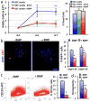
Figure 5. BMP signaling reduces proliferation and stemness of OligPCs
(A) OligPCs were grown in GSC media, media without mitogens, or media+BMP4 and total viable cells were counted using Trypan blue, shown as mean±SEM (left). **p<0.05 by two-tailed Student’s t-test. OligPCs grown in these conditions were also subjected to a live/dead assay, and the percentage of dead cells at day 7 was calculated, shown as mean+SEM (right). (B) OligPCs were grown with or without 100ng/ml BMP4 for 7 days and cell cycle status was assessed by Ki67 staining with DAPI nuclear counterstain, shown as mean+SEM at far right. Scale bar: 50μm. *p<0.01 by two-tailed Student’s t-test. (C) OligPC 40 was grown as in (B) and CD133 expression was assessed by flow cytometry, shown as mean+SEM at far right. *p<0.01 by two-tailed Student’s t-test. (D) OligPC 49 was plated at 10 cells/well in 96 well plates with or without 100ng/ml BMP4 for 10 days, and the clonogenic frequency was calculated by counting the number of spheres formed, shown as mean+SEM. **p<0.05 by two-tailed Student’s t-test.
Figure 6
Figure 6. BMP signaling induces astrocytic differentiation of OligPCs
(A) OligPCs were growth with or without 100ng/ml BMP4 for 7 days, followed by immunofluorescence for markers of oligodendroglial (O4) and astrocytic (GFAP) fate, with DAPI nuclear counterstain. Scale bar: 50μm. (B+C) Quantification of the staining shown in (A) for OligPC 49 (B) and OligPC 40 (C), shown as mean+SEM, *p<0.01 and **p<0.02 by two-tailed Student’s t-test. (D) OligPCs were grown as in (A), followed by immunofluorescence for alternate markers of oligodendroglial (CNPase) and astrocytic (S100β) fate, with DAPI nuclear counterstain (scale bar: 50μm), with quantification shown as mean+SEM at far right, *p<0.01 and **p<0.02 by two-tailed Student’s t-test.
Figure 7
Figure 7. OLIG proteins co-localize with ID2 and ID4 in BMP-treated OligPCs
(A–D) OligPCs were grown with or without 100ng/ml BMP4 for 12 hours, followed by immunostaining for OLIG1 + ID2 (A), OLIG1 + ID4 (B), OLIG2 + ID2 (C) and OLIG2 + ID4 (D), with DAPI nuclear counterstain. Scale bars: 50μm. Zoomed images of the boxed areas demonstrate nuclear (A′) and cytoplasmic (A″) Olig localization. Scale bars: 25μm. (E) OLIG1 or OLIG2 protein was immunoprecipitated from OligPCs grown as in (A–D) and Western blot analysis was performed for ID2 and ID4.

References

    1. Singh SK, Hawkins C, Clarke ID, Squire JA, Bayani J, Hide T, et al. Identification of human brain tumour initiating cells. Nature. 2004;432:396–401. - PubMed
    1. Galli R, Binda E, Orfanelli U, Cipelletti B, Gritti A, De Vitis S, et al. Isolation and characterization of tumorigenic, stem-like neural precursors from human glioblastoma. Cancer research. 2004;64:7011–21. - PubMed
    1. Das S, Srikanth M, Kessler JA. Cancer stem cells and glioma. Nat Clin Pract Neurol. 2008;4:427–35. - PubMed
    1. Dai C, Celestino JC, Okada Y, Louis DN, Fuller GN, Holland EC. PDGF autocrine stimulation dedifferentiates cultured astrocytes and induces oligodendrogliomas and oligoastrocytomas from neural progenitors and astrocytes in vivo. Genes & development. 2001;15:1913–25. - PMC - PubMed
    1. Shih AH, Dai C, Hu X, Rosenblum MK, Koutcher JA, Holland EC. Dose-dependent effects of platelet-derived growth factor-B on glial tumorigenesis. Cancer research. 2004;64:4783–9. - PubMed

Publication types