Review
. 2014 Feb:66:26-41.
doi: 10.1016/j.addr.2013.11.004. Epub 2013 Nov 21.

Nanopreparations for organelle-specific delivery in cancer

Affiliations
Review

Nanopreparations for organelle-specific delivery in cancer

Swati Biswas et al. Adv Drug Deliv Rev. 2014 Feb.

Abstract

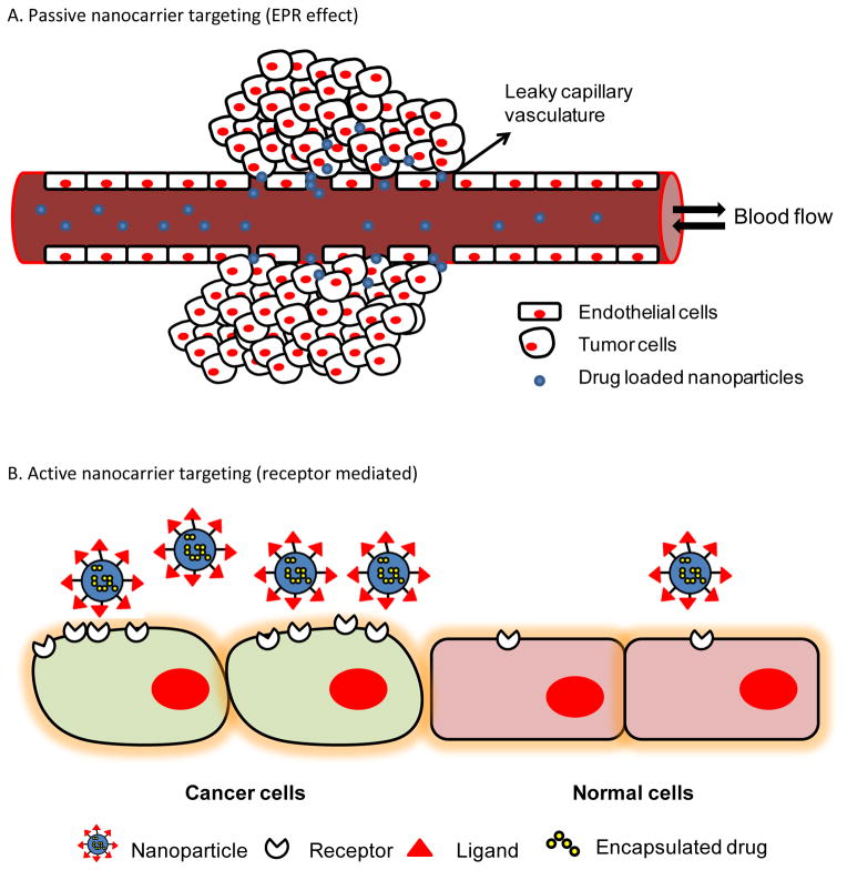
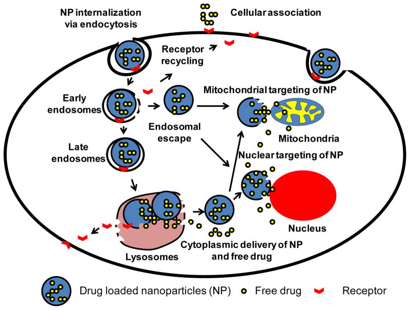
To efficiently deliver therapeutics into cancer cells, a number of strategies have been recently investigated. The toxicity associated with the administration of chemotherapeutic drugs due to their random interactions throughout the body necessitates the development of drug-encapsulating nanopreparations that significantly mask, or reduce, the toxic side effects of the drugs. In addition to reduced side effects associated with drug encapsulation, nanocarriers preferentially accumulate in tumors as a result of its abnormally leaky vasculature via the Enhanced Permeability and Retention (EPR) effect. However, simple passive nanocarrier delivery to the tumor site is unlikely to be enough to elicit a maximum therapeutic response as the drug-loaded carriers must reach the intracellular target sites. Therefore, efficient translocation of the nanocarrier through the cell membrane is necessary for cytosolic delivery of the cargo. However, crossing the cell membrane barrier and reaching cytosol might still not be enough for achieving maximum therapeutic benefit, which necessitates the delivery of drugs directly to intracellular targets, such as bringing pro-apoptotic drugs to mitochondria, nucleic acid therapeutics to nuclei, and lysosomal enzymes to defective lysosomes. In this review, we discuss the strategies developed for tumor targeting, cytosolic delivery via cell membrane translocation, and finally organelle-specific targeting, which may be applied for developing highly efficacious, truly multifunctional, cancer-targeted nanopreparations.

Keywords: Cancer; Drug delivery; Endocytosis; Intracellular; Nanopreparations; Organelle-specific.


Figures

Figure 1
Figure 1
Representation of (A) passive (via the EPR effect) and (B) active (receptor-mediated) targeting utilized for targeting nanopreparations to tumors.
Figure 2
Figure 2
Schematic drawing of the cytosolic delivery and organelle-specific targeting of drug loaded nanoparticles via receptor-mediated endocytosis. After receptor mediated cell association with nanoparticles, the nanoparticles are engulfed in a vesicle known as an early endosome. Nanoparticles formulated with an endosome disrupting property disrupt the endosomes followed by cytoplasmic delivery. On other hand, if nanoparticles are captured in early endosomes, they may make their way to lysosomes as late endosomes where their degradation takes place. Only fraction of non-degraded drug released in the cytoplasm interacts with cellular organelles in a random fashion. However, cytosolic delivery of a fraction of organelle-targeted nanoparticles via endosomal escape or from lysosomes travel to the targeting organelles to deliver their therapeutic cargo.
Figure 3
Figure 3
Summary of current strategies for mitochondrial targeting. (A) attachment of lipophilic cations such as triphenylphosphonium to small molecules or nanocarriers for targeting to the mitochondria; (B) Mitochondrial targeting Szeto-Schiller (SS) peptides containing an aromatic-cationic sequence motif selectively partition into the inner mitochondrial membrane independent of the mitochondrial membrane potential; (C) A dicationic mitochondriotropic compound, dequalinium chloride, self-assembles and forms vesicle-like aggregates called DQAsomes. These vesicles are taken up by endocytosis, fuse with the mitochondrial outer membrane and enter to the mitochondrial matrix via the mitochondrial protein import machinery; (D) MITO-porter is a liposome-based carrier that fuses with the mitochondrial membrane and releases its cargo to the intra-mitochondrial compartment; (E) Mitochondrial targeted signal peptides attached to non-mitochondrial proteins create a chimeric protein taken up by the mitochondrial matrix via the mitochondrial protein import machinery. Cargo consisting of a drug or nucleic acid attached to this chimeric protein can be selectively transferred to mitochondria.
Figure 4
Figure 4
Modification of doxorubicin-loaded commercially available Lipodox® with cell-penetrating synthetic peptide, octa-arginine, to achieve improved anticancer activity. (A) Dox-L and R8-Dox-L were prepared by incorporation of hydrophilic co-polymer polyethyleneglycol-phosphatidylethanolamine (PEG-PE) and R8-PEG-PE into the liposomal lipid bilayer. (B) The confocal laser scanning micrograph shows murine mammary carcinoma cell (4T1) treated with Dox-L and R8-Dox-L at a Dox concentration 6 μg/mL. The cells were additionally stained with a DNA stain (blue) and an endosomal marker (green) for visualization of nuclei and endosomes. The merged picture indicated that R8-Dox-L delivered its pay-load to the nucleus more efficiently than Dox-L. (Scale bar. 15 nm.) (C) Co-localization of Dox signal with DNA and endosomes quantified by Image J software. The Pearson’s coefficient of colocalization of Dox-L or R8-Dox-L with DNA and endosomes indicates that R8-Dox-L had significantly higher co-localization with DNA than endosomes, indicating an endosome-disrupting property of R8-Dox-L which resulted in more cytosolic delivery of the loaded doxorubicin compared to Dox-L.
Figure 5
Figure 5
Evaluation of efficacy of mitochondria-targeted liposomes. The liposomal surface was modified with a co-polymer, triphenylphosphonium-PEG-PE conjugate, for mitochondrial targeting (TPP-L). (A) Confocal microscopy of HeLa cells treated with rhodamine-labeled unmodified liposomes (PL) and TPP-L. Yellow dots in the merged picture indicate co-localization of red (liposome) and green (mitotracker) signals. (B) Evaluation of the cell-killing efficacy of paclitaxel-loaded TPP-L (TPP-L-PTX) on HeLa cells. The targeted delivery of paclitaxel to mitochondria resulted in a further decrease in cell viability. (C) The in vivo evaluation of the efficacy of TPP-L-PTX in reducing tumor volume. The arrows indicate the day of intravenous administration of liposomes at a paclitaxel dose of 1 mg/Kg. The significantly higher reduction in tumor volumes (p<0.01) indicated the higher therapeutic efficacy of mitochondrial-targeted PTX liposomes.
Figure 6
Figure 6
Schematic diagram of mitochondrial delivery of MITO-Porter encapsulated drugs. MITO-Porters enter cells via macropinocytosis. Disruption of macropinosomes liberates MITO-Porter, which is translocated to the mitochondria via electrostatic interaction of mitochondria with the MITO-Porter membrane component, R8. The liposomal cargo is delivered to the mitochondria via mitochondrial membrane fusion.

References

    1. Hanahan D, Weinberg RA. Hallmarks of cancer: the next generation. Cell. 2011;144:646–674. - PubMed
    1. Alexis F, Pridgen EM, Langer R, Farokhzad OC. Nanoparticle technologies for cancer therapy. Handb Exp Pharmacol. 2010:55–86. - PubMed
    1. Tiwari M. Nano cancer therapy strategies. J Cancer Res Ther. 2012;8:19–22. - PubMed
    1. Langer R. Drug delivery and targeting. Nature. 1998;392:5–10. - PubMed
    1. Farokhzad OC, Langer R. Impact of nanotechnology on drug delivery. ACS Nano. 2009;3:16–20. - PubMed

Publication types

Substances