. 2013 Dec;123(12):5052-60.
doi: 10.1172/JCI68943. Epub 2013 Nov 25.

Mice generated by in vitro fertilization exhibit vascular dysfunction and shortened life span

Mice generated by in vitro fertilization exhibit vascular dysfunction and shortened life span

Emrush Rexhaj et al. J Clin Invest. 2013 Dec.

Abstract

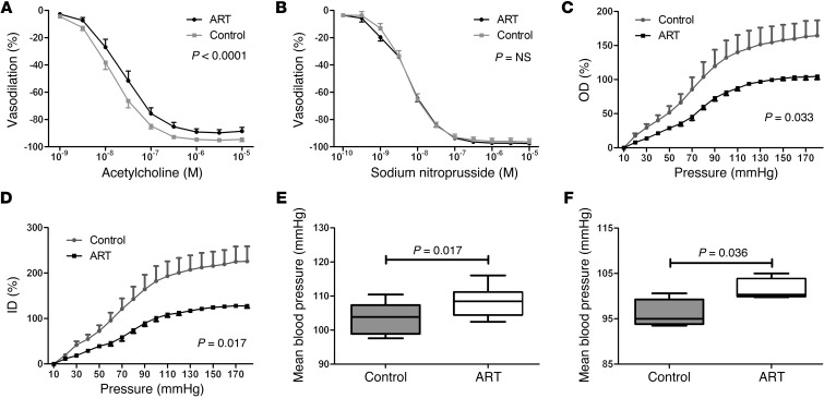
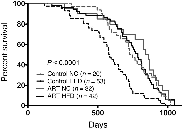
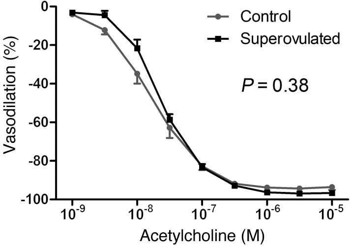
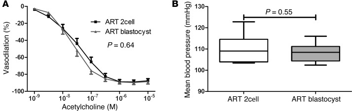
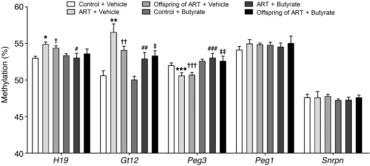
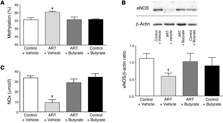
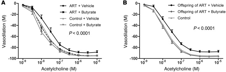
Children conceived by assisted reproductive technologies (ART) display a level of vascular dysfunction similar to that seen in children of mothers with preeclamspia. The long-term consequences of ART-associated vascular disorders are unknown and difficult to investigate in healthy children. Here, we found that vasculature from mice generated by ART display endothelial dysfunction and increased stiffness, which translated into arterial hypertension in vivo. Progeny of male ART mice also exhibited vascular dysfunction, suggesting underlying epigenetic modifications. ART mice had altered methylation at the promoter of the gene encoding eNOS in the aorta, which correlated with decreased vascular eNOS expression and NO synthesis. Administration of a deacetylase inhibitor to ART mice normalized vascular gene methylation and function and resulted in progeny without vascular dysfunction. The induction of ART-associated vascular and epigenetic alterations appeared to be related to the embryo environment; these alterations were possibly facilitated by the hormonally stimulated ovulation accompanying ART. Finally, ART mice challenged with a high-fat diet had roughly a 25% shorter life span compared with control animals. This study highlights the potential of ART to induce vascular dysfunction and shorten life span and suggests that epigenetic alterations contribute to these problems.


Figures

Figure 1
Figure 1. Mesenteric artery responsiveness, carotid artery pressure diameter relationship, and arterial blood pressure in mice generated by ART and control mice.
(A) Acetylcholine-induced and (B) sodium nitroprusside–induced mesenteric artery vasodilation in vitro (data represent mean ± SEM). (C and D) Carotid artery pressure diameter relationship in vitro. C shows outer diameter (OD) and D shows inner diameter (ID). (E) Mean arterial blood pressure in vivo obtained by short-term measurements and (F) 48-hour long-term measurements by telemetry. Horizontal lines represent the median; boxes, 25th to 75th percentiles; and T bars, 5th and 95th percentiles. Data are for at least 10 mice per group except for C and D (n = 5 per group) and F (n = 4 per group).
Figure 2
Figure 2. Kaplan-Meier survival curves of ART and control mice fed with normal chow (NC) or challenged with a HFD.
Median survival (days): Control NC = 853; control HFD = 787; ART normal chow = 736; ART HFD = 582.
Figure 3
Figure 3. Acetylcholine-induced mesenteric artery vasodilation in vitro in offspring of superovulated and control mice.
Data represent mean ± SEM for at least 7 mice per group.
Figure 4
Figure 4. Effects of embryo culture time on mesenteric artery responsiveness and blood pressure in ART mice.
Mesenteric artery responsiveness to acetylcholine (A) and arterial blood pressure (B) in ART-mice generated by implantation of 2 cell embryos and blastocysts. In A, data represent mean ± SE. In B, horizontal lines represent the median; boxes, 25th to 75th percentiles; and T bars, 5th and 95th percentiles. Data are for at least 9 mice per group.
Figure 5
Figure 5. Transmission of vascular dysfunction and arterial hypertension by mice generated by ART to their offspring.
Mesenteric artery responsiveness to acetylcholine in vitro (A) and arterial blood pressure in vivo (B) in mice generated by ART, offspring of mice generated by ART, and control mice. In A, data represent mean ± SEM. In B horizontal lines represent the median; boxes, 25th to 75th percentiles; and T bars, 5th and 95th percentiles. P values by 1-way ANOVA and Dunnett’s post hoc test. Data are for at least 10 mice per group.
Figure 6
Figure 6. Methylation of imprinted genes H19, Gtl2, Peg3, Peg1, and Snrpn in the aorta of control mice, mice generated by ART, and their progeny.
Effects of administration of the deacetylase inhibitor butyrate to control and ART mice on the methylation of these genes and on the transmission of the dysmethylation to the progeny. *P < 0.001, **P < 0.001, ***P = 0.02, ART + vehicle vs. control + vehicle; #P = 0.03, ##P = 0.03, ###P < 0.01, ART + butyrate vs. ART + vehicle; P < 0.01, ††P < 0.001, †††P = 0.014, offspring of ART + vehicle vs. control + vehicle; P = 0.04,‡‡P = 0.03, offspring of ART + butyrate vs. ART + vehicle. Data represent mean ± SEM for at least 7 mice per group.
Figure 7
Figure 7. Butyrate administration to ART mice normalizes DNA promoter methylation and expression of the eNOS gene in vascular tissue and NOx plasma concentration.
DNA methylation of the promoter of the eNOS gene in the aorta (A), eNOS expression in the carotid artery (B), and NOx plasma concentration (C), in control + vehicle, ART + vehicle, ART + butyrate, and control + butyrate mice. Data represent mean ± SEM for at least 13 mice per group. *P = 0.004, #P < 0.0001, P = 0.03 vs. control + vehicle by 1-way ANOVA and Dunnett’s post hoc test. In B, the lanes were run on the same gel but were noncontiguous.
Figure 8
Figure 8. Butyrate administration to ART mice normalizes vascular function and prevents transmission of dysfunction to offspring.
(A) Effects of butyrate administration to ART and to control mice on acetylcholine-induced mesenteric artery dilation. (B) Effects of butyrate or vehicle administration to ART mice before mating on acetylcholine-induced mesenteric artery dilation in the progeny. Data represent mean ± SEM for at least 10 mice per group.

References

    1. Barker DJ. Maternal and fetal origins of coronary heart disease. J R Coll Physicians Lond. 1994;28(6):544–551. - PMC - PubMed
    1. Barker DJ. In utero programming of cardiovascular disease. Theriogenology. 2000;53(2):555–574. doi: 10.1016/S0093-691X(99)00258-7. - DOI - PubMed
    1. Hakim TS, Mortola JP. Pulmonary vascular resistance in adult rats exposed to hypoxia in the neonatal period. Can J Physiol Pharmacol. 1990;68(3):419–424. doi: 10.1139/y90-059. - DOI - PubMed
    1. Sartori C, Allemann Y, Trueb L, Delabays A, Nicod P, Scherrer U. Augmented vasoreactivity in adult life associated with perinatal vascular insult. Lancet. 1999;353(9171):2205–2207. doi: 10.1016/S0140-6736(98)08352-4. - DOI - PubMed
    1. Jayet PY, et al. Pulmonary and systemic vascular dysfunction in young offspring of mothers with preeclampsia. Circulation. 2010;122(5):488–494. doi: 10.1161/CIRCULATIONAHA.110.941203. - DOI - PubMed

Publication types

MeSH terms