Comparative Study

ADCK4 mutations promote steroid-resistant nephrotic syndrome through CoQ10 biosynthesis disruption

Shazia Ashraf et al. J Clin Invest. 2013 Dec.

Abstract

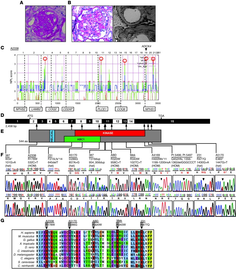
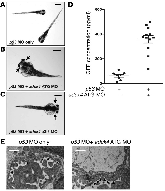
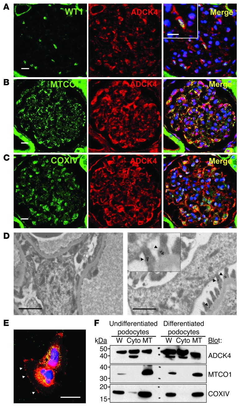
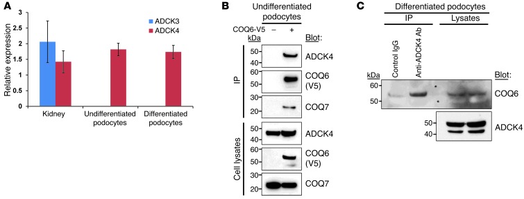
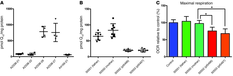
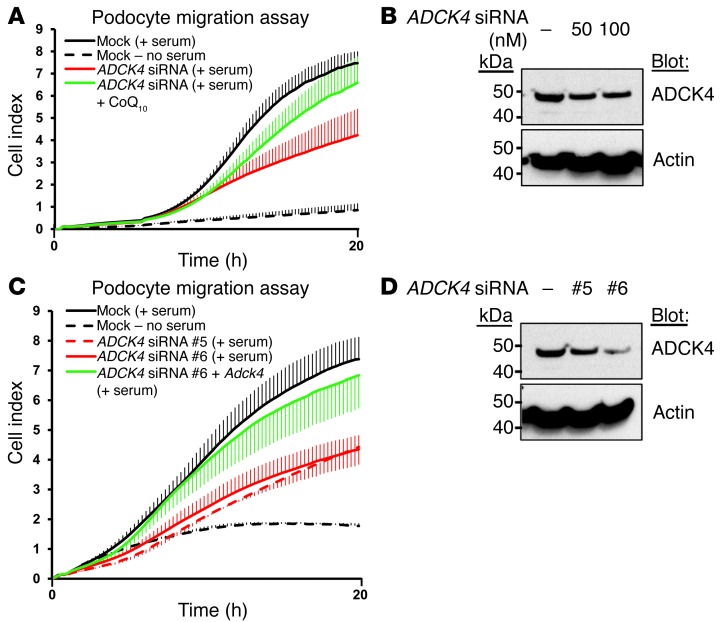
Identification of single-gene causes of steroid-resistant nephrotic syndrome (SRNS) has furthered the understanding of the pathogenesis of this disease. Here, using a combination of homozygosity mapping and whole human exome resequencing, we identified mutations in the aarF domain containing kinase 4 (ADCK4) gene in 15 individuals with SRNS from 8 unrelated families. ADCK4 was highly similar to ADCK3, which has been shown to participate in coenzyme Q10 (CoQ10) biosynthesis. Mutations in ADCK4 resulted in reduced CoQ10 levels and reduced mitochondrial respiratory enzyme activity in cells isolated from individuals with SRNS and transformed lymphoblasts. Knockdown of adck4 in zebrafish and Drosophila recapitulated nephrotic syndrome-associated phenotypes. Furthermore, ADCK4 was expressed in glomerular podocytes and partially localized to podocyte mitochondria and foot processes in rat kidneys and cultured human podocytes. In human podocytes, ADCK4 interacted with members of the CoQ10 biosynthesis pathway, including COQ6, which has been linked with SRNS and COQ7. Knockdown of ADCK4 in podocytes resulted in decreased migration, which was reversed by CoQ10 addition. Interestingly, a patient with SRNS with a homozygous ADCK4 frameshift mutation had partial remission following CoQ10 treatment. These data indicate that individuals with SRNS with mutations in ADCK4 or other genes that participate in CoQ10 biosynthesis may be treatable with CoQ10.


Figures

Figure 1
Figure 1. HM and exon capture resequencing reveal ADCK4 mutations as causes of SRNS.
(A) Renal histology of individual A2338-21 reveals global glomerulosclerosis by excess PAS staining (red). Original magnification, ×200. (B) Renal histology of individual Pt5496 shows cFSGS. PAS staining (left) reveals retraction and collapse of the capillary tuft, with numerous foam cells and groups of hyperplastic and vacuolated visceral epithelial cells. Original magnification, ×40. Electron microscopy image (right) shows foot process effacement (black arrowheads). In addition, capillary basement membranes are thickened and remodeled. Original magnification, ×15,000. (C) Nonparametric LOD (log of the odds ratio) (NPL) score profile across the human genome in 2 siblings with SRNS of consanguineous family A2338. Five maximum NPL peaks (red circles) indicate candidate regions of homozygosity by descent. ADCK4 is positioned (arrowhead) within a peak on chromosome (Chr) 19. Numbers at the bottom of the panels are measured in centimorgan (cM). (D) Exon structure of human ADCK4 cDNA. ADCK4 contains 15 exons. Positions of start codon (ATG) and of stop codon (TGA) are indicated. (E) Domain structure of ADCK4. The helical, ABC1, and kinase domains are depicted by colored bars in relation to encoding exon position. (F) Eleven different ADCK4 mutations in eight families with SRNS. Family numbers and amino acid changes (Table 1) are given above sequence traces. Arrowheads denote altered nucleotides. Lines and arrows indicate positions of mutations in relation to exon D and protein domain E. (G) For the 5 missense mutations (p.R178W, p.D286G, p.R320W, p.R343W, and p.R477Q) conservation across evolution of altered amino acid residues is shown.
Figure 2
Figure 2. Functional analysis of adck4 knockdown in zebrafish.
(A) Control zebrafish were injected with p53 MO (0.2 mM) as a control to minimize nonspecific MO effects. p53 MO was injected into fertilized eggs at the 1- to 4-cells stage and did not produce any phenotype up to 168 hpf (n >100). Scale bar: 0.5 mm. (B) Zebrafish coinjected with adck4 ATG MO (0.2 mM) targeting the translation initiation site of zebrafish adck4 and with p53 MO. At 120 hpf, adck4 morphants display the nephrosis phenotype of periorbital edema (arrows) and total body edema in 54.1% (193 out of 357) of embryos. (C) Zebrafish coinjected with adck4 e3i3 MO (0.2 mM) targeting the donor site of intron 3 of zebrafish adck4 and with p53 MO. At 120 hpf, adck4 morphants display the nephrosis phenotype (arrows) in 48.3% (274 out of 567) of embryos. Scale bar: 1 mm (B and C). (D) Proteinuria assay by ELISA against a fusion protein of vitamin D–binding protein and GFP in l-fabp::VDBP-GFP transgenic zebrafish. Note that knockdown of adck4 causes significant proteinuria compared with that in control fish injected with p53 MO only. (P < 0.001). Symbols indicate each measurements; horizontal bars indicate the average; data are presented with the average ± SEM denoted by 3 horizontal lines. (E) Electron microscopic ultrastructure of GBM and podocyte foot processes in p53 MO control and adck4 morphant zebrafish. In the control zebrafish, the foot processes are regularly spanned by slit diaphragms (black arrowheads). In contrast, the foot processes of the morphants are effaced and disorganized (black arrowheads). Scale bar: 2 μm.
Figure 3
Figure 3. ADCK4 localizes to the mitochondria and cytoplasm of podocytes in adult rat glomeruli and also in cultured human podocytes.
(AC) Coimmunofluorescence of ADCK4 with (A) WT1 as well as (B) MTCO1 and (C) COXIV in adult rat glomeruli. ADCK4 partially colocalizes to mitochondria with the 2 mitochondrial markers COXIV and MTCO1. Scale bar: 10 μm. (D) Immunogold electron microscopy of adult rat kidney displays localization of ADCK4 (black arrowheads) at podocyte foot processes of glomeruli. A control without a primary antibody is shown on the left. Scale bar: 1 μm; ×2.5 (inset). (E) Podocytes were transfected with ADCK4-RFP and stained with an anti-COXIV antibody. ADCK4 partially colocalizes to mitochondria with COXIV. Note that ADCK4 also localizes along the plasma membrane (white arrowheads) in podocytes. Scale bar: 25 μm. (F) Subcellular fractionation of ADCK4 in undifferentiated and differentiated podocytes. Mitochondrial and cytosol fractions were prepared and immunoblotted for ADCK4, MTCO1, and COXIV, respectively. Each lane was loaded with 50 μg protein. Note that ADCK4 is present in both mitochondrial (marked by MTCO1 and COXIV), and cytosolic fractions in both undifferentiated and differentiated podocytes. W, whole cell lysates; Cyto, cytosol fraction; MT, mitochondrial fraction.
Figure 4
Figure 4. ADCK4 is enriched in podocytes and interacts with COQ6 and COQ7.
(A) Relative expression of ADCK3 and ADCK4 in kidney tissue and podocytes, as measured by quantitative real-time PCR. Error bars indicate SD of 4 experiments. (B) Interaction of ADCK4 with COQ6 and COQ7 in cultured human podocytes. A C-terminally V5-tagged COQ6 construct (COQ6-V6) was transfected into the undifferentiated cultured podocytes. Coimmunoprecipitation was performed using an anti-V5 antibody and then blotting was performed with antibodies for ADCK4, COQ6, and COQ7. Note that the protein complex precipitated by COQ6 includes COQ7 and ADCK4. (C) Interaction of endogenous ADCK4 and COQ6 in differentiated podocytes. Podocyte lysates were precipitated with an anti-ADCK4 antibody or a control rabbit IgG. The precipitated proteins were separated by SDS-PAGE and blotted with an anti-COQ6 antibody.
Figure 5
Figure 5. CoQ10 content in EBV-transformed lymphoblasts and fibroblasts from SRNS families with mutations in ADCK4.
(A) Scatter plot showing the total CoQ10 content in EBV-transformed lymphoblasts derived from either healthy or affected individuals from the A2338 and A4169 families. Individuals A2338-21, A2338-22, and A4169-21 are affected with SRNS, while A2338-26 and A2338-27 have both wild-type alleles. The content of CoQ10 is presented with the average ± SD denoted by 3 horizontal lines (n = 4; except n = 6 for A4169-21). The content from A2338-26 is significantly higher than the CoQ10 content from A2338-21, A2338-22, and A4169-21 (multiple comparison, P < 0.0001). Symbols indicate individual lymphoblasts; horizontal bars indicate the average. (B) Scatter plot showing the total CoQ10 content in fibroblasts derived from either healthy parents or affected individuals (Pt5496 and Pt5497). The content of CoQ10 is presented with the average ± SD denoted by 3 horizontal lines (n = 8). CoQ10 content from #50551 is statistically lower than CoQ10 content from #50550 (multiple comparison, P = 0.0134) and significantly higher than the CoQ10 contents from #50552 and #50553 (multiple comparison, P < 0.0001). Symbols indicate individual fibroblasts; horizontal bars indicate the average. (C) In vivo maximal uncoupled OCR over the whole respiratory chain, including the Q10 transfer of electrons from mitochondrial complex I and complex II to complex III. Maximal respiration was significantly reduced in fibroblasts from Pt5496 and Pt5497 compared with that in control fibroblasts. Data shown are mean ± SD (n > 10). *P < 0.001.
Figure 6
Figure 6. Knockdown of ADCK4 decreases the migratory phenotype of podocytes.
(A) The effect of ADCK4 knockdown on podocyte migration. Podocytes transfected with ADCK4 siRNA (red) exhibited decreased migration compared with podocytes transfected with scrambled siRNA (black). The decrease in podocyte migration was partially reversed by the addition of 50 μM CoQ10 (green). Error bars are shown in only one direction for clarity and indicate SDs from 3 independent experiments. (B) The efficiency of ADCK4 siRNA used in A was confirmed by immunoblotting with an anti-ADCK4 antibody and an anti–β-actin antibody. (C) Transfection of podocytes with 2 additional ADCK4-specific siRNAs (siRNA #5 and #6) confirmed the result from A that ADCK4 knockdown in human podocytes reduces migration (red dotted and solid lines) compared with podocytes transfected with the scrambled siRNA (black). The decrease in podocyte migration by ADCK4 knockdown using ADCK4 siRNA #6 was rescued by transfection of mouse Adck4 construct (green). Mouse ADCK4 has 5 mismatches from the siRNA target sequences. Error bars indicate SDs of 3 independent experiments. (D) The efficiency of ADCK4 siRNAs used in C was confirmed by immunoblotting. 100 nM of each siRNA was transfected into podocytes.

Comment in

  • ADCK4 "reenergizes" nephrotic syndrome.
    Malaga-Dieguez L, Susztak K. Malaga-Dieguez L, et al. J Clin Invest. 2013 Dec;123(12):4996-9. doi: 10.1172/JCI73168. Epub 2013 Nov 25. J Clin Invest. 2013. PMID: 24270414 Free PMC article.

References

    1. Antignac C. Genetic models: clues for understanding the pathogenesis of idiopathic nephrotic syndrome. J Clin Invest. 2002;109(4):447–449. doi: 10.1172/JCI15094. - DOI - PMC - PubMed
    1. Hinkes BG, et al. Nephrotic syndrome in the first year of life: two thirds of cases are caused by mutations in 4 genes (NPHS1, NPHS2, WT1, and LAMB2). Pediatrics. 2007;119(4):e907–e919. doi: 10.1542/peds.2006-2164. - DOI - PubMed
    1. Kim MS, Stablein D, Harmon WE. Renal transplantation in children with congenital nephrotic syndrome: a report of the North American Pediatric Renal Transplant Cooperative Study (NAPRTCS). Pediatr Transplant. 1998;2(4):305–308. - PubMed
    1. Benoit G, Machuca E, Heidet L, Antignac C. Hereditary kidney diseases: highlighting the importance of classical Mendelian phenotypes. Ann N Y Acad Sci. 2010;1214:83–98. doi: 10.1111/j.1749-6632.2010.05817.x. - DOI - PubMed
    1. Hildebrandt F. Genetic kidney diseases. Lancet. 2010;375(9722):1287–1295. doi: 10.1016/S0140-6736(10)60236-X. - DOI - PMC - PubMed

Publication types

MeSH terms