. 2013 Dec;123(12):5071-81.
doi: 10.1172/JCI69458. Epub 2013 Nov 25.

von Willebrand factor mutation promotes thrombocytopathy by inhibiting integrin αIIbβ3

von Willebrand factor mutation promotes thrombocytopathy by inhibiting integrin αIIbβ3

Caterina Casari et al. J Clin Invest. 2013 Dec.

Abstract

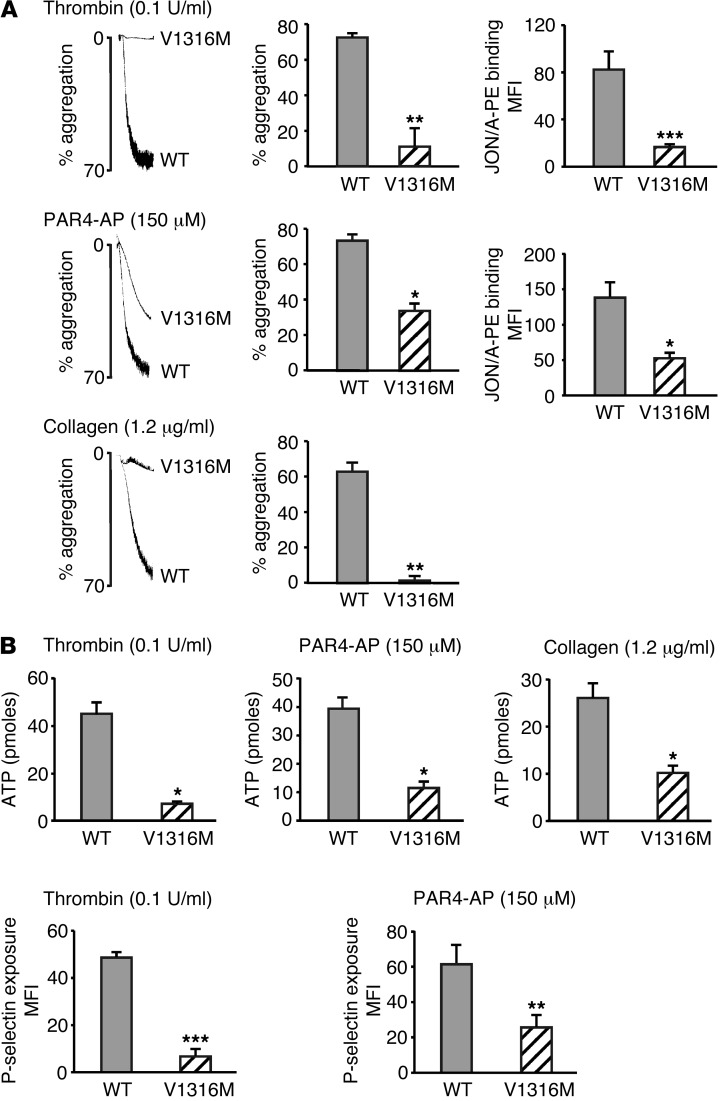
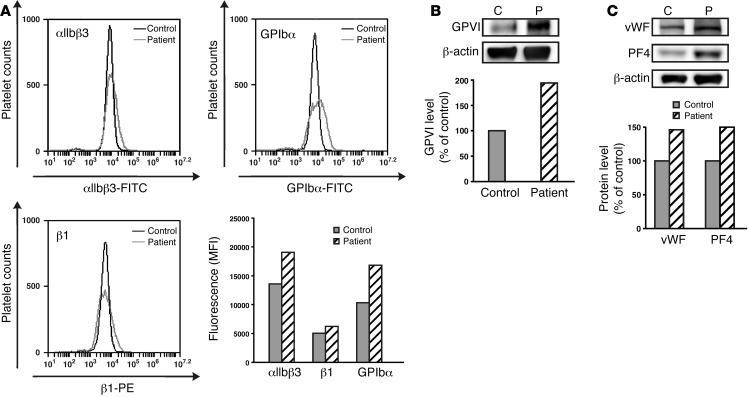
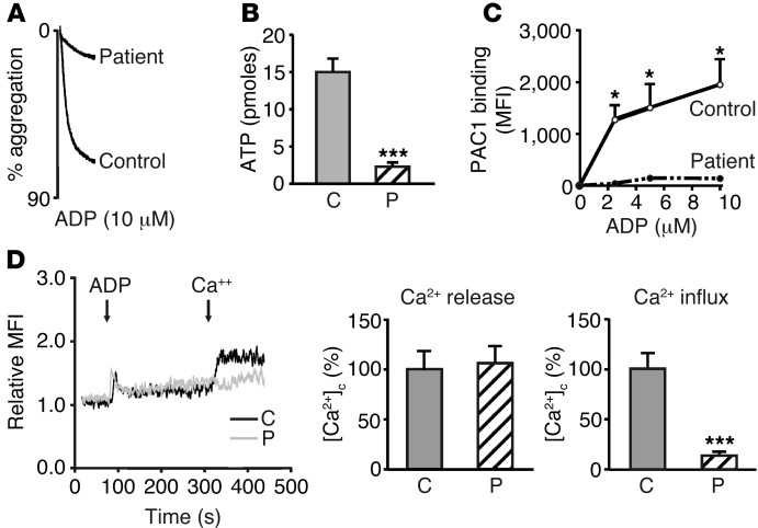
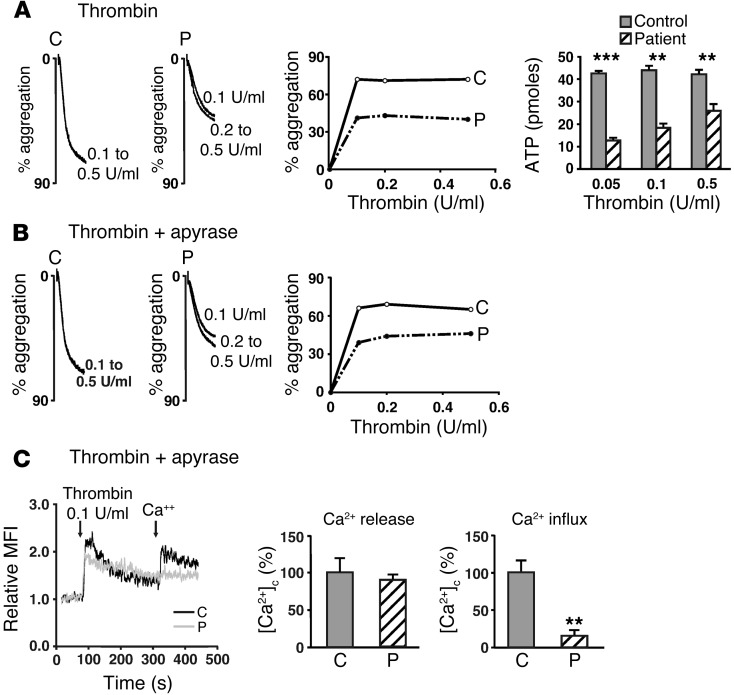
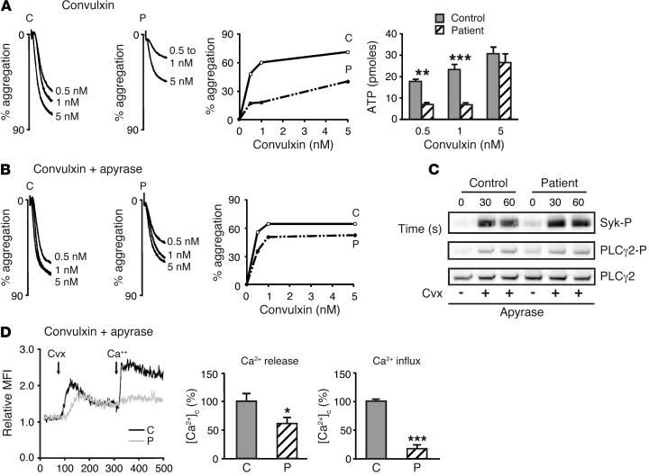
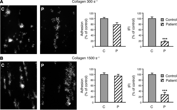
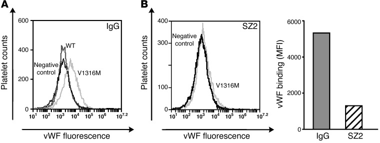
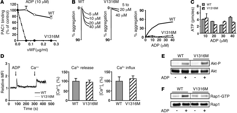
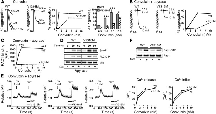
von Willebrand disease type 2B (vWD-type 2B) is characterized by gain-of-function mutations in von Willebrand factor (vWF) that enhance its binding to the glycoprotein Ib-IX-V complex on platelets. Patients with vWD-type 2B have a bleeding tendency that is linked to loss of vWF multimers and/or thrombocytopenia. In this study, we uncovered evidence that platelet dysfunction is a third possible mechanism for bleeding tendency. We found that platelet aggregation, secretion, and spreading were diminished due to inhibition of integrin αIIbβ3 in platelets from mice expressing a vWD-type 2B-associated vWF (vWF/p.V1316M), platelets from a patient with the same mutation, and control platelets pretreated with recombinant vWF/p.V1316M. Impaired platelet function coincided with reduced thrombus growth. Further, αIIbβ3 activation and activation of the small GTPase Rap1 were impaired by vWF/p.V1316M following exposure to platelet agonists (thrombin, ADP, or convulxin). Conversely, thrombin- or ADP-induced Ca2+ store release, which is required for αIIbβ3 activation, was normal, indicating that vWF/p.V1316M acts downstream of Ca2+ release and upstream of Rap1. We found normal Syk phosphorylation and PLCγ2 activation following collagen receptor signaling, further implying that vWF/p.V1316M acts directly on or downstream of Ca2+ release. These data indicate that the vWD-type 2B mutation p.V1316M is associated with severe thrombocytopathy, which likely contributes to the bleeding tendency in vWD-type 2B.


Figures

Figure 1
Figure 1. Platelet aggregation, secretion, and αIIbβ3 integrin activation in mvWF/p.V1316M.
(A) Aggregation and integrin αIIbβ3 activation of washed platelets were initiated by adding thrombin (0.1 U/ml), PAR4-AP (150 μM), and collagen (1.2 μg/ml). Aggregation was expressed as the percentage change in light transmission, with the value for the blank (buffer without platelet) set at 100%. Traces are representative of 3 independent experiments. Integrin αIIbβ3 activation was assessed by flow cytometry. Platelets were incubated with a phycoerythrin-labeled rat anti-mouse integrin αIIbβ3 mAb (JON/A) specific for the activated conformation of the mouse integrin. Flow cytometry was performed without stirring. The level of activated integrin is indicated by the MFI ± SEM from at least 3 experiments. (B) Dense and α granule secretion were assessed by measuring ATP release and P-selectin exposure, respectively. ATP was quantified after platelet aggregation induced by various agonists (thrombin, PAR4-AP, collagen) and expressed as the amount of ATP released (pmoles). P-selectin exposure was evaluated by flow cytometry using FITC-labeled rat anti-mouse P-selectin mAb. The level of P-selectin exposed at the surface is expressed as MFI ± SEM from at least 3 experiments. *P < 0.05; **P < 0.01; ***P < 0.001 (paired Student’s t test).
Figure 2
Figure 2. Analysis of adhesive receptors and granule contents of platelets from a patient with vWD-type 2B exhibiting the vWF/p.V1316M mutation.
(A and B) The expression of adhesive receptors was analyzed by flow cytometry or immunoblotting. (A) Control and patient platelet suspensions were incubated with antibodies directed against αIIbβ3 (HIP8) and α2β1 (MAR4) integrins or against GPIbα (HIP1). Antibody binding was assessed by flow cytometry. (B) GPVI expression was quantified by immunoblotting. (C) α Granule content was assessed by immunoblotting with an anti-vWF and an anti-PF4. Results are representative of 2 independent experiments.
Figure 3
Figure 3. ADP-induced activation of platelets from the patient with vWD-type 2B.
Control and patient platelet suspensions were stimulated with various concentrations of ADP. (A) Aggregation of washed platelets was initiated by adding 10 μM ADP. (B) Dense granule secretion was assessed by measuring the amount of ATP release (pmoles). C, control donor; P, patient. (C) Integrin αIIbβ3 activation was assessed by flow cytometry using integrin αIIbβ3 mAb (PAC1) specific for the activated conformation of the human integrin. The level of activated integrin is indicated by MFI. (D) Ca2+ signaling induced by 10 μM ADP was monitored by flow cytometry using the Oregon Green 488 BAPTA1-AM. Histograms represent the area under the curve of both Ca2+ store release and Ca2+ influx. Data from 1 experiment carried out in triplicate are presented as mean ± SEM. *P < 0.05; ***P < 0.001 (unpaired Student’s t test). Results are representative of 2 independent experiments.
Figure 4
Figure 4. Thrombin-induced activation of platelets from the patient with vWD-type 2B.
Washed platelets from a control donor and the patient with vWF/p.V1316M mutation were stimulated with thrombin in the (A) absence or (B and C) presence of apyrase (2 U/ml). (A) Aggregation and secretion of washed platelets were initiated by adding various concentrations of thrombin (0.1–0.5 U/ml). Dense granule secretion was assessed by measuring the amount of ATP release (pmoles). (C) Thrombin-induced Ca2+ signaling was monitored by flow cytometry using Oregon Green 488 BAPTA1-AM. Histograms represent the area under the curve of both the Ca2+ store release and Ca2+ influx. Data from 1 experiment carried out in triplicate are presented as mean ± SEM. **P < 0. 01; ***P < 0.001 (unpaired Student’s t test). Results are representative of 2 independent experiments.
Figure 5
Figure 5. Cvx-induced activation of platelets from the patient with vWD-type 2B.
Washed platelets from a control donor and patient with vWF/p.V1316M mutation were stimulated with Cvx in the (A) absence or (BD) presence of apyrase (2 U/ml). Aggregation and secretion of washed platelets were initiated by adding various concentrations of Cvx (0.5–5 nM). Dense granule secretion was assessed by measuring the amount of ATP release (pmoles). (C) Washed platelets in suspension were activated by 5 nM Cvx for 30 and 60 seconds in the absence of stirring, and then tyrosine phosphorylation of Syk (Syk-P) and PLCγ2 (PLCγ2-P) was assessed by immunoblotting with an anti–Syk-P and anti–PLCγ2-P, respectively. (D) Ca2+ signaling induced by 5 nM Cvx was monitored by flow cytometry using the Oregon Green 488 BAPTA1-AM. Histograms represent the area under the curve of both the Ca2+ store release and Ca2+ influx. Data from 1 experiment carried out in triplicate are presented as mean ± SEM. *P = 0.03; **P < 0. 01; ***P < 0.001 (unpaired Student’s t test). Results are representative of 2 independent experiments.
Figure 6
Figure 6. Thrombus formation in the patient with vWD-type 2B.
Whole blood was perfused at (A) 300 s–1 or (B) 1,500 s–1 in glass microcapillary tubes coated with type I collagen (100 μg/ml). After 5 minutes (300 s–1) and 2 minutes (1,500 s–1), platelet thrombi were observed under an epifluorescence microscope (original magnification, ×20). Total area covered by platelets and total integrated fluorescence intensity (IFI) from 1 experiment carried out in triplicate are presented as the mean ± SEM. ***P < 0.001 (unpaired Student’s t test). Results are representative of 2 independent experiments.
Figure 7
Figure 7. Binding of recombinant hvWF/p.V1316M to human control platelets.
Binding of hvWF/p.V1316M or WT was monitored by flow cytometry using vWF polyclonal antibody. Human control platelet suspensions were pretreated for 5 minutes with (A) mouse IgG (10 μg/ml) or (B) GPIbα mAb (SZ2; 10 μg/ml) before addition of recombinant hvWF/p.V1316M or WT for 30 minutes. Results are representative of 3 independent experiments.
Figure 8
Figure 8. ADP-induced activation of human control platelets pretreated with recombinant hvWF/p.V1316M.
Washed human control platelets pretreated for 10 minutes with recombinant hvWF/p.V1316M or WT were stimulated with ADP. (A) Integrin αIIbβ3 activation was assessed by flow cytometry using integrin αIIbβ3 mAb (PAC1) specific for the activated conformation of the human integrin. Data are mean ± SEM of 3 independent experiments carried out in triplicate. (B) Aggregation of washed platelets was initiated by adding various concentrations of ADP. (C) Dense granule secretion was assessed by measuring the amount of ATP release (pmoles). Data are mean ± SEM of 3 independent experiments carried out in triplicate. (D) Ca2+ signaling induced by 10 μM ADP was monitored by flow cytometry using the Oregon Green 488 BAPTA1-AM. Histograms represent the area under the curve of both Ca2+ store release and Ca2+ influx. Data from 1 experiment carried out in triplicate are presented as mean ± SEM. Results are representative of 2 independent experiments. (E) Akt-P after 5 minutes of stimulation with ADP (10 μM) in the absence of stirring was assessed by immunoblotting with anti–Akt-P. Total Akt and Akt-P blots were obtained by running equal amounts of sample on a parallel gel; the lanes of the Akt-P blot were derived from the same gel but were noncontiguous, as indicated by the black line. (F) Rap1 activity was measured by pull-down assay after 30 seconds of stimulation with ADP (10 μM) in the absence of stirring. The lanes of the blots were derived from the same gel but were noncontiguous, as indicated by the black lines. Results are representative of 3 independent experiments.
Figure 9
Figure 9. Cvx-induced activation of human control platelets pretreated with recombinant hvWF/p.V1316M.
Washed human control platelets pretreated for 10 minutes with recombinant hvWF/p.V1316M or WT were stimulated with Cvx in the (A) absence or (BE) presence of apyrase (2 U/ml) or (F) in the presence of antagonists of ADP receptors (AR-C69931MX, 10 μM; MRS 2179, 10 μM). (A and B) Aggregation and secretion of washed platelets were initiated by adding concentrations of thrombin (0.02–0.5 U/ml). Dense granule secretion was assessed by measuring the amount of ATP release. (C) Integrin αIIbβ3 activation induced by Cvx (1–10 nM) was assessed by flow cytometry using the integrin αIIbβ3 mAb PAC1 specific for the activated conformation of the human integrin. The level of activated integrin is indicated by MFI. (D) Washed platelets in suspension were activated by 4 nM Cvx for 30 and 60 seconds in the absence of stirring, and tyrosine Syk-P and PLCγ2-P was assessed by immunoblotting with anti–Syk-P and anti–PLCγ2-P, respectively. (E) Ca2+ signaling was monitored using Oregon Green 488 BAPTA1-AM. Curves represent the area under the curve of Ca2+ store release and Ca2+ influx. (F) Rap1 activity was measured by pull-down assay after 30 seconds of stimulation with Cvx (4 nM) in the absence of stirring and identified by immunoblotting with anti-Rap1 antibody. The presence of comparable amounts of Rap1 in aliquots of the all platelet samples is shown in a parallel gel; lanes of the Rap1-GTP blot were derived from the same gel but were noncontiguous, as indicated by the black line. Data from 1 experiment carried out in triplicate are presented as mean ± SEM. *P < 0. 05, ***P < 0.001 (unpaired Student’s t test). Results are representative of 3 independent experiments.

Comment in

References

    1. Ruggeri ZM. Von Willebrand factor: looking back and looking forward. Thromb Haemost. 2007;98(1):55–62. - PubMed
    1. Sadler JE. Von Willebrand factor, ADAMTS13, and thrombotic thrombocytopenic purpura. Blood. 2008;112(1):11–18. doi: 10.1182/blood-2008-02-078170. - DOI - PMC - PubMed
    1. Ginsburg D, Bowie EJ. Molecular genetics of von Willebrand disease. Blood. 1992;79(10):2507–2519. - PubMed
    1. Ginsburg D, Sadler JE. von Willebrand disease: a database of point mutations, insertions, and deletions. For the Consortium on von Willebrand Factor Mutations and Polymorphisms, and the Subcommittee on von Willebrand Factor of the Scientific and Standardization Committee of the International Society on Thrombosis and Haemostasis. Thromb Haemost. 1993;69(2):177–184. - PubMed
    1. Federici AB, et al. Clinical and molecular predictors of thrombocytopenia and risk of bleeding in patients with von Willebrand disease type 2B: a cohort study of 67 patients. Blood. 2009;113(3):526–534. doi: 10.1182/blood-2008-04-152280. - DOI - PubMed

Publication types

MeSH terms